PD catheter placement techniques Professor Paul Daher Chief


 • Percutaneous Needle-Guidewire Technique • Open Surgical Dissection •")


 • Open technique: -safe (low risk")

- Slides: 27
PD catheter placement techniques Professor Paul Daher Chief of department of pediatric surgery Faculty of Medicine-St joseph University IPNA-ISN 2019
Introduction • PD is a renal-replacement therapy. Essentials elements: -viable peritoneal cavity lined by a functional membrane -attained with a catheter device through which dialysis solutions can be exchanged -Less infection and mechanical complications (tissue attachment, tip migration, and pericatheter leaks) Best method of PD catheter placement and our experience about 30 cases
CATHETER TYPES
• The material of the catheter is either silicone rubber or polyurethane • No significant difference in functionality has been shown convincingly between straight and coiled-tip catheters with or without a preformed arc bend. Akyol AM, Porteous C, Brown MW. A comparison of two types of catheters for continuous ambulatory peritoneal dialysis (CAPD). Perit Dial Int. 1990; 10: 63 -6. Ouyang CJ, Huang FX, Yang QQ, Jiang ZP, Chen W, Qiu Y, et al. Comparing the incidence of catheter-related complications with straight and coiled Tenckhoff catheters in peritoneal dialysis patients—a single-center prospective randomized trial. Perit Dial Int. 2015; 35: 443 -9.
• Advantages of Coiled-tip catheters: provide for better dispersion of the dialysate during inflow remains speculative. Shape of curled hold visceral peritoneal surface away from side holes Better patency and outflow Does not impinge on rectum no rectal discomfort Catheters equipped with two cuffs provide better immobilization of the tubing within the abdominal wall Most utilized: double cuff with coiled tip.
CHOOSING PD CATHETER SIZE
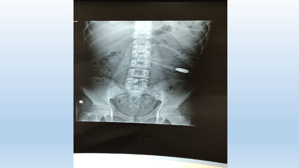
PD CATHETER INSERTION • The deep cuff is best implanted between the posterior sheath and fascia transversalis • The superficial cuff is positioned in the subcutaneous tissues 2 to 4 cm from the exit site to prevent debris and bacteria • Upwardly directed exit sites should be avoided to prevent pooling of cutaneous bacteria and debris, perspiration, and shower water in the exit sinus, predisposing the patient to exit site and tunnel infections Peritoneum
PD CATHETER INSERTION PROCEDURES (3) • Percutaneous Needle-Guidewire Technique • Open Surgical Dissection • Surgical Laparoscopy
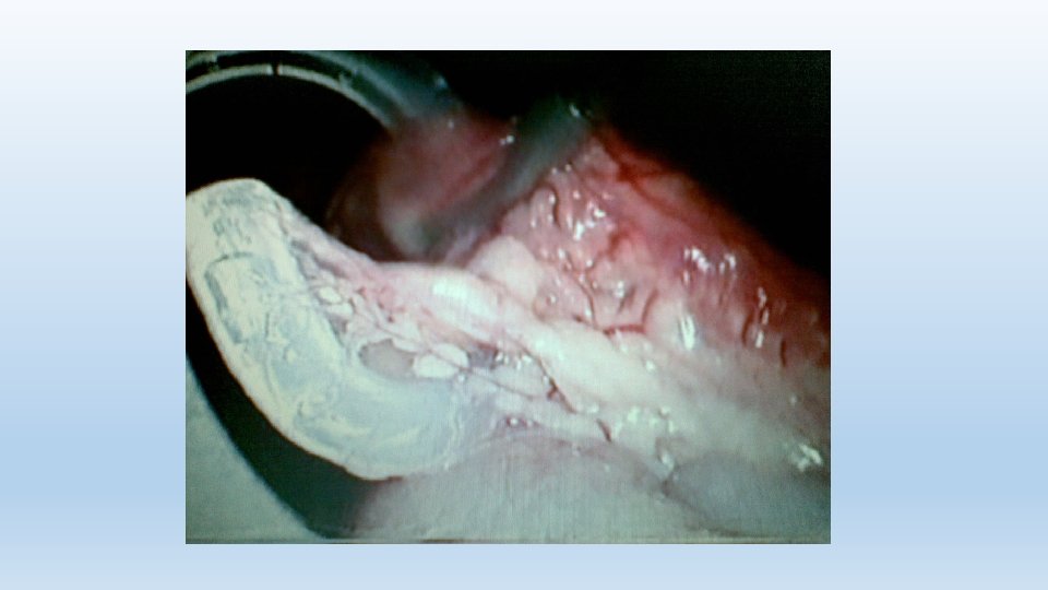
Open Surgical Dissection • Omentectomy is performed systematically • The peritoneum and posterior rectus sheath are closed with the internal cuff secured in between • The catheter then is directed to the desired skin exit site placed subfascia.
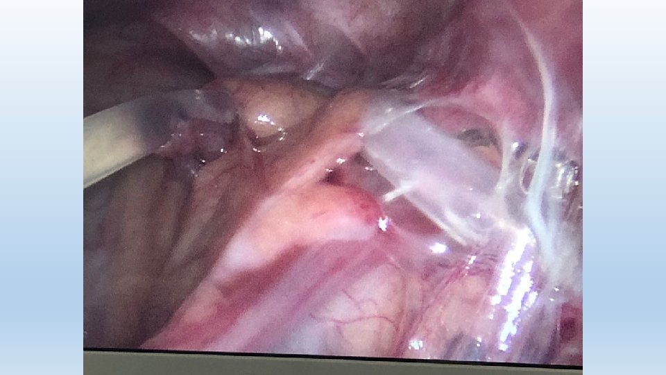
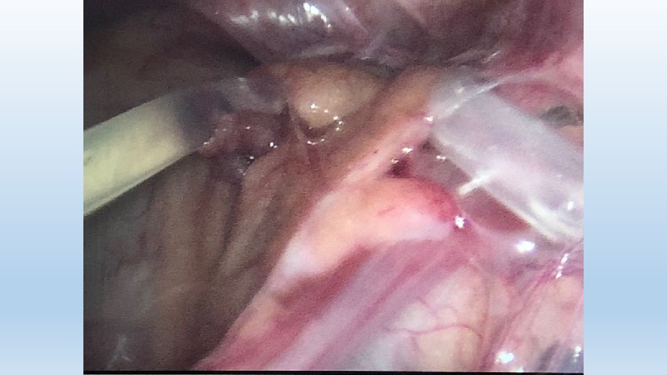
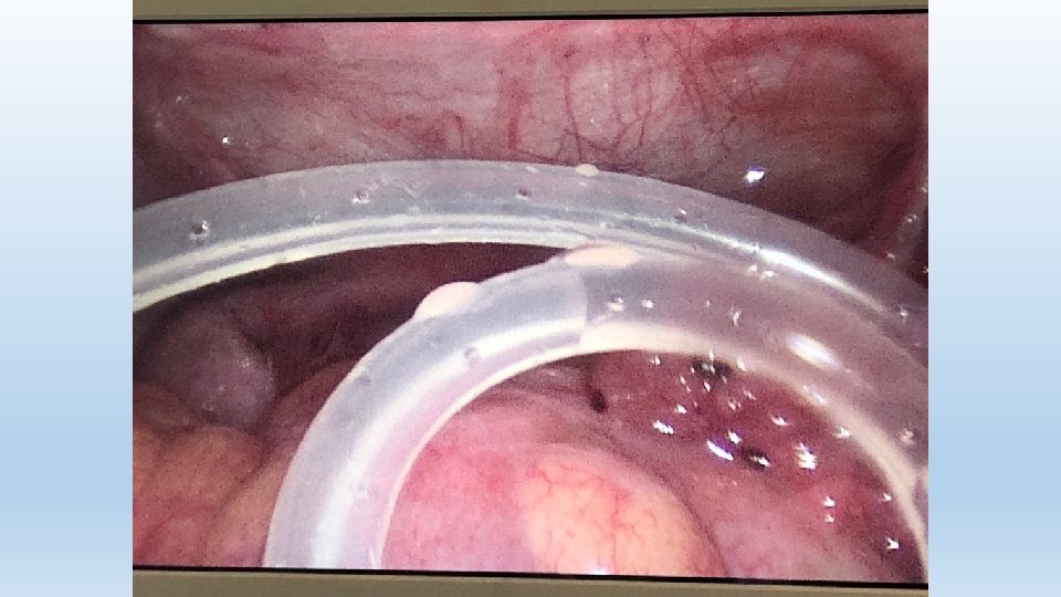
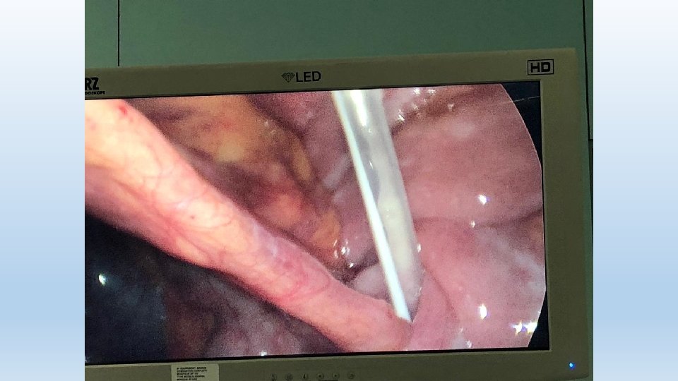
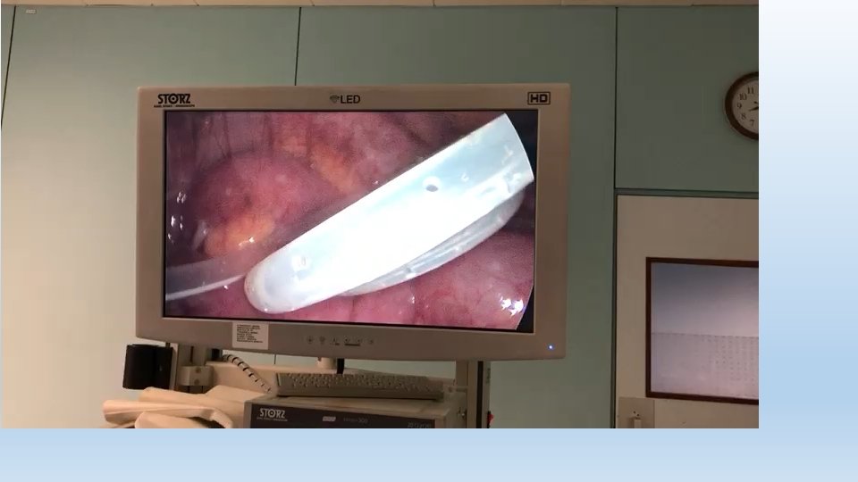
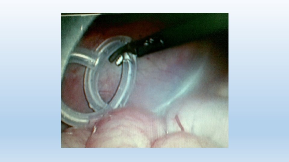
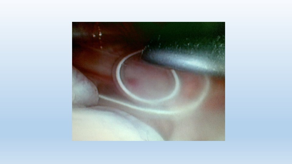
Surgical Laparoscopy • minimally invasive approach with complete visualization of the peritoneal cavity during the catheter implantation procedure. • Umbilical port for introducing the PD catheter and for the Camera • A right paramedian port is then introduced for manipulation • The catheter end is retrieved via a left introduced grasping forceps • The internal cuff is left in between the muscle and the peritoneum • The external cuff is then retrieved in the same manner of the open technic • The catheter tip is surely well placed in the pelvis and the mechanical outflow is checked by direct vision.
Open vs Laparoscopic
Open vs Laparoscopic
Open vs Laparoscopic
Open vs Laparoscopic • It has been stated that no single implantation approach has been shown to produce superior outcomes
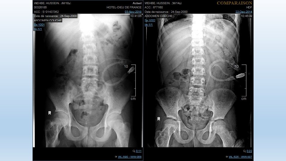
Mechanical Flow Dysfunction secondary to: • Inside the peritoneum, bowel trapping , omental wrapping, and ensnaring by adhesions, or uterine tubes can result in outflow failure, secondary to catheter infection (drainage volume being significantly less than the inflow volume). • Within the catheter lumen, blood clots or fibrin can lead to slow inflow and outflow rates. • Migration of the catheter : Excessive bending, poor immobilization and fixation of the transmural segment
Based on our experience ( Take Home Message) • Open technique: -safe (low risk of injury to viscera) -Catheter placed using guide wire in douglas with or without fluoroscopy -Ommentectomy is manadatory and is best done by open surgery to prevent complications -Less expensive • the advantage of laparoscopy is principally reserved for recurrence procedures due to mechanical flow dysfunction • In both techniques it’s advisable to keep some fluid in the peritoneal cavity.