Pattern recognition DR KIRAN VEERA ED PHYSICIAN AND


RBBB")


- Slides: 28
Pattern recognition DR KIRAN VEERA ED PHYSICIAN AND CO-DEMT BENDIGO HEALTH
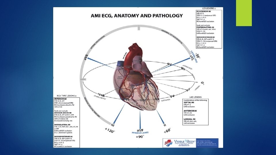
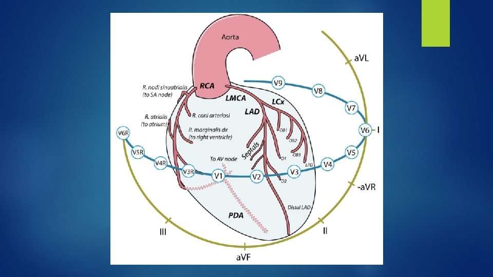
Rate Rhythm Axis Intervals Chamber enlargements Ischaemia
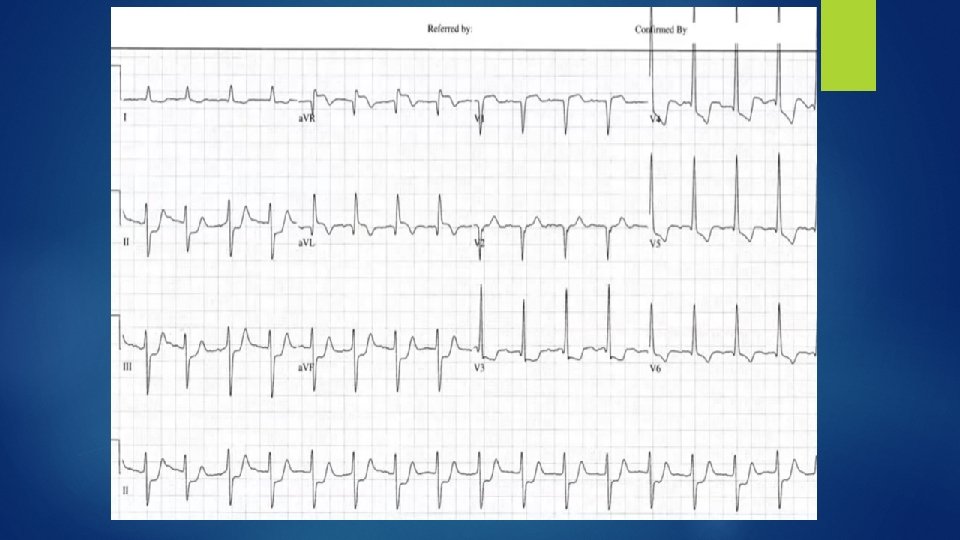
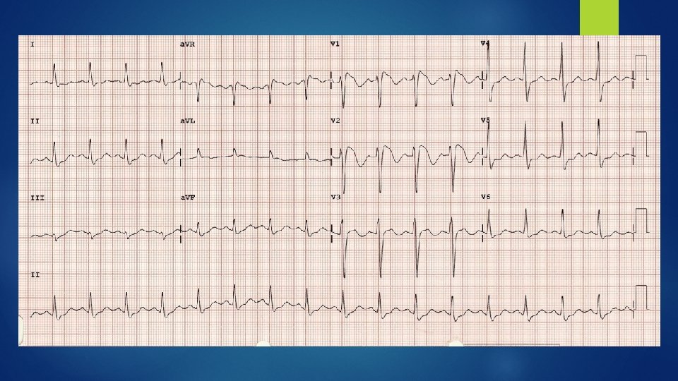
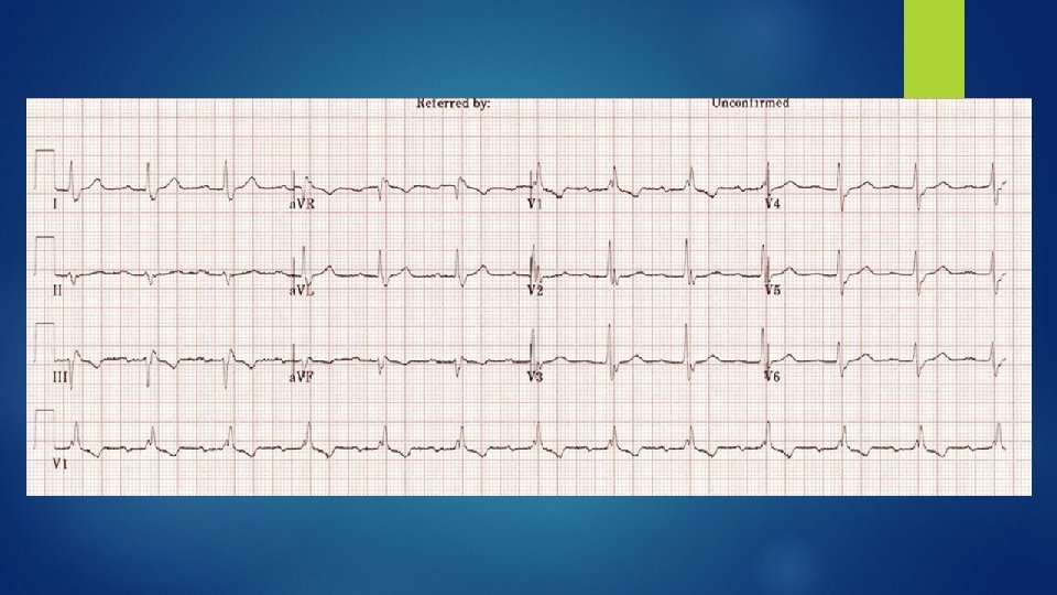
75 yo male with HTN, DM-2 complains of angina What does this ECG show?
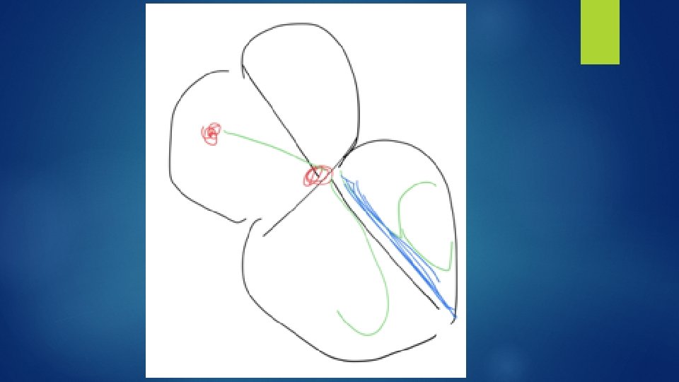
LMCA Obstruction
LMCA Obstruction Widespread horizontal ST depression ST elevation in a. VR ≥ 1 mm ST elevation in a. VR ≥ V 1
Also seen in Prox LAD obstruction Severe Triple vessel disease Diffuse subendocardial ischaemia PE, LVH with strain, LBBB(including PPM), SVTs, hypok+, aortic dissection, Na ch pathology (TCA, Brugada etc), severe anaemia
In the presence of anginal symptoms, STE in a. VR + STE in V 1 - Highly predictive of LMCA or Prox LAD obstruction STE in a. VR > STE in V 1 - almost always indicates a LMCA obstruction (81% sensitive and 80% specific)
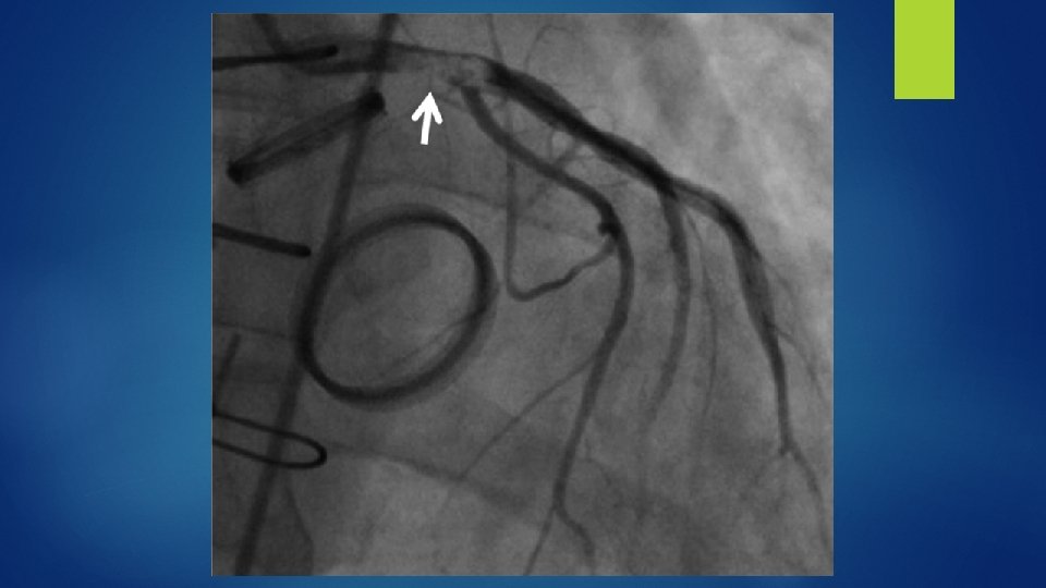
Patient had a severe ostial LAD thrombus that was close to the left main.
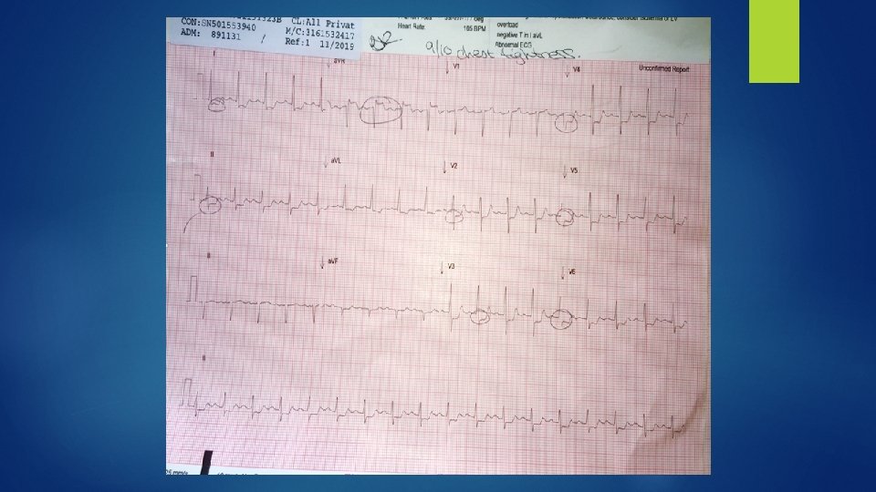
Another classic example of the LMCA / 3 VD ECG pattern
I would treat a patient with LMCA obstruction with all the following except: Aspirin Clopidogrel Heparin Early Cath lab
40 yo female with anxiety, palpitations and pseudoseizures
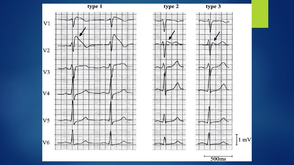
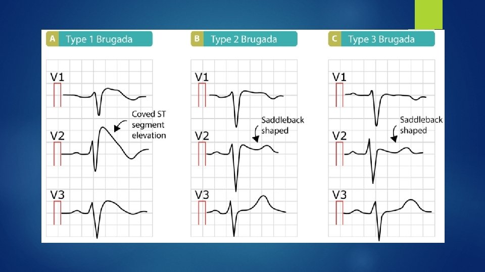
Brugada Syndrome
• RBBB-like pattern with secondary R’ wave following the QRS complex. • ST elevation at the J point > 2 mm with a “coved” • T wave inversion
Diagnosis ECG plus one of the following: Documented VF or VT Family history of SCD at <45 years old Coved-type ECGs in family members Syncope Nocturnal agonal respiration Only proven therapy is ICD
Take Home points Consider Brugada syndrome in any patient presenting after syncope ECG: (I)RBBB + STE in V 1 - V 2 Coved STE is most concerning Discuss/ refer to electrophysiologist
50 yo male with syncope
Trifascicular block
But AVN is not a fascicle - why is it a trifascicular block?