
Patient Vital Signs and Medical Emergencies Orientation Fall
- Slides: 42
Patient Vital Signs and Medical Emergencies Orientation Fall 2011
Homeostasis • A constancy in the internal environment of the body • Naturally maintained by adaptive responses that promote healthy survival • Primary mechanisms: – – – Heartbeat Blood pressure Body temperature Respiratory rate Electrolyte balance
Vital Signs • • • Body Temperature Respiratory Rate Pulse / Heart Rate Blood Pressure Sensorium (mental alertness)
Importance of Vital Signs • Indicates the patient’s immediate condition • Can show improvement due to treatment • Can show a decline in condition
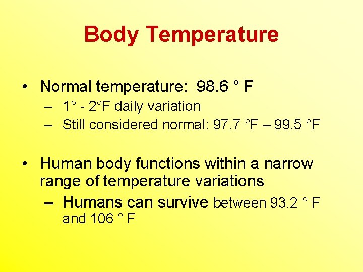
Body Temperature • Normal temperature: 98. 6 ° F – 1° - 2°F daily variation – Still considered normal: 97. 7 °F – 99. 5 °F • Human body functions within a narrow range of temperature variations – Humans can survive between 93. 2 ° F and 106 ° F
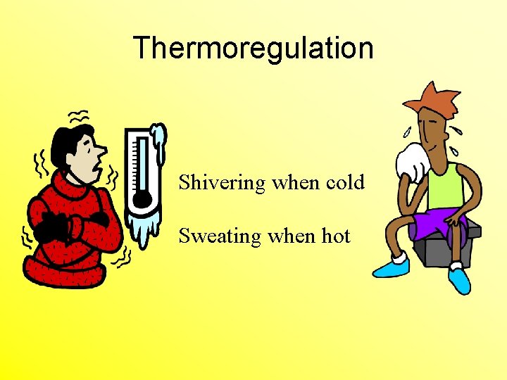
Thermoregulation Shivering when cold Sweating when hot
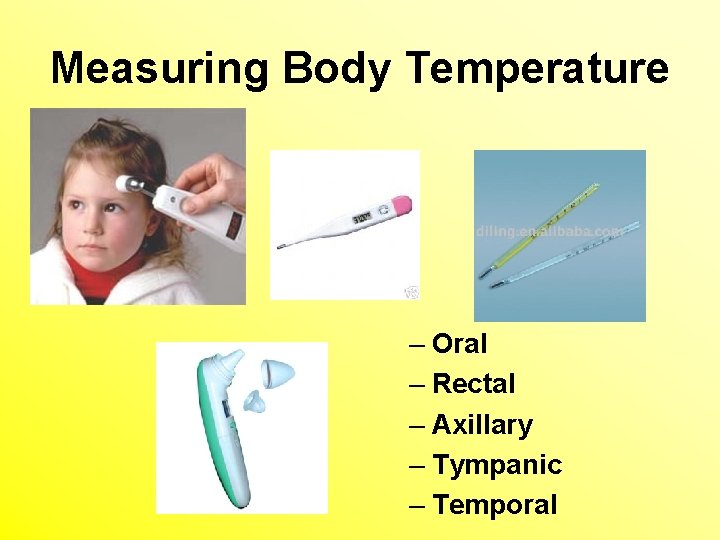
Measuring Body Temperature – Oral – Rectal – Axillary – Tympanic – Temporal
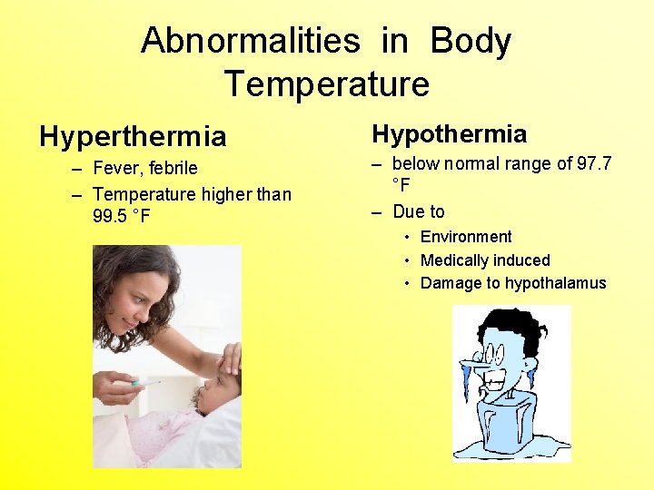
Abnormalities in Body Temperature Hyperthermia – Fever, febrile – Temperature higher than 99. 5 °F Hypothermia – below normal range of 97. 7 °F – Due to • Environment • Medically induced • Damage to hypothalamus
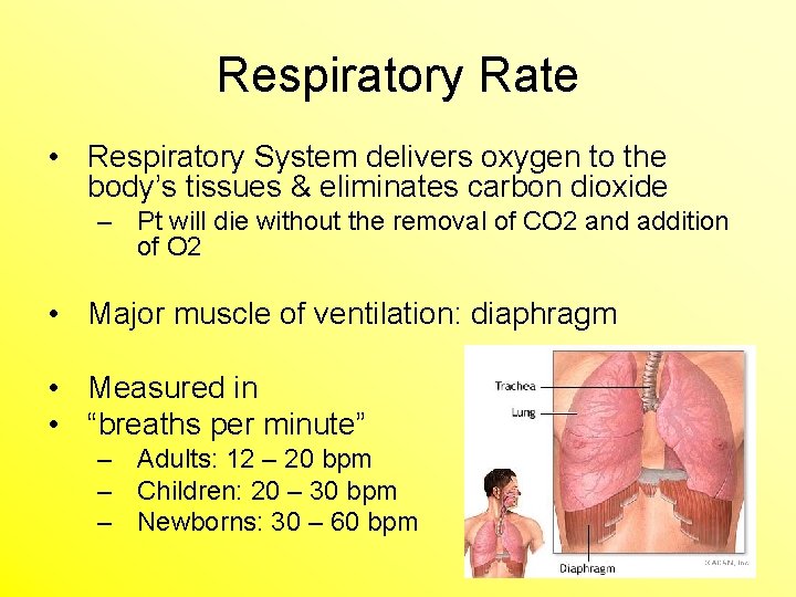
Respiratory Rate • Respiratory System delivers oxygen to the body’s tissues & eliminates carbon dioxide – Pt will die without the removal of CO 2 and addition of O 2 • Major muscle of ventilation: diaphragm • Measured in • “breaths per minute” – Adults: 12 – 20 bpm – Children: 20 – 30 bpm – Newborns: 30 – 60 bpm
Abnormalities of Respiratory Rate • Tachypnea – Greater than 20 breaths per minute (adult) • Bradypnea – decrease is breathing • Dyspnea- difficulty breathing • Apnea- no breathing
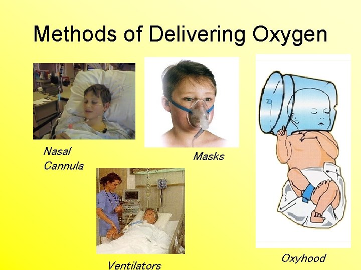
Methods of Delivering Oxygen Nasal Cannula Masks Ventilators Oxyhood
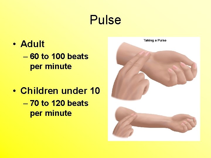
Pulse • Adult – 60 to 100 beats per minute • Children under 10 – 70 to 120 beats per minute
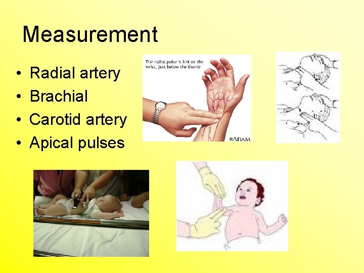
Measurement • • Radial artery Brachial Carotid artery Apical pulses
Abnormalities of Pulse Rate • Tachycardia – Pulse rate increases by more than 20 bpm in resting adult – Greater than 100 bpm • Bradycardia – Decrease in heart rate
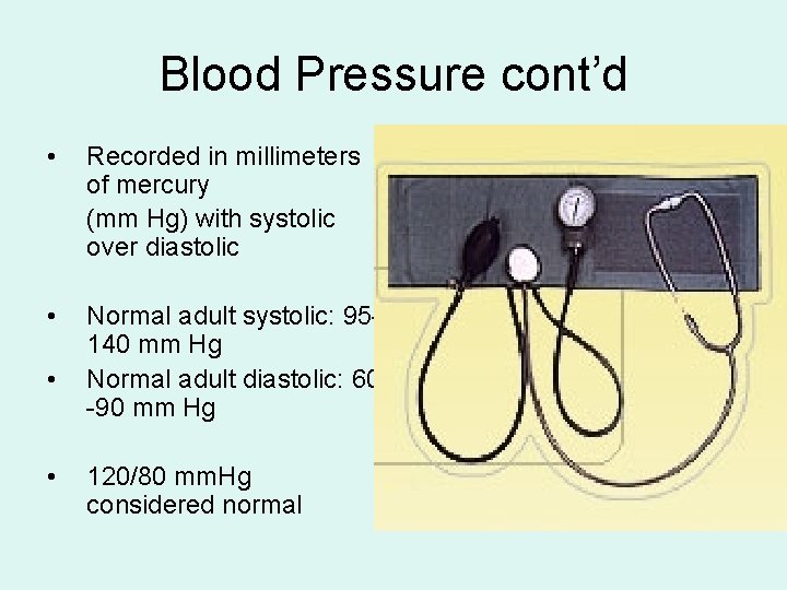
Blood Pressure • Measure of the force exerted by blood on the arterial walls during contraction & relaxation. • Measured pressure when the heart is relaxed: Diastolic • Measured pressure when the heart is contracted: Systolic • Measured with a Sphygmomanometer
Blood Pressure cont’d • Recorded in millimeters of mercury (mm Hg) with systolic over diastolic • Normal adult systolic: 95140 mm Hg Normal adult diastolic: 60 -90 mm Hg • • 120/80 mm. Hg considered normal
Abnormalities of Pulse Rate • Hypertension – Persistent elevation above 140/90 mm. Hg • Hypotension – Persistent less than 95/60 mm. Hg
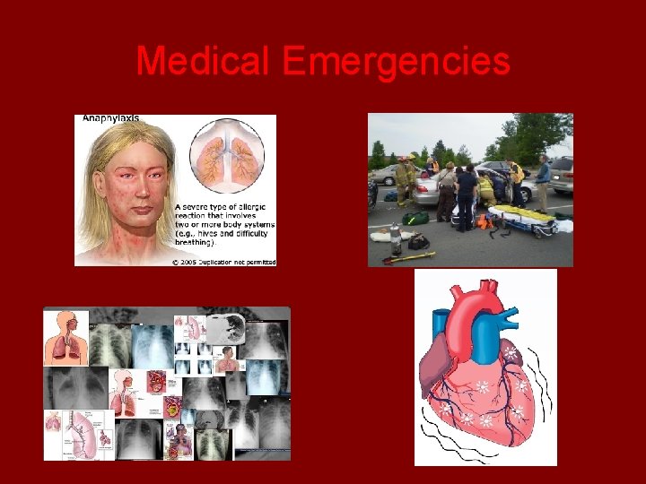
Medical Emergencies
Medical Emergencies • What a Radioilogic technologist should know • Common Radiology Emergencies
Medical Emergencies • Sudden change in medical status requiring immediate action. • For RT’s medical emergencies are rare – Recognize emergencies – Remain calm and confident – Avoid additional harm to the patient – Obtain appropriate medical assistance quickly – Know where crash cart is, emergency phone and code blue buttons
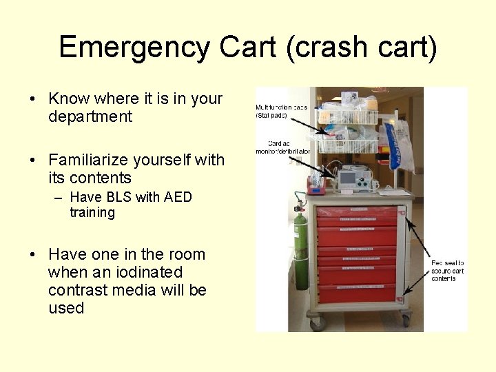
Emergency Cart (crash cart) • Know where it is in your department • Familiarize yourself with its contents – Have BLS with AED training • Have one in the room when an iodinated contrast media will be used
Major Medical Emergencies 1. 2. 3. 4. 5. 6. 7. ALOC Shock Anaphylactic shock Diabetic Crisis Respiratory Distress Cardiac Arrest Cerebrovascular accident
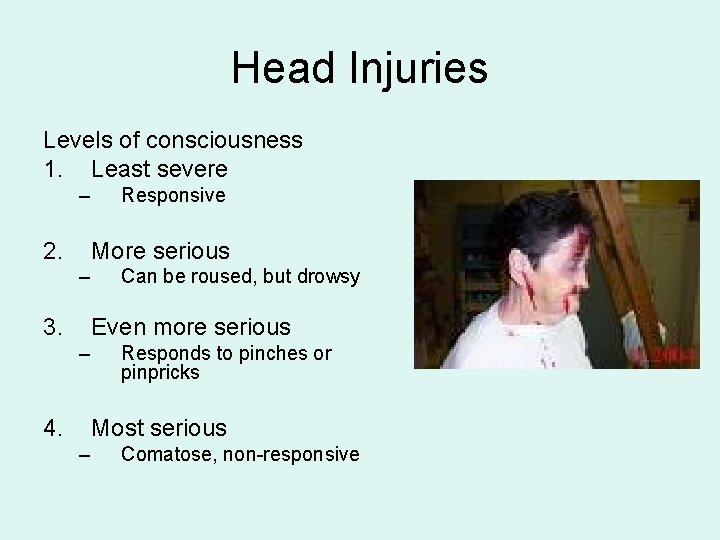
Head Injuries Levels of consciousness 1. Least severe – 2. Responsive More serious – 3. Can be roused, but drowsy Even more serious – 4. Responds to pinches or pinpricks Most serious – Comatose, non-responsive
Shock • Hypovolemic – Loss of blood or tissue • Cardiogenic – Cardiac disorders • Neurogenic – Spinal anesthesia or damage to spinal cord • Vasogenic – Caused by sepsis, deep anesthesia or anaphylaxis
Anaphylactic Shock • An allergic reaction to contrast media – Iodinated • Can happen quickly or have a delayed reaction – Requires prompt recognition and treatment from the technologist – More severe usually have quick onset – Less severe takes longer for reaction
Water Soluble Iodine • High atomic # 53 • Radiopaque • Used to radiograph – – Vessels Arteries Veins Function of internal organs
Prevention and Signs - Symptoms • Restlessness • Maintain normal body temperature • Apprehension • Handle pt’s gently • Tachycardia • RT should work calmly and confidently • Sudden blood pressure drop – Anxiety • Cold –clammy skin – pallor
Diabetic Crisis Hypoglycemia 2. Hyperglycemia 1.
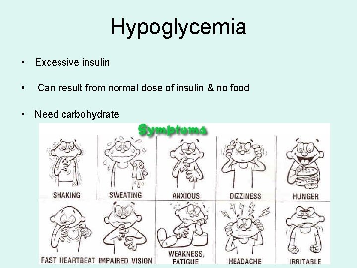
Hypoglycemia • Excessive insulin • Can result from normal dose of insulin & no food • Need carbohydrate
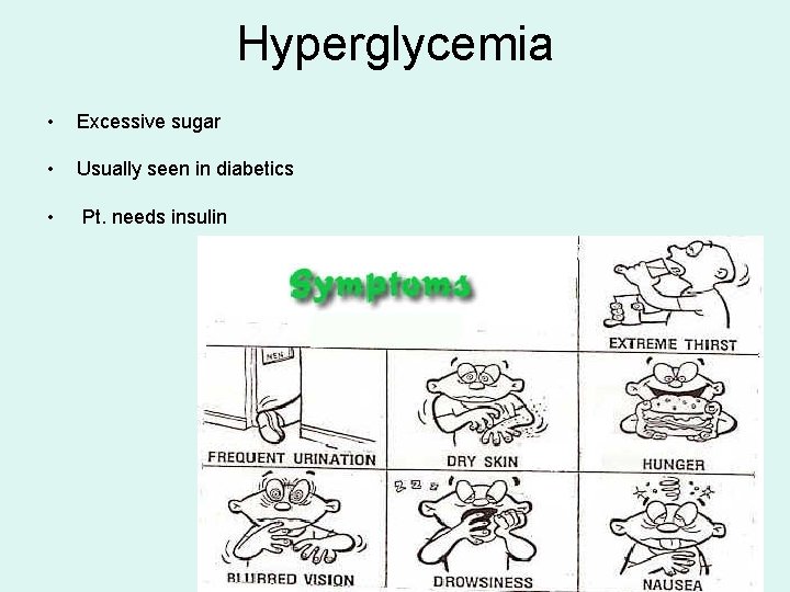
Hyperglycemia • Excessive sugar • Usually seen in diabetics • Pt. needs insulin
Respiratory Distress Asthma 2. Choking 1.
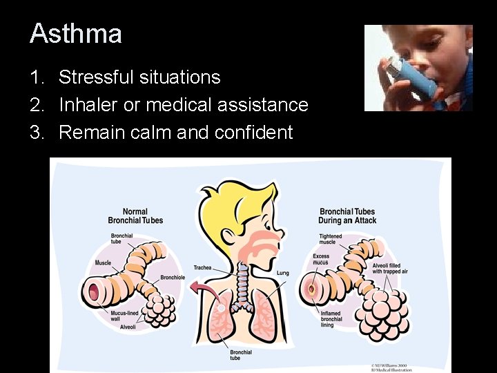
Asthma 1. Stressful situations 2. Inhaler or medical assistance 3. Remain calm and confident
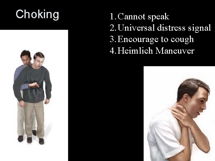
Choking 1. Cannot speak 2. Universal distress signal 3. Encourage to cough 4. Heimlich Maneuver
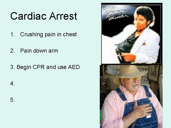
Cardiac Arrest 1. Crushing pain in chest 2. Pain down arm 3. Begin CPR and use AED 4. 5.
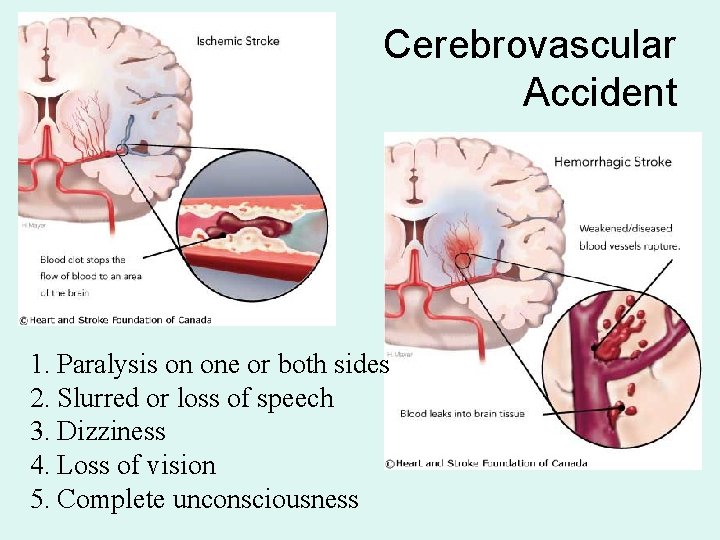
Cerebrovascular Accident 1. Paralysis on one or both sides 2. Slurred or loss of speech 3. Dizziness 4. Loss of vision 5. Complete unconsciousness
Minor Medical Emergencies 1. 2. 3. 4. 5. 6. 7. Nausea and vomiting Epistaxis Vertigo and syncope Seizures Falls Wounds Burns
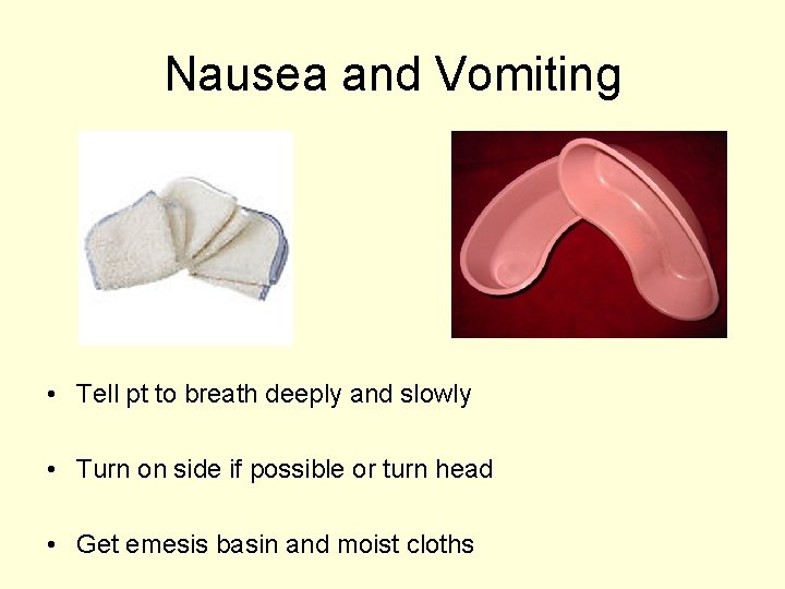
Nausea and Vomiting • Tell pt to breath deeply and slowly • Turn on side if possible or turn head • Get emesis basin and moist cloths
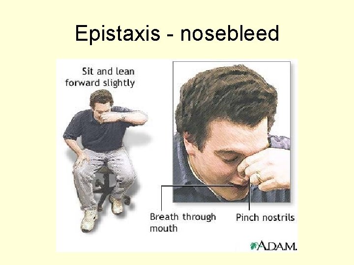
Epistaxis - nosebleed
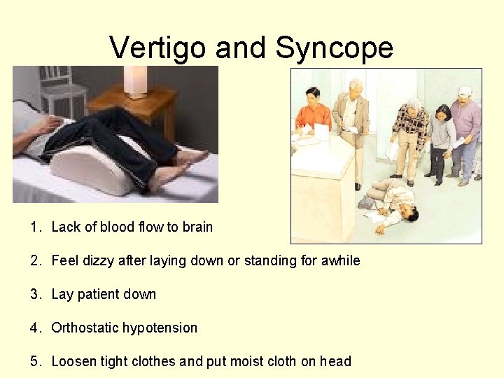
Vertigo and Syncope 1. Lack of blood flow to brain 2. Feel dizzy after laying down or standing for awhile 3. Lay patient down 4. Orthostatic hypotension 5. Loosen tight clothes and put moist cloth on head
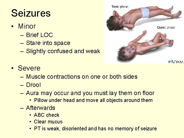
Seizures • Minor – Brief LOC – Stare into space – Slightly confused and weak • Severe – Muscle contractions on one or both sides – Drool – Aura may occur and you must lay them on floor • Pillow under head and move all objects around them – Afterwards • ABC check • Clear mucus • PT is weak, disoriented and has no memory of seizure
Falls, wounds and burns • Falls – Get appropriate help as needed and report incident to supervisor and get a medical assessment of pt • Wounds – – Do not remove dressing Pay attn to any changes in dressing Place extremity above level of heart Apply pressure • Burns – Maintain sterile precautions – Be extra gentle
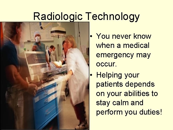
Radiologic Technology • You never know when a medical emergency may occur. • Helping your patients depends on your abilities to stay calm and perform you duties!