PATHOPHYSIOLOGY OF PRIMARY AND SECONDARY HEMOSTASIS FIBRINOLYSIS May

PATHOPHYSIOLOGY OF PRIMARY AND SECONDARY HEMOSTASIS. FIBRINOLYSIS. May 9, 2017

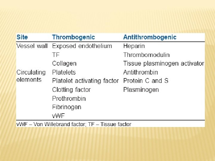
HEMOSTASIS The normal physiological response that prevents significant blood loss following vascular injury is called haemostasis. 6 Familiarity with haemostasis lays the groundwork for a thorough understanding of the major disease states associated with thrombosis, such as venous thromboembolism (VTE), atherothrombosis (thrombosis triggered by plaque rupture), and cardioembolic stroke.

HEMOSTASIS Subendothelial matrix Hemostatic plug Endothelial cell WBC Platelets Fibrin RBC WBC

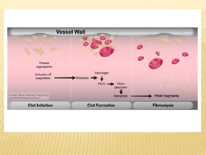
CLOT FORMATION Platelet Red Blood Cell Fibrin

ABNORMAL HAEMOSTASIS Excessive coagulation leads to the formation of a thrombus, potentially obstructing blood flow. This is a common problem, especially in hospitalised or immobilised patients. Venous thromboembolic disease, for example, is a major problem in the European Union, where it causes more than one million events or deaths every year. Excessive bleeding results when certain coagulation factors are lacking, as in patients with haemophilia.

BLOOD VESSEL INJURY Blood vessel injury triggers the following sequence: The vessel constricts to reduce blood flow Circulating platelets adhere to the vessel wall at the site of trauma Platelet activation and aggregation, coupled with an intricate series of enzymatic reactions involving coagulation proteins, produces fibrin to form a stable haemostatic plug This finely tuned process serves to maintain the integrity of the circulatory system. However, the process can go out of balance, leading to significant morbidity and mortality.
 COLLAGEN THROMBIN ADP Aggregation Gp. IIb/IIIa Platelet G p. I")
PLATELET ACTIVATION PATHWAYS (1) COLLAGEN THROMBIN ADP Aggregation Gp. IIb/IIIa Platelet G p. I Adrenaline b Adhesion v. WF Endothelium Exposed Collagen

Platelet Activation Pathways ADP Platele t Thrombin Tx. A 2 Fibrinogen Binding Site Fibrinogen Platelet Aggregation Herbert. Exp Opin Invest Drugs 1994; 3: 449455. (2)

THE COAGULATION CASCADE Coagulation involves a complex set of protease reactions involving roughly 30 different proteins. The final result of these reactions is to convert fibrinogen, a soluble protein, to insoluble strands of fibrin. Together with platelets, the fibrin strands form a stable blood clot.

Palta S et al. , Indian J Anaesth, 58: 515 -523, 2014
TF) and a")
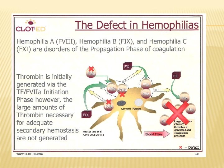
„CELL-BASED MODEL“ This model identifies membranes of cell presenting tissue factor )TF) and a surface of platelets as places of activation of specific coagulation factors. The model supposes the model of zhree phases: initiation, amplificatopn (propagation) and the proper action of thrombinthrombus formation. Initiation = formation of complex TF-FVIIa which is leading to avtivation of a small amount of thrombin. Propagation = activation of platelets by thrombin and formation of complex FIXa-FVIIIa with subsequent activation of factor Xa. Thrombus formation = formation of prothrombinase complex anf of large amount of thrombin which is leading to formation of thrombus.
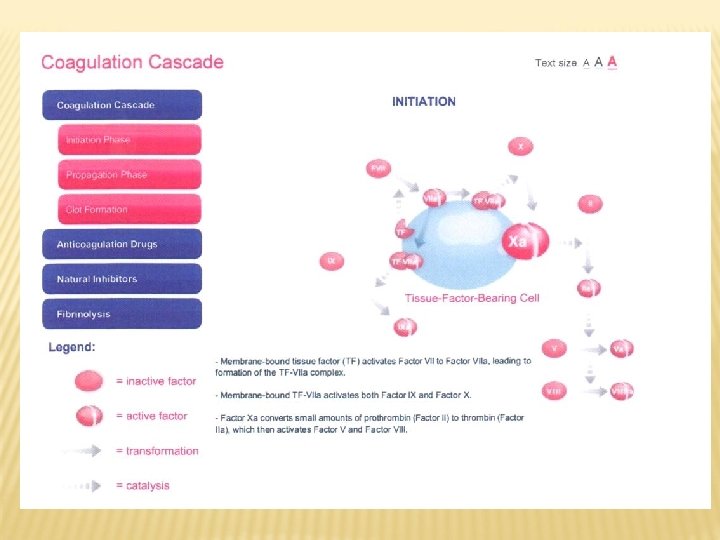

INICIATION PHASE OF COAGULATION Palta S et al. , Indian J Anaesth, 58: 515 -523, 2014

INITIATION PHASE OF COAGULATION Coagulation cascade is activated when defect of vessel wall enables contact of the blood with cells with TF. Platelets membrane bound tissue factor TF activates FVII to VIIa which is leding to formation of complex TF-VIIa. The complex binding on platelets membranes activates Factor IX(a) and Factor X(a). Factor Xa converts small amount of prothrombin (Faktor II) on trombin (Factor IIa) which can activate Factor V on FVa and Factor VIII on FVIIIa.

PROPAGATION PHASE OF COAGULATION Palta S et al. , Indian J Anaesth, 58: 515 -523, 2014

PROPAGATION OF COAGULATION: CENTRAL ROLE FOR FACTOR XA Factor Xa together with activated Factor V (Va) as cofactor support coagulation by thrombin formation (Factor IIa ) from prothrombin (Factor II). Factor Xa is primary pount for propagation of the porcess; one molecule of Factor Xa catalyses formation of about 1, 000 molecules of thrombin.

FINAL STEP: FIBRIN FORMATION In the final step, sequence of serin proteinases reactions which lead to formation of blood clot, thrombin will convert soluble fibrinogen to insoluble fibrin. Thrombin also activates Factor XIII (stabilizing fibrin) which can stabilize clot by crosslinking of fibrin. Stabilized fibrin is able to retain cellular components (red blood cells, platelets or both).

Fibrinolýza

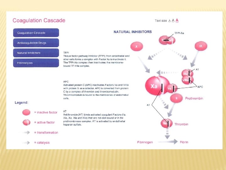
Palta S et al. , Indian J Anaesth, 58: 515 -523, 2014

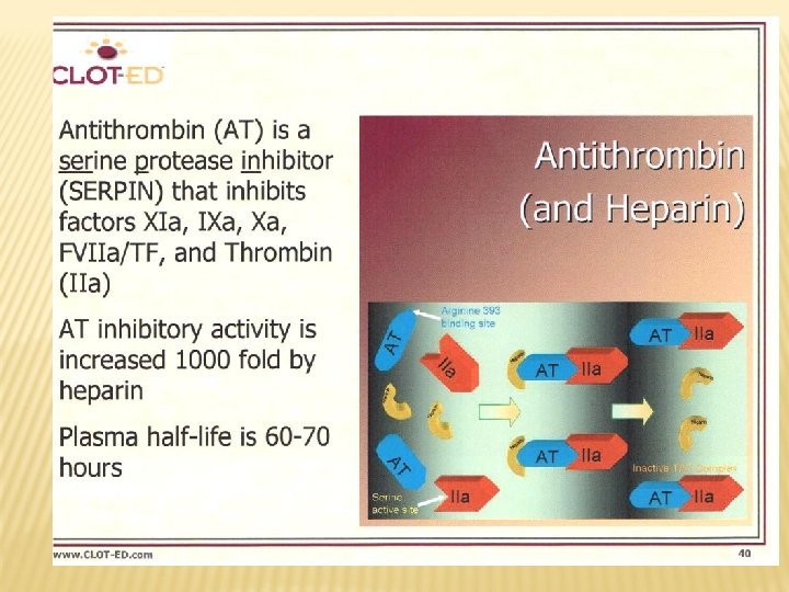
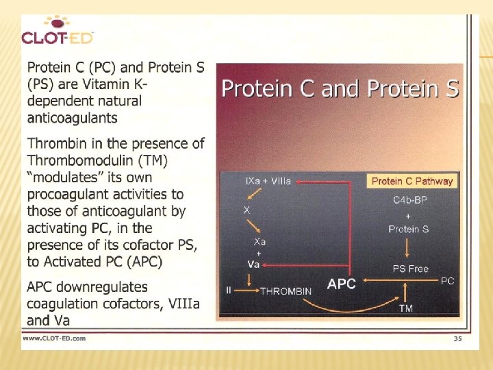
NATURAL INHIBITORS OF COAGULATION „Tissue factor plasminogen inhibitor“ –produced by endothelial cellls. It inhibits complex TF-VIIa. Antithrombin (previously AT III) – binds activated vitamin K dependent coagulation factors (can be activated by heparin which increases its binding capacity) „Protein Z dependent protease inhibitor/ protein Z (PZI)“ produced by liver. It inhibits FXa in presence PZ and Ca++.

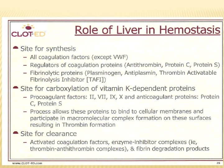
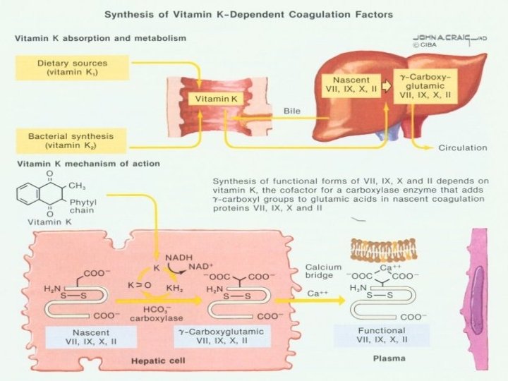
COAGULATION FACTORS -STATE Non activated-after their synthesis in liver Posttranslationally modified –vitamin K dependent coagulation factors = serin proteázy Activated –activated serin proteases, other activated factors (Va, VIIIa)
 Partial thromboplastin time (PTT)")
TESTS Screening tests Bleeding time Platelet count Prothombin time (PT) Partial thromboplastin time (PTT) Thrombin time (TT) More specific tests

SAMPLING Venous blood Excessive stress and exercise cause changes in blood clotting. and fibrinolysis. Whenever possible, venous samples should be collected without a pressure cuff (to avoid haemoconcentration, increase of fibrinolysis, platelet release, and activation of some clotting factors. To minimize the effect of contact activation plastic or polypropylene, siliconized glass, syringes and containers should be used. Thoroughly mixing the blood with the anticoagulant by inverting the containers several time. The sample should be brought to the laboratory as soon as possible. Labeling the patient sample is very important.

SAMPLING Anticoagulant trisodium citrate 3. 2 % in a ratio of 1 : 9. Time of sample collection is very important factor in the interpretation of results. Centrifugation and preparation of platelets poor plasma - 4000 rpm in a cooling centrifuge. P. T & Factor VII kept at room temperature. Other assays at 4 o. C. Testing should preferably be completed within 2 hours of the collection.

BLEEDING TIME Time taken for bleeding to cease from a small superficial wound Affected - by Platelet count and function Vessel wall - Normal range Ivy’s method: 2 -7 min -

PLATELET COUNT Normal A platelet count = 150 -400 x 109 l. L part of complete blood picture (CBC) Performed by electronic counters or manually (inherent error)

PROTHROMBIN TIME Indicates the overall efficiency of extrinsic ex pathway of blood coagulation (FVII, FVII FII, FV, X) Normal range: 10 -14 sec

PROTHROMBIN TIME Causes - - - of prolonged PT Liver disease Vit K deficiency (FII, V, VII, IX are Vit k dependent) Deficiency of factors involved in extrinsic pathway DIC Oral anticoagulants

PARTIAL THROMBOPLASTIN Indicates the overall efficiency of intrinsic in pathway of blood coagulation (FVIII, FVIII FIX, FIX FXI, FXII, FV, X) Normal range: 30 -40 sec

PARTIAL THROMBOPLASTIN Causes - - of prolonged PTT Deficiency of factors involved in intrinsic pathway (coagulation factors other than FVII) Liver disease DIC Massive transfusion (labile FV, FVIII) Heparin

PT & PTT Prolonged PT + normal PTT= extrinsic ex pathway defect Prolonged PTT + normal PT= intrinsic in pathway defect Prolonged PT and PTT= PTT common pathway defect or combined factor deficiencies

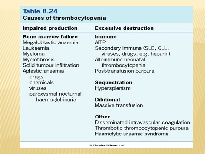
THROMBOCYTOPENIA Platelet count below 150 x 109/L Causes: - Congenital - Acquired failure of production Increased destruction (ITP) - Splenic sequestration (hypersplenism)

IDIOPATHIC THOMBOCYTOPENIC PURPURA ITP is immune thrombocytopenia due to formation of antibodies against platelets and BM megakaryocytes. Clinical picture: re spontaneous bleeding purpuric eruptions. BT: BT prolonged Platelet count: count thrombocytopenia PT, PTT: PT, PTT normal BM: increased megakaryocytes with poor platelet separation

QUALITATIVE PLATELET DEFECT Platelet function defect + normal plt count Causes: - Hereditary (Glanzmann’s disease, Bernard. Soulier syndrome) - Acquired (drugs as aspirin, aspirin uremia)

QUALITATIVE PLATELET DEFECT Clinical picture: picture spontaneous bleeding purpuric eruptions. BT: BT prolonged Platelet count: normal or slightly decreased PT, PTT, TT: TT normal Platelet function: abnormal depending on the defect (defective aggregation in Glanzmann’s disease and Bernard-Soulier syndrome)

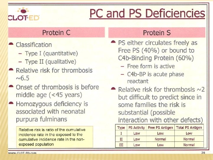
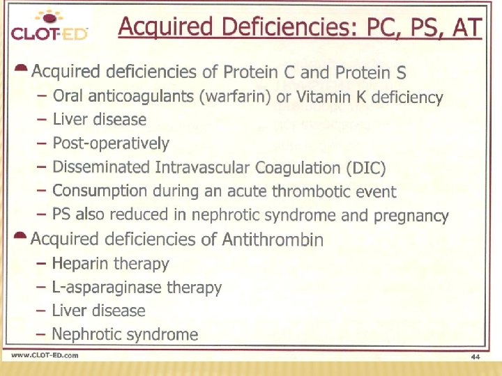
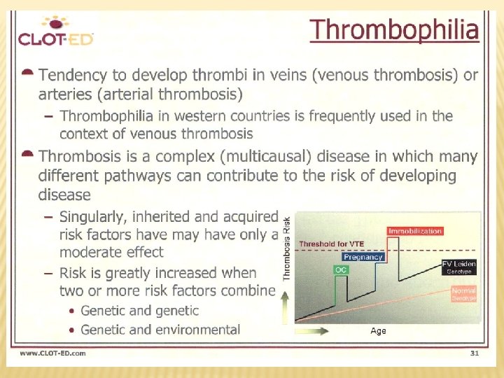
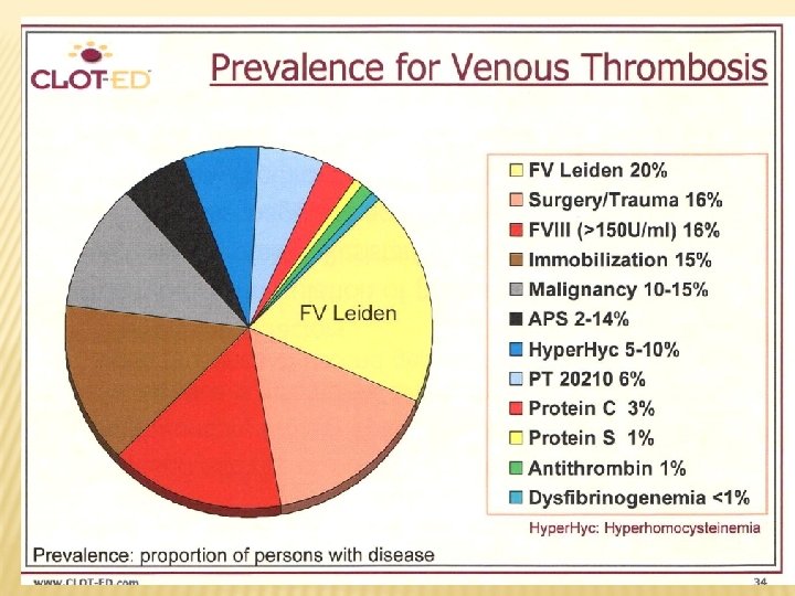
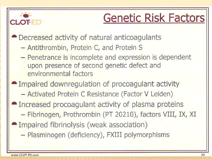
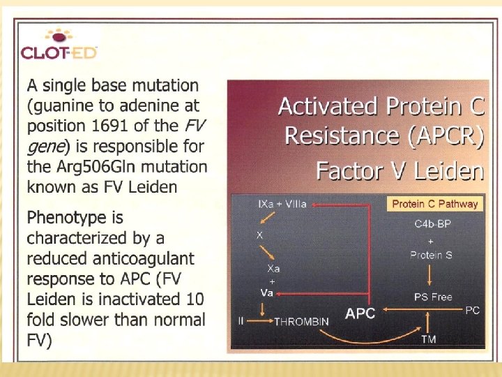
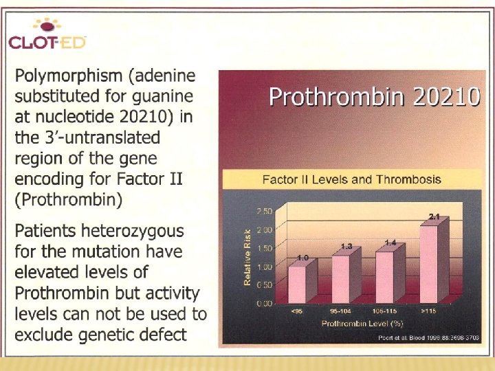
HEREDITARY THROMBOPHILIA Hereditary thrombofilia AT deficiency Protein C deficiency Protein S deficiency Factor V Leiden Prothrombin polymorphism (G/A 20210 in 3´ area of the gene)

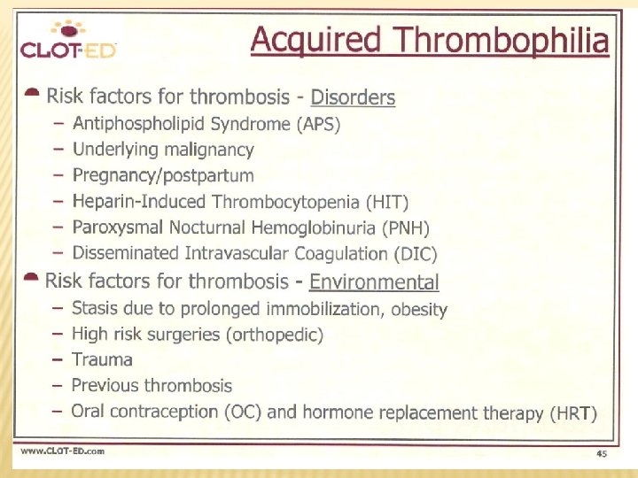
ACQUIRED THROMBOTIC DISORDERS Sy of antiphospholipid antibodies Increased levels of factors VIII, IX, XI and fibrinogen Fibrinolysis defects

HEPARIN/LMWH—ADVERSE EFFECTS Heparin Bleeding n Thrombocytopenia Osteoporosis Hypersensitivity LMWH • • • Bleeding Thrombocytopenia Hypersensitivity

WARFARIN—ADVERSE EFFECTS Fatal or non-fatal hemorrhage from any tissue or organ Necrosis of skin and other tissues Other adverse reactions reported less frequently include: Systemic cholesterol microembolization Alopecia Purple toes syndrome, urticaria, dermatitis including bullous eruptions
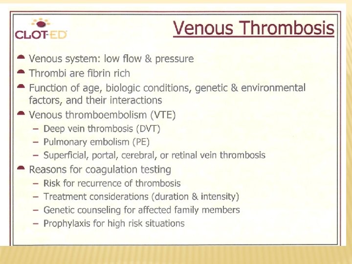


THROMBOSIS AND AF is the most common arrhythmia seen in clinical practice. Without appropriate anticoagulant treatment, most patients with AF are at increased risk of cardioembolic stroke.

THROMBOSIS AND CORONARY ARTERY DISEASE Cardiovascular disease is the leading cause of death in industrialised countries. Coronary artery disease (CAD) is the most common form of cardiovascular disease. In CAD, atherosclerosis damages the coronary artery wall, predisposing to thrombus formation. The symptoms and severity of acute coronary syndromes (unstable angina and myocardial infarction) vary depending on the degree to which thrombi occlude the coronary arteries.

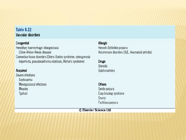
VASCULAR DISORDERS Pattern of bleeding: purpura Causes…… - Screening tests for hemostasis: BT: prolonged Platelet count: normal - PT, PTT, TT: normal
")
BLEEDING DISORDERS Abnormal - bleeding may result from Vascular disorders Thrombocytopenia ( platelet count) Defective platelet function (qualitative defect) Coagulation disorders

HEREDITARY BLEEDING DISEASES Von Willebrand´s disease Hemophilia A Hemophilia B Hemophilia C Factor V deficiency Factor VII deficiency Factor XIII deficiency Prothrombin deficiency Afibrinogenemia

ACQUIRED BLEEDING DISORDERS Consumption coagulopathies DIC-diseminated intravascular coagulation Microangiopathic hemolytic anemia Vitamin K deficinecy Liver diseases
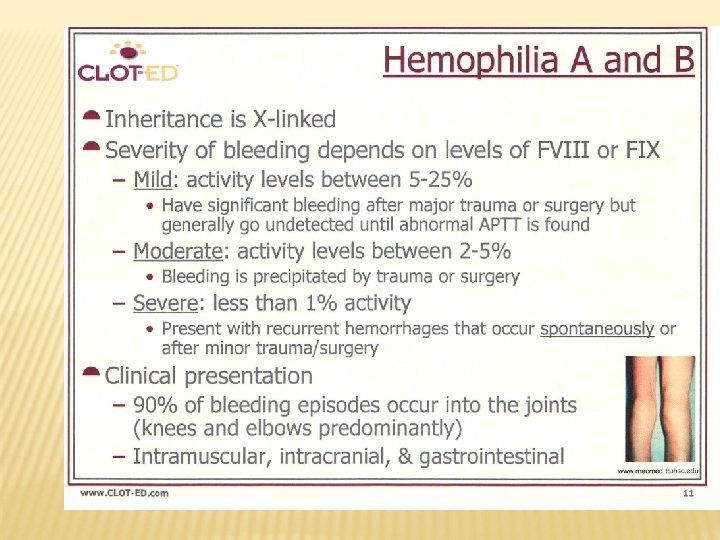

HEMOPHILIA A X-linked disorder Quantitative or qualitative disorder of factor VIII Screening tests: BT: normal Platelet count: normal PTT: prolonged Platelet count: normal Specific test: test FVIII assay: assay decreased activity
 Factor VIII synthesis. (b) Hemofilia A has a defect synthesis of VIIIc. (c)")
(a) Factor VIII synthesis. (b) Hemofilia A has a defect synthesis of VIIIc. (c) von Willebrand ´s disease has a reducted synthesis of v. WF

HEMOPHILIA B Also called Chritmas disease Compared to hemophilia A: - Less common - same presentation - Same screening tests results - Specific test: FIX assay: decreased activity
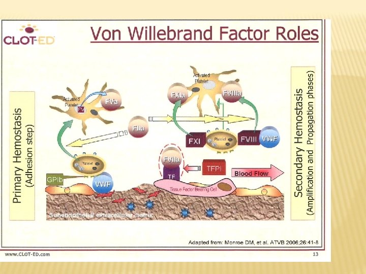
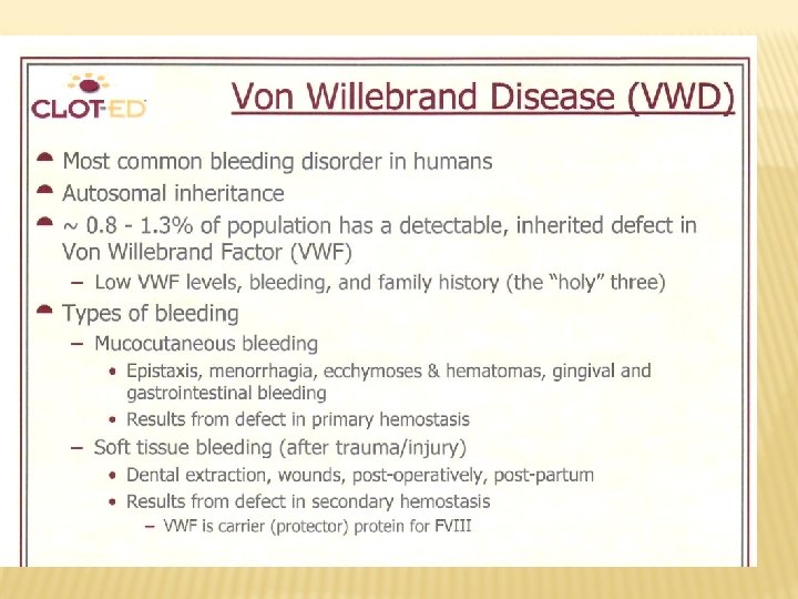

VON WILLEBRAND DISEASE Autosomal dominant disease Quantitative or qualitative disorder of v. WF Von Willebrand factor acts as a carrier for FVIII Acts as an essential cofactor for platelet adhesion and aggregation

VON WILLEBRAND DISEASE Screening tests: BT : prolonged. Platelet count: normal PTT: prolonged Specific tests: Platelet aggregation: aggregation defective with ristocetin FVIII assay: assay decreased activity v. WF antigen : reduced
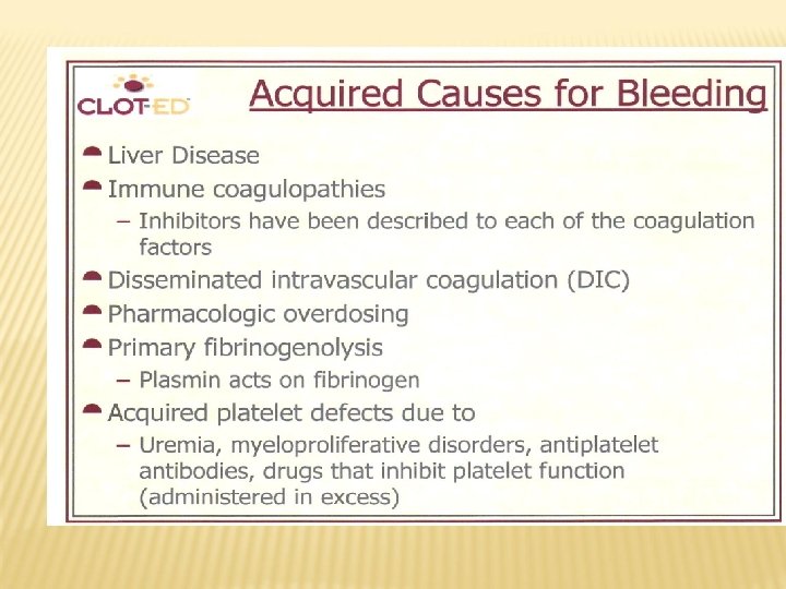

DIC (DISSEMINATED INTRAVASCULAR COAGULATION Release of tissue factor, TF. TF is expressed on many cell types (endothelial, macrophages, monocytes). Contact with blood after damage of vessel wall (the effects of cytokines and endotoxins). TF is binding to coagulation factors which is leading to activation of both pathways of coagulation cascades.
 Due to extensive coagulation followed by fibrinolysis with consumption of")
DISSEMINATED INTRAVASCULAR COAGULATION (DIC) Due to extensive coagulation followed by fibrinolysis with consumption of hemostatic factors. Causes: infection, malignancy, obstetric complications, liver disease

DIAGNOSIS OF DIC BT: prolonged Platelet count: decreased PT: prolonged PTT: prolonged Fibrinogen level: reduced FDPs (D dimer): increased Red cell fragmentation in the blood film

BT PT PTT Platelet count Platelet function Other tests ITP P N N Glanzman P N N N Hemoph A N N P N FVIII assay Hemoph B N N P N FIX assay v. WD P N DIC P P P Defect aggreg FVIII, v. WF Fibrinogen FDPs

THANK YOU FOR YOUR ATTENTION
- Slides: 80