
Pathophysiology L 7 blood lymphatic vessels fakeralani 2000yahoo
Patho-physiology L 7 blood & lymphatic vessels fakeralani 2000@yahoo. com
Vascular Diseases 1. Diseases of arteries 2. Diseases of veins & lymphatics 3. Tumors
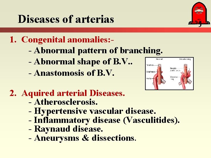
Diseases of arterias 1. Congenital anomalies: - Abnormal pattern of branching. - Abnormal shape of B. V. . - Anastomosis of B. V. 2. Aquired arterial Diseases. - Atherosclerosis. - Hypertensive vascular disease. - Inflammatory disease (Vasculitides). - Raynaud disease. - Aneurysms & dissections.
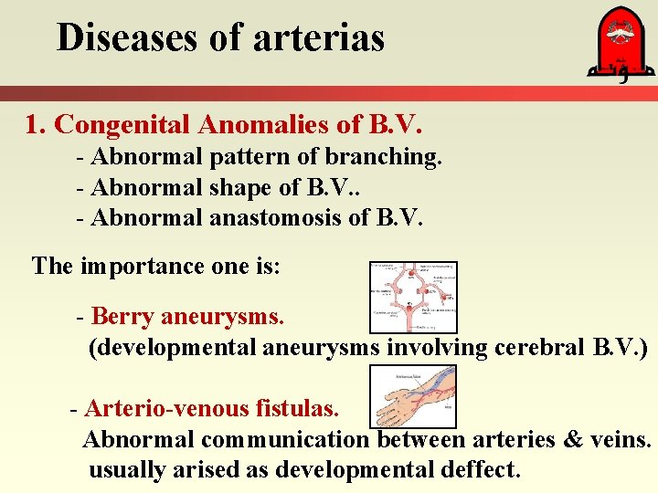
Diseases of arterias 1. Congenital Anomalies of B. V. - Abnormal pattern of branching. - Abnormal shape of B. V. . - Abnormal anastomosis of B. V. The importance one is: - Berry aneurysms. (developmental aneurysms involving cerebral B. V. ) - Arterio-venous fistulas. Abnormal communication between arteries & veins. usually arised as developmental deffect.
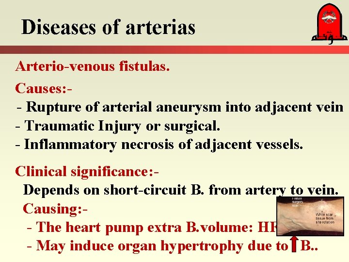
Diseases of arterias Arterio-venous fistulas. Causes: - Rupture of arterial aneurysm into adjacent vein - Traumatic Injury or surgical. - Inflammatory necrosis of adjacent vessels. Clinical significance: Depends on short-circuit B. from artery to vein. Causing: - - The heart pump extra B. volume: HF - May induce organ hypertrophy due to B. .
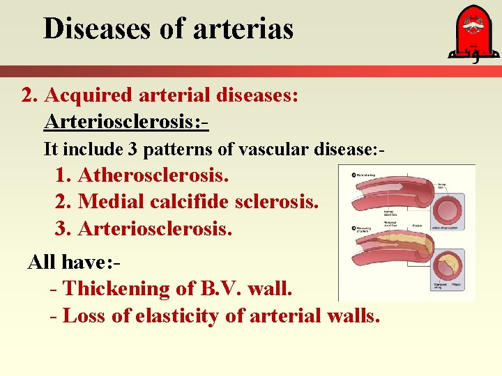
Diseases of arterias 2. Acquired arterial diseases: Arteriosclerosis: - It include 3 patterns of vascular disease: - 1. Atherosclerosis. 2. Medial calcifide sclerosis. 3. Arteriosclerosis. All have: - Thickening of B. V. wall. - Loss of elasticity of arterial walls.
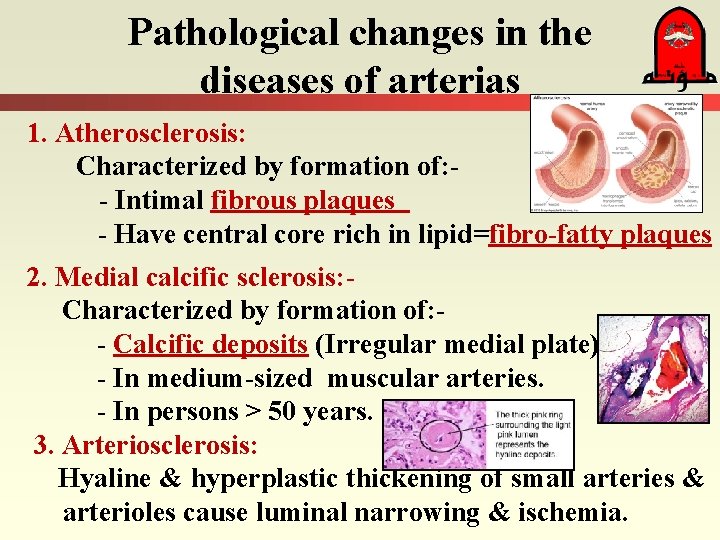
Pathological changes in the diseases of arterias 1. Atherosclerosis: Characterized by formation of: - Intimal fibrous plaques - Have central core rich in lipid=fibro-fatty plaques 2. Medial calcific sclerosis: Characterized by formation of: - Calcific deposits (Irregular medial plate) - In medium-sized muscular arteries. - In persons > 50 years. 3. Arteriosclerosis: Hyaline & hyperplastic thickening of small arteries & arterioles cause luminal narrowing & ischemia.
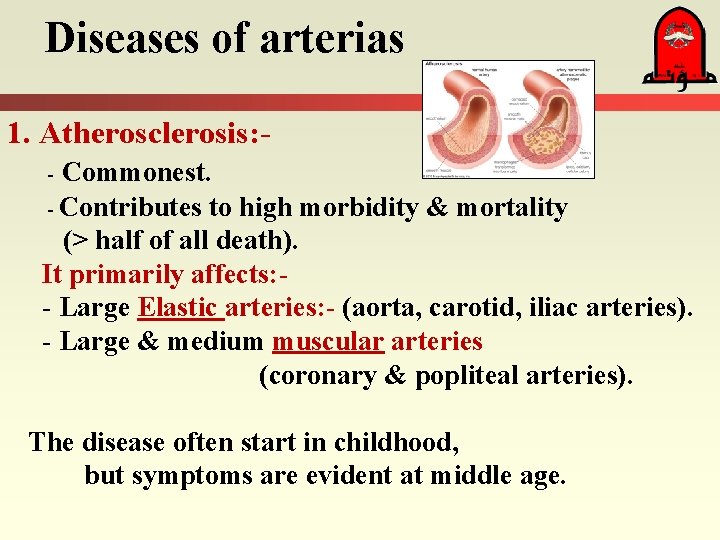
Diseases of arterias 1. Atherosclerosis: - Commonest. - Contributes to high morbidity & mortality (> half of all death). It primarily affects: - Large Elastic arteries: - (aorta, carotid, iliac arteries). - Large & medium muscular arteries (coronary & popliteal arteries). The disease often start in childhood, but symptoms are evident at middle age.
Symptom of atherosclerotic disease Related to arteries supplying the: - Heart. = MI - Brain. = Stroke - Kidneys. = Kidney failure. - Lower extremities = gangrene. - Small intestine = gangrene.
Consequences of Atherosclerosis Consequences: - Aortic aneurysms. - Gangrene of the legs. - Gangrene of intestine (Mesenteric occlusions) - Ischemic encephalopathy. - Chronic IHD. - Sudden death.
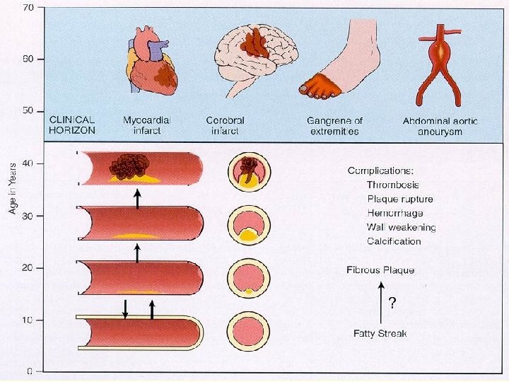
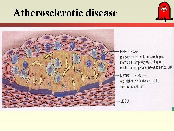
Atherosclerotic disease
Atherosclerotic disease Risk Factors 1. Age & Sex (advanced age, male, postmenopausal) 2. Genetics (Family history, genetic abnormality in the lipoprotein metabolism) ( LDL, Cholesterol) 3. Diet, life style, personal habit. 4. Hyperlipidemia. 5. Hypertension. 6. Smoking. 7. Diabetes mellitus, 8. Elevated plasma homocysteine. 9. Factors affecting hemostasis & thrombosis.
Hyperlipidemia: It is a major risk factor. Most implicated is hypercholesterolemia (hypertriglyceridemia is a less significant, but its effect may be greater in women than men). Total serum cholesterol include: - VLDL. - (HDL). - Cholesterol. - TG.
Lipoproteins HDL-cholesterol is called as the “good cholesterol“ Mobilize cholesterol from atheroma to the liver for excretion in the bile. The various classes of blood lipids are transported as lipoproteins complexed to specific apoproteins. Lipoproteins have many functions including: - Activation & inhibition of enzymes. - Facilitating transmembrane transport of lipoproteins. - Serving as ligands to high affinity cellular receptors that guide the lipoproteins to specific sites of catabolism.
Dyslipoproteinemias: Causes: 1. Genetic mutations: - Involving apolipoproteins genes. 2. Other underlying disorder: Like nephrotic syndrome, alcoholism, diabetes mellitus hypothyreoidism. Types of lipoprotein abnormalities: 1. LDL / cholesterol level. 2. HDL / cholesterol level. 3. Chylomicron remnants & intermediate-density lipoproteins (IDL). 4. level of an abnormal lipoprotein Lp(a).
Other Risk Factors Of arterial Diseases Hypertension: Strong risk factor > hypercholesterolemia after age 45 y. - B. Pr. > 169/90 mm Hg have 5 X > risk of IHD than those with B. Pr. of 140/90 mm Hg. Smoking: One or more packs of cigarettes /day for several years, increases the death rate from IHD twice. Diabetes mellitus: DM induces hypercholesterolemia atherosclerosis. The incidence of MI is 2 X, strokes is 4 X greater, and the
Other Risk Factors Of arterial Diseases Elevated Plasma Homocysteine: serum homocysteine levels Artery disease. (IHD, peripheral vascular disease, stroke, & DVT). Homocysteine (Pathogenesis of B. V. Diseases): 1. Formation of reactive oxygen species. 2. Inhibit vasodilator & antithrombotic functions of NO. Factors Affecting Hemostasis & Thrombosis: Markers of hemostatic & thrombotic function are potent predictors of risk for major atherosclerotic events (plasminogen activator inhibitor-1 level, plasma fibrinogen level, C-reactive protein level).
Pathogenesis of atherosclerosis: Atherosclerosis is a chronic inflammatory response of arterial wall initiated by some form of endothelial injury. There are described the following steps: 1. Chronic injury of endothelium. 2. Deposit of lipoproteins (LDL & VLDL) into the vessel wall. 3. Oxidation of these lipoproteins. 4. Adhesion of monocytes to the endothelium. 5. Migration of monocytes into the intima. 6. Transforamation of monocytes into macrophages & foam cells. 7. Adhesion of platelets to focal areas of adherent leukocytes.
Pathogenesis of atherosclerosis 9. Release of factors from activated platelets & Macrophage 10. Migration of smooth muscle from media into intima. 11. Proliferation of smooth muscle in the intima & elaboration of extracellular matrix. 12. Accumulation of collagen & proteoglycans. 13. Enhanced accumulation of lipids within macrophages, smooth muscle and extracellularly.
C. Hypertensive Vascular Disease Most important risk factors in both IHD & CVA. It may also lead to: - Cardiac hypertrophy & heart failure. - Aortic dissectin. - Renal failure.
Hypertension Types of hypertension: 1. Essential Hypertension: - - 90 -95% of hypertension. - Idiopathic & apparenly primary. 2. Secondary Hypertension: - 5 -10% - (Renal disease, renal artery stenosis, Cushing Ds. , Hyper-aldosteronism, pheochromocytoma). 3. Malignant Hypertension: - 5% of hypertension. - Rapidly rising B. Pr. , if untreated, leads to death within 1 - 2 years.
Hypertensive Vascular Disease Hypertension atherogenesis & changes in the structure of the wall of B. V. that potentiate both aortic dissection & CV hemorrhage. Hypertension is associated with 2 small B. V. disease: - – Hyaline arteriolosclerosis : - (in old & Diabetics) A homogenous, pink, hyaline thickening of the walls of arterioles with narrowing of the lumen). - Hyperplastic arteriolosclerosis: - (in patients with severe hypertension) Typical onion-skin, concentric thickening of arteriole walls with progressive narrowing of lumens).
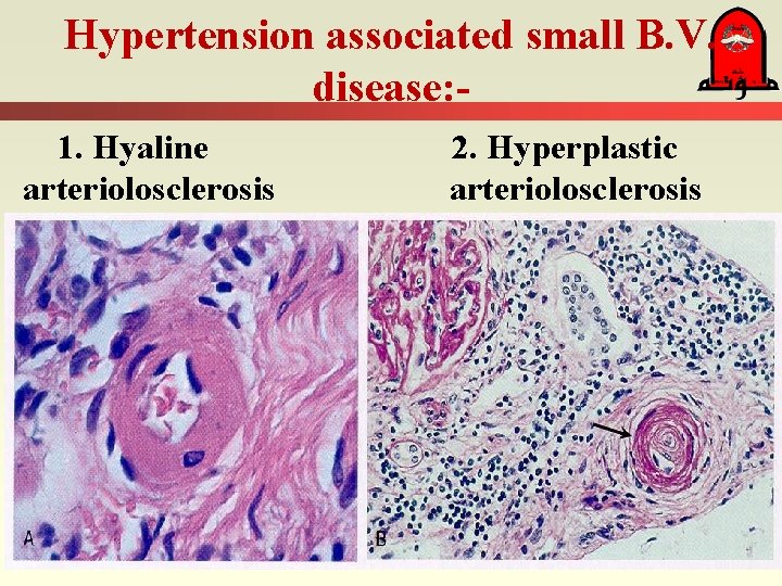
Hypertension associated small B. V. disease: - 1. Hyaline 2. Hyperplastic arteriolosclerosis
Inflammatory Disease – (Arteritides, Vasculitides) Infectious Vasculitides: bacterial: - Neisseria, Rickettsial, Spirochetal. Fungal & Viral. Non-Infectious Vasculitides: - Systemic necrotizing vasculitides (affecting aorta, medium-sized vessels). - Small vessel vasculitides (affecting arterioles, venules and capillaries). Pathogenesis involves: Immune compexes, antineutrophil cytoplasmic antibodies (ANCA) & antibodies to endothelial cells. (Classification depend on size of B. V. )
Raynaud Disease Paroxysmal pallor or cyanosis of the digits of the hands or feet and infrequently the tips of the nose or ears. Caused by: - Intense vasospasm of small arteries or arterioles. - Typically in young healthy women. - No organic changes are present in the arterial walls except late, when intimal proliferation can appear.
Aneurysms and Dissection Aneurysm: – localized abnormal dilatation of B. V. that occurs most commonly in the aorta or the heart. It can be true: (attenuated arterial wall components) In aorta it is due to atherosclerosis & cystic medial degeneration. or syphilitic aneurysm (tertiary stage of syphilis). False: (extravascular hematoma that communicates with the intravascular space).
Aneurysms and Dissection Aortic Dissection (Dissecting Hematoma): Characterized by: - Dissection of wall & penetration of blood in between & along the laminar planes of the media, with the formation of a blood-filled channel within the aortic wall (Aneurysm). - It can rupture causing massive hemorrhage. - It is not associated with marked aortic dilatation. (Not like atherosclerotic & syphilitic aneurysms)
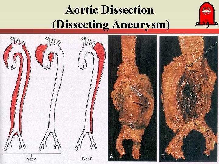
Aortic Dissection (Dissecting Aneurysm)
Diseases of veins and lymphatics A) Varicose Veins B) Thrombophlebitis & Phlebothrombosis C) Syndrome of vena cava superior D) Syndrome of vena cava inferior E) Lymphangitis and Lymphedema
A) Varicose Veins Abnormally dilated, tortuous veins. Produced by: - Prolonged, increased intraluminal pressure. - Loss of support of the vessel wall. Clinically: - Superficial veins of the leg are usually the involved. - Marked variation in the thickness of the wall. - Intraluminal thrombosis & valvular deformities are frequently found. - Embolism is rare despite thrombosis of superficial varicose veins.
A) Varicose Veins Sites of varicosity formation: 1. Esophagus: In patients with liver cirrhosis & portal hypertension. - Ruptura of an esophageal varix followed by hemorrhage may be very serious). 2. Anorectal junction: - In patient with hemorrhoids. - Result from prolonged pelvic congestion. - It is a source of bleeding & site of thrombosis).
B. Thrombophlebitis & Phlebothrombosis Venous thrombosis due to: - - Inflammation. - Hypercoagulability. - Immobilization, - Operation. - Pregnancy. - DVT account for > 90% of thrombophlebitis. - DVT have a tendency to embolization into lung.
C) Syndrome of vena cava superior Compression or invading the v. cava superior: - Occasionally by: - aortic aneurysm. - But usually caused by neoplasms: Primary bronchogenic carcinoma Mediastinal lymphoma. The consequent obstruction produces: - Cyanosis. - Marked dilation of the veins of the head, neck, & arms. - Commonly compression of the pulmonary vessels, causing resipartory distress.
D) Syndrome of vena cava inferior Compression or invading the v. cava inferior: Caused by: Hepatocellular carcinoma. Renal cell carcinoma. Obstruction induces: - Marked edema of the legs. - Distention of the superficial collateral veins of the lower abdomen. - Massive proteinuria due to renal veins involvment.
Lymphangitis & Lymphedema Lymphangitis: Bacterial infections spreading into lymphatics. - Primary diseases: - Uncommon. - Secondary processes: - In association with inflammation or cancer. Obstructive lymphedema: Occlusions of lymphatic drainage by: - Tumor, Postradiation fibrosis, Filariasis, - Trauma or by inflammatory thrombosis
Tumors of B. V. A) Benign tumors & tumor-like lesions B) Bordeline tumors C) Malignant tumors
A. Benign tumors & tumor-like lesions Hemangioma: In the skin, Liver - Capillary Hemangioma : - Cavernous Hemangioma: - Lymphangioma: - Simple lymphangioma: - skin of head & neck, axilla. - Cavernous lymphangioma: - children, neck & axilla.
B. Borderline tumors Kaposi sarcoma: Frequent occurence in patients with AIDS. - Multiple red-to-purple skin plaques or nodules. - Primarily in the distal lower extremities. - Slowly growing & spreading to more proximal sites. - It is composed of irregular B. V. - It is difficult to distinguish from granulation tissue
Malignant tumors Angiosarcoma: More common in old adults. Anywhere in the body. Commen sites: – skin, soft tissues, breast & liver).
- Slides: 40