Pathophysiology L 5 A Cardiovascular Regulation Prof Fakhir


 Ø")


 contractility (Inotropic) Norepinephrine General vasoconstrictor")


: Norepinephrine general vasoconstrictor Sympathetic nerves (cholingergic): Acetylcholine")


- Slides: 29
Pathophysiology L 5 A Cardiovascular Regulation Prof. Fakhir Al-Ani fakeralani 2000@yahoo. com
Regulation of the CVS Heart Rate Regulation Blood Flow Regulation
Heart Rate Regulation The heart is regulated by: - Intrinsic mech. (situated within the heart) - Extrinsic mech. (originating outside the heart) The intrinsic mech. include: - SA node & AV node = regulate the heart rate. - Starling low regulate force of contraction. The extrensic mech. include: - Sympathetic. - Parasympathetic.
Intrinsic Regulation of HR Sino atrial node: pacemaker
Intrinsic Regulation Ø Depolarization SA node membrane creates an A. P. (electrical impulse) Ø Impulse travels through the heart in an established pathway Ø The normal pathway: -
Normal Route of Depolarization S-A Node Atria A-V Node Bundle of His Purkinje Fibers Ventricles
Intrinsic Heart Rate ü SA node rate approximately 90 beat/m. ü Parasympathetic innervation slows rate – Referred to as parasympathetic tone – Training increases parasympathetic tone
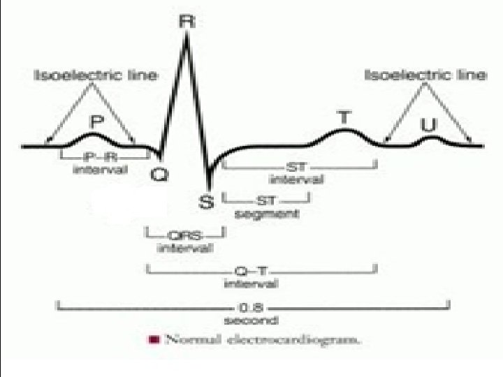
Electrocardiogram The ECG: Recorded by placing electrodes on body surface connected to an amplifier & recorder. ECG wave shape is related to: specific electrical change in the heart. ECG monitor: - Heart rate - Rhythm. - Wave.
Electrocardiogram EEG wave related to different cardiac places - P wave: - atrial depolarization - PR interval: - Delay at the AV node - QRS complex: - ventricular depolarization masks atrial repolarization - ST segment: - Delay between depolarization & repolarization of the ventricles - T wave: - ventricular repolarization
Abnormal Rhythem ECG Arrhythmias PACs- premature atrial contraction § PVCs- premature ventricular contraction § Ventricular fibrillationcardiovert
Extrinsic Regulation of HR Neural Influences override intrinsic rhythm Sympathetic: catecholamines Epinephrine Norepinephrine Parasympathetic Acetylcholine - Cortical Input - Peripheral Input
Neural Regulation of HR Sympathetic influence Epinephrine HR (Tachycardia) contractility (Inotropic) Norepinephrine General vasoconstrictor Parasympathetic influence Acetylcholine HR (Bradycardia)
Cardiac Accelerator Nerves Sympathetic Fibers - Innervate SA node & ventricles Heart rate Contractility Pressure
Vagus Nerve Parasympathetic Nerve - Innervates SA node & AV node heart rate pressure
Cortical Influences on Heart Rate Cerebral cortex impulses & Emotional state affects cardiovascular response change heart rate & B. Pr.
Peripheral Influences on HR Peripheral receptors monitor state of active muscle; modify vagal or sympathetic: - Chemoreceptors Monitor p. CO 2, H+, p. O 2 - Mechanoreceptors Heart and skeletal muscle mechanical receptors - Baroreceptors
Peripheral Influence on HR Baroreceptors in carotid sinus & aortic arch. B. Pr. → HR & contractility
Blood Flow Regulation During exercise: - local arterioles dilate - & venous capacitance vessels constrict. B. flow is regulated according to Poiseuille’s Law: Flow = pressure X resistance.
Blood Flow Regulation Flow = Pr. gradient x Vessel radius vessel length x viscosity Blood flow Resistance Factors 1. Viscosity or blood thickness 2. Length of conducting tube 3. Radius of blood vessel
Blood Flow Regulation At rest : 1 / 30 or 40 capillaries is open in muscle During exercise: Opening “dormant” capillaries B. flow to muscle Speed of blood flow Increases surface area for gas exchange
Local Factors Resulting in Dilation tissue O 2 produces potent vasodilation in skeletal and cardiac muscle. CO 2 temperature p. H ADP Nitric Oxide (NO) Ions of Mg+2 & K+ Acetylcholine
Blood Flow Neural Factors Sympathetic nerves (adrenergic): Norepinephrine general vasoconstrictor Sympathetic nerves (cholingergic): Acetylcholine vasodilation in skeletal & cardiac muscle.
Blood Flow Humoral Factors Sympathetic nerves to adrenal medulla causes release of epinephrine & norepinephrine into blood (humor).
Blood Flow Humoral Factors Sympathetic Nerves to Adrenal Medulla epi & norepi in blood vasoconstriction except in skeletal muscle
Neural Factors of Flow Control
Integrated Response
Regulation from Rest to Exercise - Rapid in heart rate, SV, cardiac output – withdrawal of parasympathetic stimuli – input from sympathetic nerves Continued in heart rate – temperature. – feedback from proprioceptors – accumulation of metabolites
Integrated Response in Exercise