Pathophysiology BMS 243 Lecture I Continued Introduction The
 Dr. Aya M.")
Pathophysiology BMS 243 Lecture I Continued Introduction; The Cardiovascular System (CVS) Dr. Aya M. Serry 2016
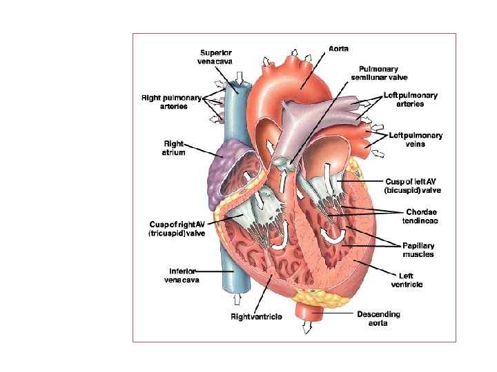
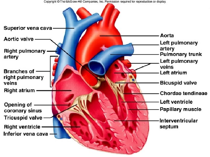

Cardiac valves and their functions The human heart contains four valves: ØTwo Atrioventricular valves (AV valves) between the atria and the ventricles: 1. Tricuspid valve between the right atrium and the right ventricle. 2. Mitral valve between the left atrium and the left ventricle.

Cardiac valves and their functions The human heart contains four valves: ØTwo Atrioventricular valves (AV valves) between the atria and the ventricles: 1. Tricuspid valve between the right atrium and the right ventricle. 2. Mitral valve between the left atrium and the left ventricle.

Cardiac valves and their functions ØTwo semilunar valves: Aortic valve between the left ventricle and the aorta. 2. Pulmonary valve between the right ventricle and the pulmonary artery. 1.

Functions of the cardiac valves The cardiac valves allow the blood to pass only in one direction The AV valves : Allow for the blood to pass from the atria into the ventricles during ventricular diastole. During ventricular systole, the AV valves close to prevent back flow of blood from the ventricles into the atria.

Functions of the cardiac valves The semilunar valves: Allow for the blood to pass from the ventricles into the arteries during ventricular systole. During ventricular diastole, these valves prevent back flow of blood from the arteries into the ventricles (as these valves become closed during ventricular diastole).

Preload Vs. Afterload Preload • The preload represents the volume work of the heart. It is usually considered the end-diastolic pressure when the ventricle has been filled. • It is called the preload because it is the work or load imposed on the heart before the contraction begins. • Preload represents the amount of blood that the heart must pump with each beat, It is largely determined by the venous return to the heart and the accompanying stretch of the cardiac muscle fibers.

Preload Vs. Afterload • The afterload is the pressure in which the muscle exerts its contractile force in order to move blood into the aorta. • It is called the afterload because it is the work presented to the hea after the contraction. • The systemic arterial blood pressure is the main source of afterload work on the left heart, and the pulmonary arterial pressure is the main source of afterload work on the right heart. • The afterload work of the left ventricle is also increased with narrowing (i. e. , stenosis) of the aortic valve.

THANK YOU…. .
- Slides: 11