Pathology of the Urogenital Tract Male Genital Tract


")


 is within normal limits for a man this")


- Slides: 45
Pathology of the Urogenital Tract Male Genital Tract March 24, 2014
Case 1
Q 1 Name the organ. Describe the normal histology.
Q 2. Name the organ. Describe the normal histology
Q 3 Describe the normal histology (high power)
Case 2
Case 2 HISTORY: A 32 -year-old previously healthy man presents with a painless mass in his left testicle. He noticed it about 1 month ago. It was not getting smaller so he sought medical attention. VITAL SIGNS: 135/80 HR 80 RR 15 T 98° PHYSICAL EXAMINATION: Palpable, mobile 3 cm non-tender mass is present in the left testicle. Exam is otherwise unremarkable. There is no inguinal lymphadenopathy.
Q 1: What is the main clinical problem and differential diagnosis?
LAB TESTS Chest X-ray is normal Serum AFP and HCG are normal
Q 2: Describe gross findings
Q 3: Describe the pathologic changes
Q 4: What is your diagnosis?
Q 5: List the key clinical and pathologic features of this tumor.
Case 3
Case 3 HISTORY: 67 -year-old male has nocturia, urinary hesitancy (difficulty in starting and stopping urine flow), “weak” urine stream, and dribbling at the end of urination. PHYSICAL EXAMINATION: On digital rectal exam the prostate gland is enlarged and non-tender. There are no palpable masses.
Q 1: What is the clinical problem and list your differential diagnosis?
LAB TESTS: Prostate specific antigen (PSA) is within normal limits for a man this age.
Q 2: Describe gross findings
Q 3: Identify organ and describe the pathologic changes
Q 4: What is your diagnosis?
Q 5: What complications may occur because of this problem?
Q 6: What hormone is related to this process?
Case 4
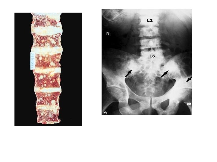
Case 4 • HISTORY: A 72 -year-old man presents with back pain. It is constant and exacerbated by movement. The pain often keeps him up at night. • PHYSICAL EXAMINATION: There is tenderness over the lower spine. Neurologic exam is normal. A single, hard prostatic nodule is palpated on digital rectal exam. No lymphadenopathy is noted.
Q 1: What are the main clinical problems and differential diagnosis?
• RADIOGRAPHY: Osteoblastic vertebral lesions are noted on x -rays of the spine. • LAB TESTS: PSA 353. 46 H ( 0. 0 - 4. 0 NG/ML)
Transrectal Biopsy of Prostate
Q 2: Describe the gross findings
Q 3: Identify organ and describe the pathologic changes
Q 4. What is your diagnosis?
Q 5: What is a Gleason grade?
Q 6: What is a Gleason score?
Q 7: Which genetic changes may occur in this process?
Q 8: Correlate the clinical and radiographic findings with the pathologic diagnosis:
Case 5
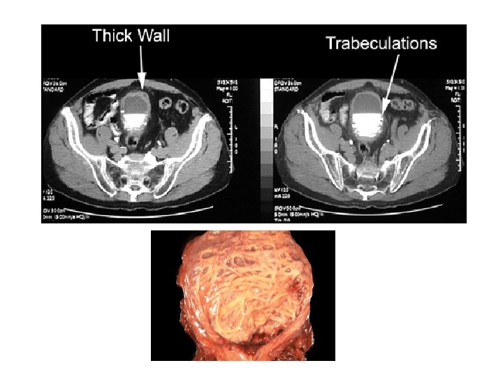
Case 5 • HISTORY: 62 -year-old man presents with hematuria. He has no difficulty in voiding and no flank or back pain. He has a history of smoking. • PHYSICAL EXAMINATION: Unremarkable. • LAB TESTS: • Urinalysis 4+ blood 1+protein 50 -100 red blood cells/high power field
Q 1: What is the main clinical problem?
Q 2: What is the clinical differential diagnosis?
Q 3: Describe the following findings: Cystoscopic Findings Urine Cytology
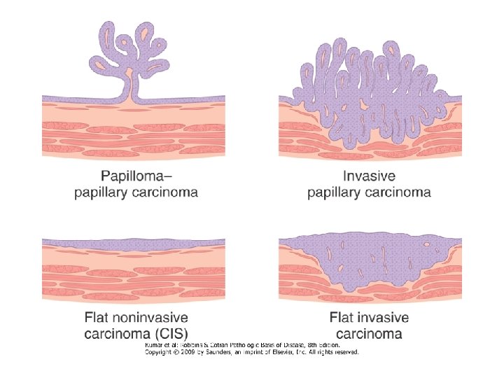
Q 4: Identify organ and describe the pathologic changes
Q 5: What is your diagnosis?
Q 6: What are the epidemiologic predisposing factors to this process?