Pathology of inflammation Lab Inflammation can be divided

Pathology of inflammation ﺩ ﻫﺒﺔ ﺍﺣﻤﺪ ﻏﻴﺪﺍﻥ Lab

Inflammation can be divided into two basic types; • Acute inflammation : • Chronic inflammation:

Signs of acute inflammation

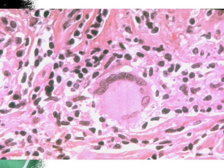
The main acute inflammatory cells are : Neutrophils and macrophages
 with multilobed nuclei")
Acute inflammation with densely packed (PMNs) with multilobed nuclei
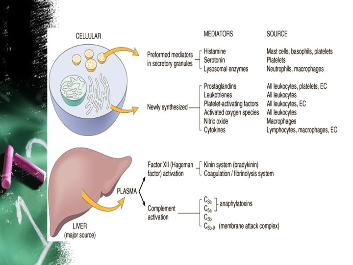

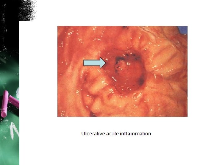
Morphological patterns of inflammation: Several types of inflammation are recognized, which vary in their morphology and clinical correlates. 1 - Serous inflammation 2 - Fibrinous inflammation 3 - Suppurative (purulent) inflammation 4 -Pseudomembranous inflammation of mucous membranes 5 - Catarrhal inflammation 6 -Hemorrhagic inflammation 7 - Necrotizing (gangrenous) inflammation: 8 -Ulcerative inflammation
")
skin blister (serous inflammation)

Fibrinous exudate

acute suppurative appendicitis gross

abscess is the skin furuncle

Pseudomembranous inflammation Diphtheria

Chronic inflammation: Types of chronic inflammatory cells 1 1
. 2. Parasitic")
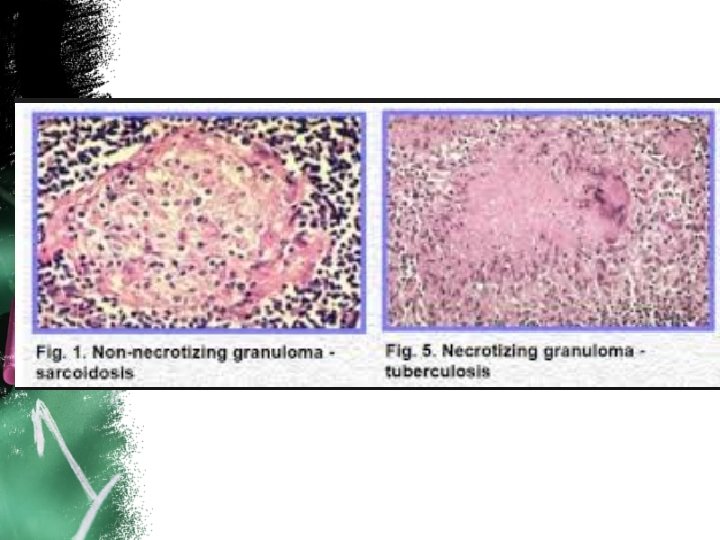
Chronic Granulomatous inflammation: Causes of Granuloma: 1. Bacterial causes: Tuberculosis, leprosy, syphilis). 2. Parasitic causes: Schistosomiasis. 3. fungal causes: Histoplasmosis, cryptococccus neoformans. 4. Inorganic metals or dusts: Silicosis, Berylliosis. 5. Foreign body: suture material, breast prosthesis. 6. Unknown: Sarcoidoisis.

Epitheloid cell

Langhans giant cell

Foreign body giant cell
")
caseating granuloma ( necrotizing granuloma)
")
caseating granuloma ( necrotizing granuloma)
: Granulation tissue Gross: Pink, soft, granular Mic. : small blood vessels,")
Tissue Repair (healing): Granulation tissue Gross: Pink, soft, granular Mic. : small blood vessels, fibroblasts, edematous stroma & macrophages
")
Complications of wound healing: These complications can be grouped into three general categories: (1) deficient scar formation (2) excessive formation of the repair components (3) formation of contractures.

Dehiscence or rupture of a wound

Keloid. A, Excess collagen deposition in the skin forming a raised scar known as keloid. B, Note thick connective tissue deposition in the dermis

Keloid

Wound contracture. Severe contracture of a wound after deep burn injury

Thank you
- Slides: 29