
Pathology of hard dental tissue Classification and etiological



- Slides: 26
Pathology of hard dental tissue. Classification and etiological factors. Diagnostics. Methods of orthopedic treatment using non-removable dentures. Types of dentures, restoring the anatomical shape of the tooth.
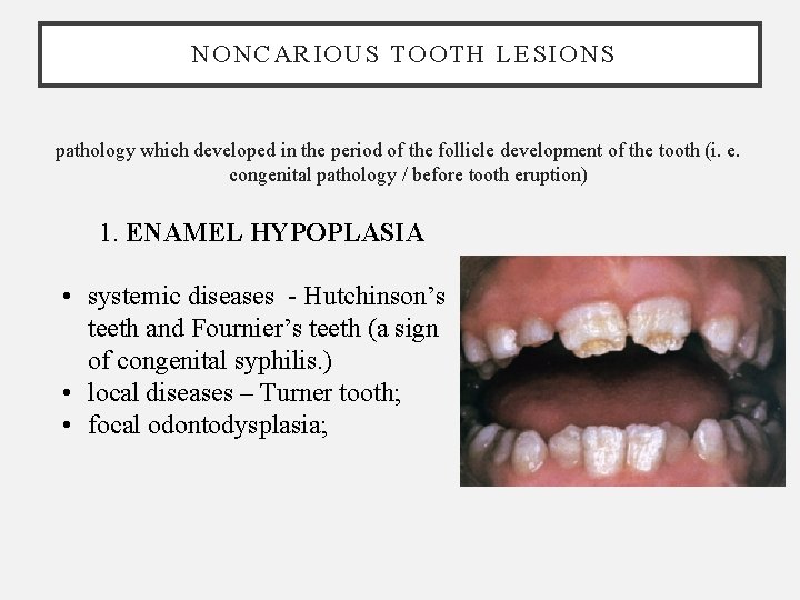
NONCARIOUS TOOTH LESIONS pathology which developed in the period of the follicle development of the tooth (i. e. congenital pathology / before tooth eruption) 1. ENAMEL HYPOPLASIA • systemic diseases - Hutchinson’s teeth and Fournier’s teeth (a sign of congenital syphilis. ) • local diseases – Turner tooth; • focal odontodysplasia;
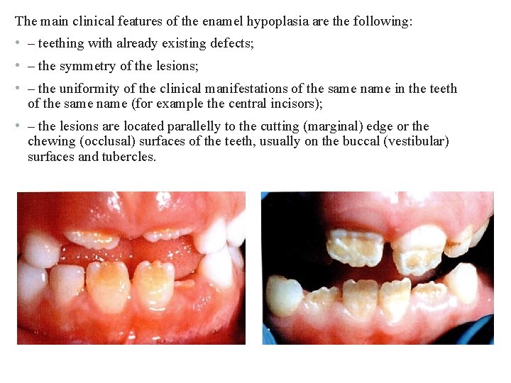
The main clinical features of the enamel hypoplasia are the following: • – teething with already existing defects; • – the symmetry of the lesions; • – the uniformity of the clinical manifestations of the same name in the teeth of the same name (for example the central incisors); • – the lesions are located parallelly to the cutting (marginal) edge or the chewing (occlusal) surfaces of the teeth, usually on the buccal (vestibular) surfaces and tubercles.
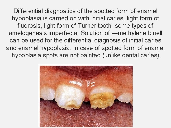
Differential diagnostics of the spotted form of enamel hypoplasia is carried on with initial caries, light form of fluorosis, light form of Turner tooth, some types of amelogenesis imperfecta. Solution of ―methylene blue‖ can be used for the differential diagnosis of initial caries and enamel hypoplasia. In case of spotted form of enamel hypoplasia spots are not painted (unlike dental caries).
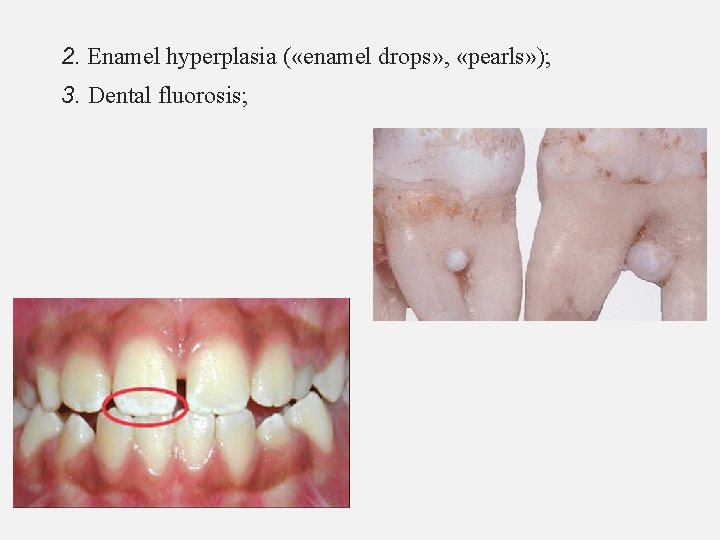
2. Enamel hyperplasia ( «enamel drops» , «pearls» ); 3. Dental fluorosis;
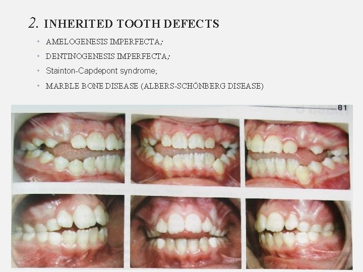
2. INHERITED TOOTH DEFECTS • AMELOGENESIS IMPERFECTA; • DENTINOGENESIS IMPERFECTA; • Stainton-Capdepont syndrome; • MARBLE BONE DISEASE (ALBERS-SCHÖNBERG DISEASE)
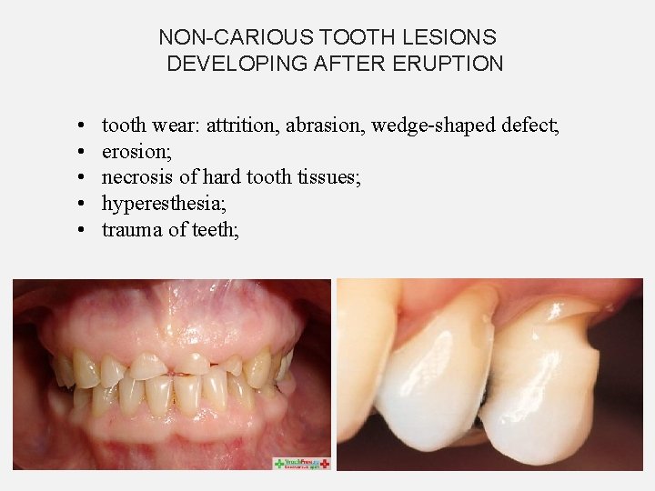
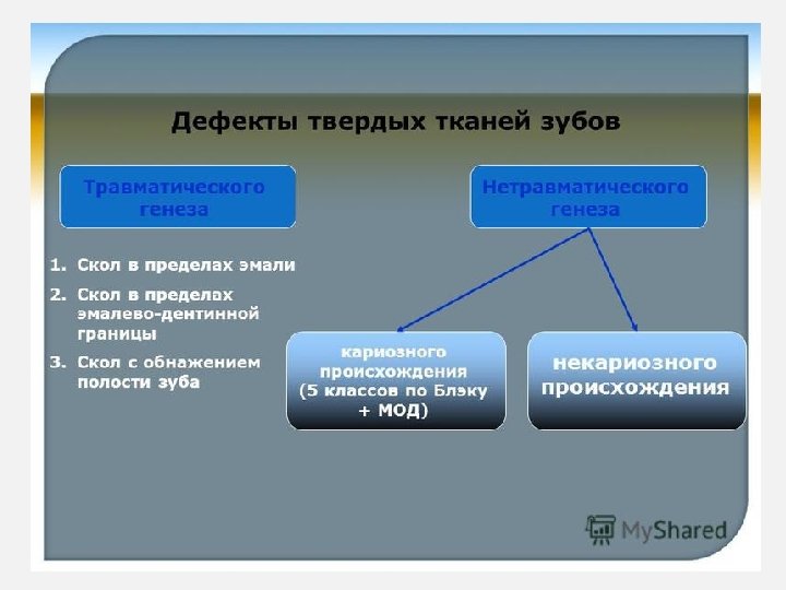
NON-CARIOUS TOOTH LESIONS DEVELOPING AFTER ERUPTION • • • tooth wear: attrition, abrasion, wedge-shaped defect; erosion; necrosis of hard tooth tissues; hyperesthesia; trauma of teeth;
The term “wedge-shaped defect” figuratively describes a hard substance defect that often appears in the mouth at the neck of teeth. It can primarily be found in prominently positioned teeth, i. e. in canines and premolars; in rare cases even in all teeth. First, the tiniest, groove-shaped usures appear at the transition from the enamel to the dentin, which grow larger and deeper over time. The surface of the defects looks smoothly polished and hard. Macroscopic symptoms of caries are missing, although a regular carious process may also appear in later stages due to increased paque retention.
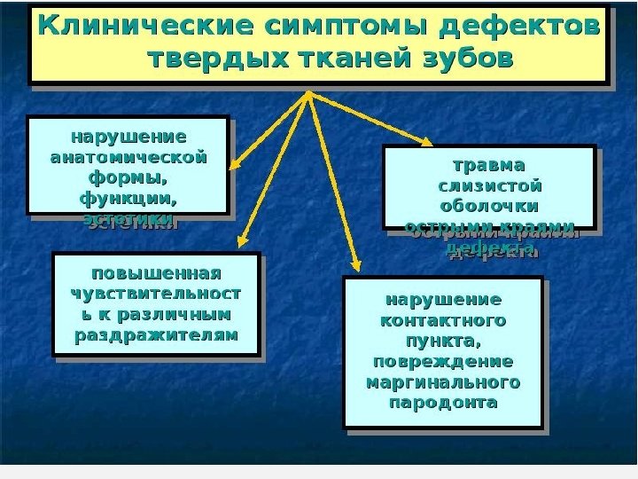
LONG-EXISTING DEFECTS CAN LEAD TO: • Deformation of the dentition and changes in the function of the masticatory muscles and temporomandibular joint; • Unilateral chewing, the associated change in the function of the masticatory muscles and the asymmetry of the facial skeleton
LONG-EXISTING DEFECTS CAN LEAD TO: • Damage to the oral mucosa sharp-edged teeth; • Violation of aesthetics, and in connection with this and a change in the psyche of the patient; • Complete destruction of the tooth crown.
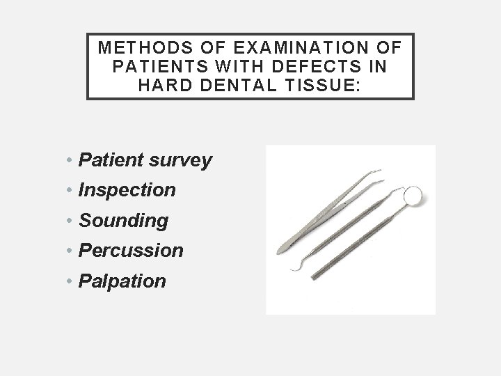
METHODS OF EXAMINATION OF PATIENTS WITH DEFECTS IN HARD DENTAL TISSUE: • Patient survey • Inspection • Sounding • Percussion • Palpation
AUXILIARY EXAMINATION METHODS - electrical diagnostics - electrometry - X-ray examination (fluoroscopy, radiography - inside - and extraoral, stereoscopic radiography, tomography, panoramic X-ray, dental computed radiography (TFR)). - Vital staining - Transillumination
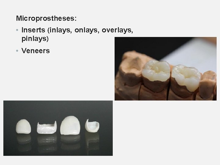
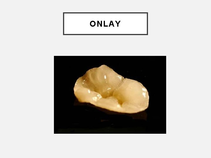
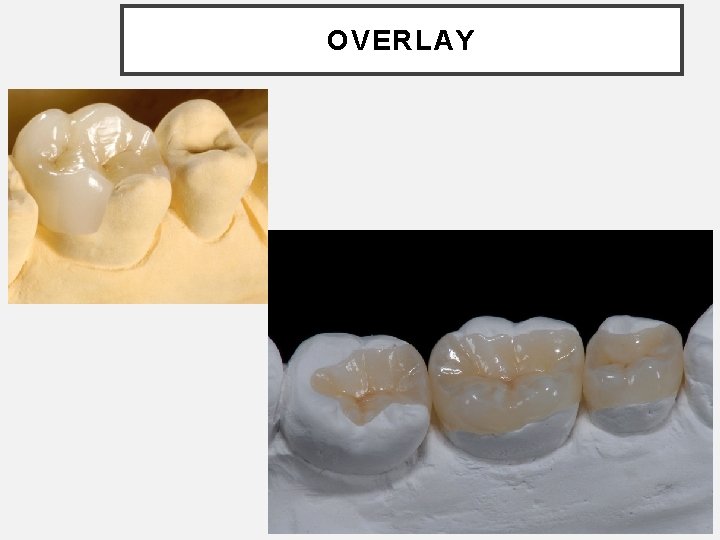
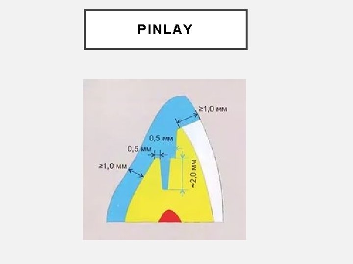
Microprostheses: • Inserts (inlays, overlays, pinlays) • Veneers
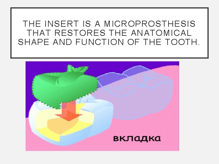
THE INSERT IS A MICROPROSTHESIS THAT RESTORES THE ANATOMICAL SHAPE AND FUNCTION OF THE TOOTH.
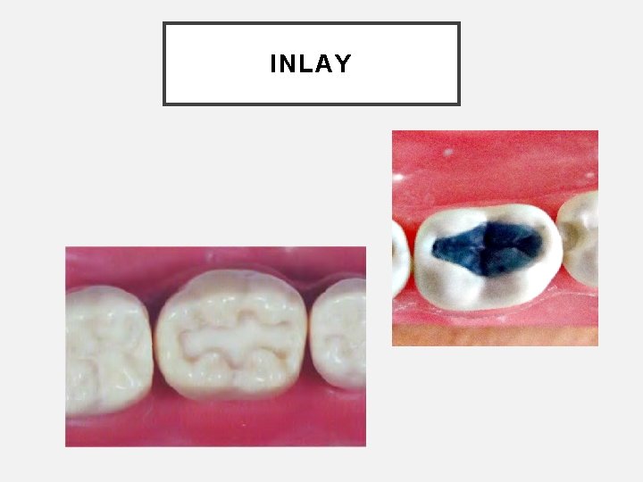
INLAY
ONLAY
OVERLAY
PINLAY
GENERAL PRINCIPLES OF FORMATION OF CAVITIES FOR INSERTS 1. The cavities are given the most appropriate form, such that the insert can be freely displayed. 2. When forming a cavity, prophylactic expansion is carried out to prevent recurrence of caries. 3. The bottom and walls of the cavity must be resistant to chewing pressure. 4. The cavity must have sufficient depth, sink into the dentin and not shift under the influence of chewing pressure. 5. The cavity must be asymmetric or have additional recesses, which serve as a guide when inserting the insert. 6. Prevention of spallation of the thinned edges of the enamel is carried out by creating a bevel along the edge of the cavity or molten chewing surface
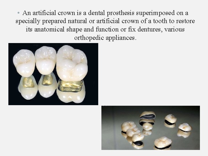
• An artificial crown is a dental prosthesis superimposed on a specially prepared natural or artificial crown of a tooth to restore its anatomical shape and function or fix dentures, various orthopedic appliances.
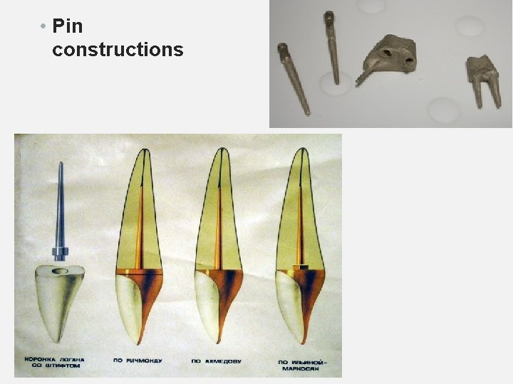
• Pin constructions
THANK YOU FOR ATTENTION!