PATHOLOGY OF ENDOCRINE SYSTEM PITUITARY GLAND Dr Bushra

PATHOLOGY OF ENDOCRINE SYSTEM PITUITARY GLAND Dr. Bushra Al. Tarawneh, MD Anatomical pathology Mutah University School of Medicine- Department of Laboratory medicine & Pathology Endocrine system lectures 2021

The Endocrine system is divided into : 1 -Endocrine organs which are entirely dedicated to production of hormones e. g pituitary, thyroid , parathyroid & adrenal. 2 - Endocrine components in clusters in organs having mixed functions, e. g. pancreas, ovary & testes. 3 -Diffuse endocrine system, comprising scattered cells within organs or tissues acting locally on adjacent cells without entry into blood stream ( Paracrine).

Classification of Hormones A- Hormones that trigger biochemical signals upon interacting with cell surface receptors: Leads to an increase in intracellular molecules, termed second messenger (c. AMP) , production of mediators from membrane phospholipid in the intracellular calcium proliferation, differentiation, survival, functional activity of the cells. 1 - peptide hormones: Growth hormones, and insulin. 2 - small molecules: epinephrine.

B- Hormones that diffuse across the plasma membrane and interact with intracellular receptors: Lipid -soluble hormones include: steroids ( estrogen, progesterone, glucocorticoids), retinoids, thyroxine.
")
Disease divided into : 1 - Diseases of overproduction of secretion ( Hyperfunction ) 2 - Diseases of underproduction ( Hypofunction ) 3 - Mass effects ( Tumors ) N. B. Correlation of 1 - Clinical picture. 2 - Hormonal assays , 3 - Biochemical findings , together with 4 -Pathological picture are of extreme importance in most conditions.

PITUITARY GLAND

� The pituitary lies in sella turcica, & weighs about 0. 5 gm. It is connected to the HYPOTHALAMUS through its stalk, and composed of : A-ADENOHYPOPHYSIS- (80%) developed from Rathke’s pouch. Its blood supply is through venous plexus from hypothalamus. It is controlled under Hypothalamic-Hypophyseal feed back control. Produce GH, PROLACTIN, ACTH, FSH, LH, TSH. B- NEUROHYPOPHYSIS developed from the floor of the third ventricle &consists of modified glial cells & axons from cell bodies in hypothalamus. It has its own blood supply. Produce oxytocin &ADH
 and prolactin (PRL) The dark purple")
. The pink acidophils secrete growth hormone (GH) and prolactin (PRL) The dark purple basophils secrete corticotrophin (ACTH), thyroid stimulating hormone (TSH), and gonadotrophins follicle stimulating hormone-luteinizing hormone (FSH and LH). The pale staining chromophobes have few cytoplasmic granules, but may have secretory activity. 8

The neurohypophysis shown here resembles neural tissue, with glial cells, nerve fibers, nerve endings, and intra-axonal neurosecretory granules. The hormones vasopressin (antidiuretic hormone, or ADH) and oxytocin made in the hypothalamus (supraoptic and paraventricular nuclei) are transported into the intraaxonal neurosecretory granules where they are released. 9

10
 1 - Somatotrophs from")
CELLS & SECRETIONS : A- Anterior pituitary ( Adenohypophysis ) 1 - Somatotrophs from acidophilic cells → Growth H. 2 - Lactotrophs from acidophilic cells → Prolactin 3 - Corticotrophs from basophilic cells → ACTH, POMC derived peptides. 4 - Thyrotrophs from pale basophilic cells → TSH 5 - Gonadotrophs from basophilic cells → FSH, LH B- Posterior pituitary ( Neurohypophysis ) 1 - Oxytocin 2 - ADH

HYPERPITUITARISM & PITUITARY ADENOMA In most cases, excess is due to ADENOMA arising in the anterior lobe. Less common causes include : * Hyperplasia * Carcinoma * Ectopic hormone production * Some hypothalamic disorders

Incidence of pituitary adenomas: � 10% of all intracranial neoplasms � 25% are incidental � 3% occur with MEN syndrome � Most occur between 30 -50 years of age

Behaviour of pituitary adenomas : � Primary pituitary adenomas usually benign. � Radiological changes in sella turcica. � May or may not be functional(20%). If functional (80%), the clinical effects are secondary to the hormone produced. � More than one hormone can be produced from the same cell ( monoclonal ).

CLINICAL FEATURES of PITUITARY ADENOMA: 1 - Symptoms of hormone production. 2 - Visual field abnormalities (pressure on optic chiasma above sella tursica ). 3 - Elevated intracranial pressure (blockage of CSF flow ): Headache , nausea , vomiting. 4 - Hypopituitarism ( result from pressure on adjacent pituitary ): Diabetes insipidus. 5 -Cranial nerve palsy ( invasion to brain ).

16

Mass effect of pituitary adenoma Downloaded from: Robbins & Cotran Pathologic Basis of Disease (on 4 December 2005 01: 50 PM)

Morphology of pituitary adenomas : � Well circumscribed, invasive in up to 30% � Size 1 cm. or more, specially in nonfunctioning tumor � Hemorrhage & necrosis seen in large tumors. Microscopic picture: � Uniform cells, one cell type (monomorphism) � Absent reticulin network � Rare or absent mitosis

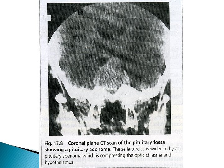
Sella turcica with pituitary adenoma

Normal pituitary gland

Uniform cells of pituitary adenoma Downloaded from: Robbins & Cotran Pathologic Basis of Disease (on 4 December 2005 01: 50 PM)

Types of Pituitary Adenomas � Previously classified according to histological picture e. g : Acidophilic Adenoma � Now according to immunohistochemical findings & clinical picture …. . e. g. Growth hormone secreting adenoma

24

1 - PROLACTINOMA : � 30% of all adenomas, chromophobe or w. acidophilic � Functional even if microadenoma , but amount of secretion is related to size � Mild elevation of prolactin does NOT always indicate prolactin secreting adenoma ! � Other causes of prolactin include : ◦ ◦ ◦ estrogen therapy pregnancy certain drugs, e. g reserpine (dopamin inhibitor). hypothyroidism mass in suprasellar region ?

� Any mass in the suprasellar region may interfere with normal prolactin inhibition Prolactin ( STALK EFFECT )

Symptoms : � Galactorrhea � Amenorrhea � Decrease libido � Infertility Treatment : Bromocreptine (dopamine agonist ); cause shrinkage of neoplasm & regression of hyperplasia in most causes.

2 - Growth hormone secreting adenoma : � 40% Associated with GNAS 1 gene mutation � Persistent secretion of GH stimulates the hepatic secretion of insulin-like growth factor I (IGF-I) many of clinical effects � Initial investigation : measurement of GF & IGF-I which is increased. � Confirm by failure to suppress GH production in response to an oral load of glucose.

Structure : Composed of granular ACIDOPHILIC cells and may be mixed with prolactin secretion. Symptoms : May be delayed so adenomas are usually large Produce GIGANTISM (children) or ACROMEGALLY (adults). Diabetes, arthritis, large jaw & hands, osteoporosis, BP, HF…. . etc

3 - Corticotroph cell adenoma � Usually microadenomas � Higher chance of becoming malignant � Chromophobe or basophilic cells � Functionless or Cushing ‘s Disease ( ACTH ) � Bilateral adrenalectomy or destruction may result in aggressive adenoma: Nelson’s Syndrome � ICP

4 - Non functioning adenoma, 20% silent or null cell , nonfunctioning & produce mass effect only. 5 - Gonadotroph producing LH &FSH, ( 10 -15%)- Function silent or is minimal , late presentation mainly mass effect produced. Produce gonadotrophin subunit, β- FSH & β-LH. 6 - TSH producing, (1%) rare cause of hyperthyroidism. 7 - Pituitary carcinoma, Extremely rare, diagnosed only by metastases.

HYPOPITUITARISM : of 75% of ant. Pituitary Symptoms � Congenital or acquired, intrinsic or extrinsic � Acquired causes include : � Loss 1 - Nonsecretory pituitary adenoma 2 - SHEEHAN’S SYNDROME 3 - Ischemic necrosis e. g. sickle cell anemia, DIC… 4 - Pituitary apoplexy… 5 - Iatrogenic by radiation or surgery 6 - Autoimmune ( lymphocytic) hypophysitis 7 - Hypothalamic mass 8 - Inflammatory e. g sarcoidosis or TB …. .

9 - Empty Sella Syndrome : Radiological term for enlarged sella tursica, with atrophied or compressed pituitary. May be primary due to downward bulge of arachnoid into sella floor compressing pituitary. Secondary is usually surgical. 10 - Infiltrating diseases in adjacent bone e. g. Hand Schuller – Christian Disease Metastatic tumors 11 - Craniopharyngioma
 in children. ◦ Effect of individual hormone")
� Symptoms of hypopituitarism: Dwarfism (Pituitary Dwarf) in children. ◦ Effect of individual hormone deficiencies. ◦ Amenorrhea & no lactation ◦ Loss of MSH → Decreased pigmentation ◦

Craniopharyngioma : * 1 -5 % of intracranial neoplasms * Derived from remnants of Rathke’s Pouch * Suprasellar or intrasellar , often cystic with calcification * Children or adolescents most affected * Symptoms may be delayed ≥ 20 yrs( 50%) * Symptoms of hypofunction or hyperfunction of pituitary and /or visual disturbances, diabetes insipidus * Benign & slow growing

POSTERIOR PITUITARY SYNDROMES: A- ADH deficiency : Diabetes Insipidus Polyuria, polydipsia, hypernatremia & dehydration. Urine is dilute, due to inability to reabsorb water from the collecting tubules. Causes : Head trauma, tumors & inflammations in pituitary or hypothalamus…etc.
: • Part of paraneoplastic Syndrome : Small")
B- Syndrome of inappropriate ADH secretion (SIADH): • Part of paraneoplastic Syndrome : Small Cell CA of Lung • Causes excessive resorption of water hyponatremia, cerebral edema. C-Abnormal oxytocin secretion : Abnormalitis of synthesis & release have not been associated with any significant abnormality.
- Slides: 37