PATHOLOGY Chapter CELL INJURY for first year medical
 [Topics in")
 The students should: A. Understand the")


, and everything looks")


 § Fat necrosis")
 §")


 and apoptosis (right). Apoptosis is different from necrosis.")
 Reduced (shrinkage) Nucleus Pyknosis Karyorrhexis karyolysis Fragmentation")
- Slides: 34
PATHOLOGY Chapter: CELL INJURY (for first year medical students in 3 lectures) [Topics in 3 lectures: cell injury, free radical injury, necrosis and apoptosis, cellular accumulations, pathological calcification, adaptation to cell injuries] (lecture 2) Dr. Sufia Husain Associate Prof & Consultant Department of Pathology College of Medicine, KSU, Riyadh. 29 th September 2020 Reference: Robbins & Cotran Pathology and Rubin’s Pathology
Objectives for Cell Injury Chapter (in 3 lectures) The students should: A. Understand the concept of cells and tissue adaptation to environmental stress including the meaning of hypertrophy, hyperplasia, atrophy, hypoplasia and metaplasia with their clinical manifestations. B. Is aware of the concept of hypoxic cell injury and its major causes. C. Understand the definitions and mechanisms of free radical injury. D. Knows the definition of apoptosis, tissue necrosis and its various types with clinical examples. E. Able to differentiate between necrosis and apoptosis. F. Understand the causes of and pathologic changes occurring in fatty change (steatosis), accumulations of exogenous and endogenous pigments (carbon, silica, iron, melanin, bilirubin and lipofuscin). G. Understand the causes of and differences between dystrophic and metastatic calcifications.
Lecture 2 outline A. Types of necrosis : Coagulative, Liquefactive, Caseous, gangrenous, fibrinoid and fat necrosis. B. Apoptosis : definition, morphologic features, regulation of apoptosis C. Comparison between necrosis and apoptosis.
Normal cell Stress/increased demand Adaptation e. g. hypertrophy Unable to adapt Injurious stimuli e. g. hypoxia Cell injury No more injurious stimuli injury is reversible complete repair Cells/ tissue back to normal Restoration of normal organ function. Persistent or strong injurious stimuli 2 possibilities 1) tissue repair but with diminished capacity impaired cell function 2) injury becomes irreversible cell death (necrosis or apoptosis)
NECROSIS. § Necrosis is a type of cell death, in which there is enzymatic digestion of the dying cell. In necrosis, enzymes leak out of lysosomes, enter the cytoplasm and digest the cell. § Necrosis is always a pathologic process, associated with pathologic cell injury. § It occurs in irreversible cell injury. It is usually associated with inflammation in the surrounding tissue. § It involves the death of a group of cells in one area. § In necrosis: Øthere is loss of function of the involved tissue/organ. Øthere is an inflammatory response ØThe necrotic cell releases certain enzymes into the blood and these enzymes can be detected in blood tests. The level of these enzymes can be used as markers to diagnose the injury and also can help determine the time and the extent of injury e. g. Detection of cardiac enzymes in myocardial infarction (heart attack).
NOTE: The enzymes used in this degradation of a cell come from either the lysosomes of the dying cell itself referred to as autolysis or from lysosomes of neighboring leukocytes referred to as heterolysis. Autolysis is the death/disintegration of cells or tissues by it’s own enzymes. Autolysis is seen in cells in necrosis and after death/ post mortem.
Types of necrosis § There are 5 types of necrosis: Ø coagulative necrosis Ø liquefactive necrosis Ø caseous necrosis Ø fat necrosis Ø fibrinoid necrosis
Coagulative necrosis is characteristically seen when blood flow to an organ is affected leading to ischemic, hypoxic death of cells in that organ. Coagulative necrosis is seen in all organs except the brain. It is not seen in the brain. Coagulative necrosis in heart is called as myocardial infraction, in kidney is called as renal cortical necrosis or renal infarct, in spleen splenic infarct in liver hepatic infarct etc. Grossly: the affected organ looks pale and firm/solid. It looks like cooked meat or boiled egg. The tissue is firm and architecture is maintained for days after cell death. Microscopy: in the beginning there is preservation of the general tissue architecture. The basic ghost outline of the affected/coagulated cell remains preserved for a few days but the nucleus is lost. The cell cytoplasm is eosinophilic. Ultimately, the necrotic cells are removed by phagocytosis by the macrophages (they act like vacuum cleaners) and the affect area is replaced by fibrosis.
Kidney: coagulative necrosis Gross: tissue is firm
Kidney: coagulative necrosis Micro: Cell outlines are preserved (cells look ghostly), and everything looks red
Liver coagulative necrosis
Liquefactive necrosis § is a type of necrosis which results in transformation of the tissue into a liquid viscous material. § Liquefactive necrosis is characteristically seen in: a) Ischemic (hypoxic) cell death in the brain/ central nervous system b) And it is also seen in the necrosis that results from infections especially suppurative bacterial infection (suppurative = pus or abscess producing). § In infections: the affected tissue is softened/liquefied by the action of hydrolytic (digestive) enzymes released from the lysosomes of neutrophils. The affected area becomes soft and liquefied with a creamy yellow center containing necrotic cells and neutrophils and is called pus/abscess. § Note: the reason for liquefactive necrosis following ischemic injury in the brain is poorly understood. § Ultimately, the necrotic cells are phagocytosed.
Liquefactive necrosis
Infection: Liquefactive necrosis (center labeled one is necrosis and surrounded by neutrophils.
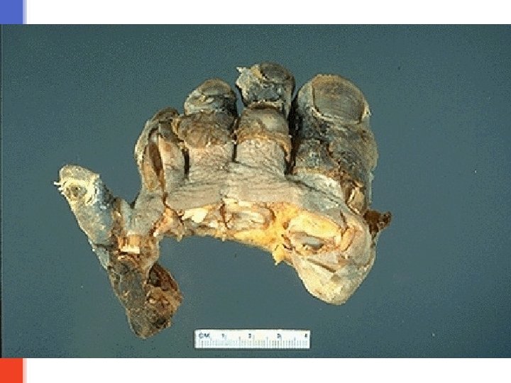
Caseous necrosis § is a type of coagulative necrosis classically seen in tuberculosis (infection by mycobacterium tuberculi). § Grossly: it is white, soft, curdy, cheesy-looking “caseous” material. § On microscopic examination, the necrotic area appears as amorphous pink granular debris surrounded by a collar of epitheloid cells (they are modified macrophages), lymphocytes and giant cells. This is known as granuloma. § Here the tissue architecture is completely obliterated.
Tuberculous lung with a large area of caseous necrosis. The caseous debris is yellow-white and cheesy
Fat necrosis § It is necrosis of fat cells. § Typically, it is seen in acute pancreatitis in which the injured pancreatic cells release the lipase enzyme into the surrounding fat in the abdominal cavity and cause enzymatic digestion of fat cells. § The released lipase breaks down the fat cells into glycerol and free fatty acids. The produced fatty acids combine with calcium circulating in the blood to produce calcium soaps which looks like chalky white spots in the necrotic fat. This process is called as fat saponification. § Microscopy: the outlines of necrotic/dead fat cells can be seen. Inflammation is minimal. § Fat necrosis can also be seen in breast fat and other fatty areas due to traumatic injury. © Putz/Pabst: Sobotta. Atlas der Anatomie des Menschen, 21. Aufl. Urban & Fischer, 2000
Figure 1 -21 Foci of fat necrosis with saponification in the mesentery. The areas of white chalky deposits represent calcium soap formation at sites of lipid breakdown. Downloaded from: Robbins & Cotran Pathologic Basis of Disease (on 4 September 2005 10: 51 AM) © 2005 Elsevier
§ Normal fat (adipose tissue) § Fat necrosis
Fibrinoid necrosis § Is necrosis in the blood vessels (arteries, arterioles and capillaries) § There is deposition of fibrin material in the arterial walls, which appears smudgy and acidophilic/eosinophilic. § It is seen in immune mediated diseases (autoimmune diseases) and also seen in malignant hypertension.
Fibrinoid necrosis
Gangrenous necrosis It is a term commonly used in clinical practice by surgeons. It can be dry or wet. v. Dry gangrene/ mummification: it is coagulative necrosis typically seen in a limb with inadequate blood supply (ischemia of that limb). Dry gangrene is non-infected ischemic coagulative necrosis of tissue. It is without superadded infection. It is seen as a complication of peripheral artery disease e. g. atherosclerosis and diabetes mellitus. The affected part is dry, shrunken and dark reddish-black. v. Wet/ infected gangrene: it is dry gangrene (coagulative necrosis) with superadded bacterial (putrefactive) infection resulting in liquefactive necrosis. So, initially there is coagulative necrosis and then there is superadded infection associated liquefactive necrosis. Wet gangrene usually develops rapidly. The bacteria is usually saprogenic (i. e. it lives in the gut or the soil and it can thrive in low oxygen states) e. g. gram-positive Clostridia or Bacillus fusiformis. It has a poor prognosis compared to dry gangrene because the infection can spread to the rest of the body (septicemia) and be life threatening (death). The limb becomes foul smelling and black and starts decomposing. Treatment of gangrene: amputation. NOTE: Diabetes mellitus is a risk-factor for dry gangrene and for wet gangrene (as elevated serum glucose creates a favorable environment for bacterial infection).
§ When a cell is deprived of growth factors or the cell's DNA or proteins are damaged beyond repair, the cell kills itself by another type of death, called apoptosis, which is characterized by nuclear dissolution without complete loss of membrane integrity. Apoptosis is an active, energy-dependent, tightly regulated type of cell death that is seen in some specific situations. Apoptosis serves many normal functions and is not necessarily associated with pathologic cell injury.
APOPTOSIS Apoptosis is programmed cell death. Apoptosis means “falling off”. It is a type of cell suicide. Is results from activation of ‘death pathway genes’. It is a pathway of cell death in which cells destined to die activate their own enzymes to degrade their own nuclear DNA and proteins. When a cell is deprived of growth factors or the cell's DNA or proteins are damaged beyond repair, the cell kills itself by apoptosis. Apoptosis is characterized by nuclear dissolution without complete loss of membrane integrity. It is an active, energy-dependent, tightly regulated type of cell death. It can be Physiological/ adapative. Pathologic. NOTE: Apoptosis and necrosis can sometimes coexist.
Apoptosis in Physiologic Situations § The programmed destruction of cells during embryogenesis. § Hormone-dependent: e. g. endometrial cell breakdown during the menstrual cycle, the regression of the lactating breast after weaning, and prostatic atrophy after castration (adaptive atrophy). § Apoptosis in proliferating cells e. g. intestinal epithelial lining is always being replaced. § Cells that after performing their function undergo apoptosis e. g. neutrophils and lymphocytes in inflammation. § Sometimes body produces harmful lymphocytes and they are also destroyed by apoptosis.
Apoptosis in Pathologic Conditions § Cell death produced by injury e. g. radiation. § In certain diseases e. g. in viral hepatitis the infected hepatocytes undergo apoptosis (acidophilic bodies) or injury of skin epidermal cells (keratinocytes) leads to apoptosis of keratinocytes (Civatte bodies). § Pathologic atrophy in organs e. g. pancreas, parotid gland, and kidney § Corticosteroid induced atrophy of the neonatal thymus § Cell death in tumors (usually accompanied by necrosis).
Mechanism of Apoptosis § The death pathway genes are activated which trigger apoptosis. § Cell shrinkage. § Chromatin condensation in the nucleus: This is the most characteristic feature of apoptosis. The nucleus may break up into fragments. § Formation of cytoplasmic blebs and apoptotic bodies: The apoptotic cell first shows surface blebbing, then fragments into membrane-bound apoptotic bodies. The apoptotic bodies contain cytoplasmic content with or without nuclear material. § The cell's plasma membrane remains intact. The plasma membrane of the apoptotic cell sends signal to macrophages, inviting the macrophages to phagocytose it. § Phagocytosis of apoptotic bodies by the macrophages. Because, during the entire process, the apoptotic body is bound by plasma membrane, there is no release of the cytoplasmic content into the surrounding tissue and therefore there is no inflammation.
Morphology of Apoptosis § On histology apoptosis involves single cells or small clusters of cells. The apoptotic cell appears as a round or oval mass of intensely eosinophilic cytoplasm with dense nucleus. There is no inflammation. apoptosis in liver cell apoptosis in epidermis.
Important enzymes of apoptosis 1. Cysteine proteases named caspases 2. Ca 2+ dependent endonucleases 3. Mg 2+ dependent endonucleases Regulation of apoptosis It is mediated by a number of genes and their products e. g: bcl-2 gene inhibits apoptosis bax genes facilitates apoptosis p 53 facilitates apoptosis by inhibiting bcl 2 and promoting bax genes.
The changes seen in necrosis (left) and apoptosis (right). Apoptosis is different from necrosis. In necrosis there is loss of membrane integrity, enzymatic digestion of cells, and frequently an inflammatory reaction. Apoptosis and necrosis sometimes coexist.
Feature Necrosis Apoptosis Cell size Enlarged (swelling) Reduced (shrinkage) Nucleus Pyknosis Karyorrhexis karyolysis Fragmentation into nucleosome size fragments Plasma membrane Disrupted Intact; altered structure, especially orientation of lipids Cellular contents Enzymatic digestion; may leak out of cell Intact; may be released in apoptotic bodies Adjacent Frequent inflammation No Physiologic Invariably pathologic or pathologic (culmination of irreversible role cell injury) Often physiologic, means of eliminating unwanted cells; may be pathologic after some forms of cell injury, especially DNA damage