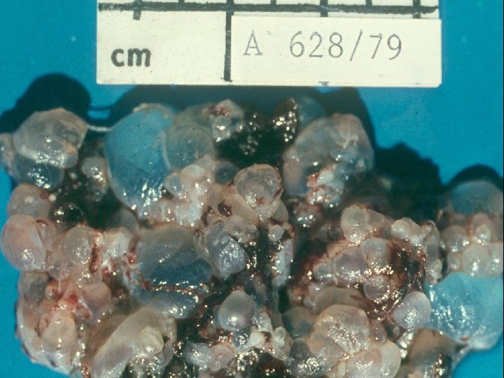
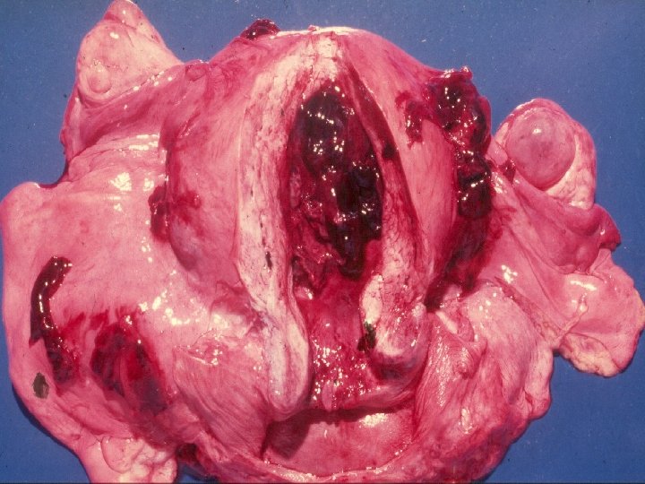
Pathologic diagnosis of mola hydatidosa Hydatidiform mole 1977


")
- Slides: 6
Pathologic diagnosis of mola hydatidosa Hydatidiform mole 1977 - morphology Complete mole Partial mole Diploid Triploid 1990 s - imprinting Androgenetic Biparental 2000 s – diagnostic refinement Early diagnostic criteria Mimics, diploid variants 1980 s - ploidy
Hydatidiform mole: genetic differences Gestation type Ploidy M M P 1 P 2 Normal Partial mole 2 n 3 n Complete mole (90%) (10%) 2 n 2 n (Duplic. 1 n x 2) Genome p. GTN / CC risk Biparental Paternal Low High
Histology of early / first trimester hydatidiform mole Complete mole Partial mole Villous morphology Smooth, contoured with “toe-like” budding, pseudoinclusions irregular, large and ovoid Irregular “dentate” with scalloped outline, pseudoinclusions regular small and round Villous stroma Hypercellular with karyorrhectic debris, mucoid, mild oedema Scattered villi with mild oedema, fibrosis Villous blood vessels Absent to severely attenuated, collapsed and empty Numerous, with many nucleated RBC, “pseudoangiomatoid” Trophoblast hyperplasia Prominent and multifocal / circumferential, extravillous pleomorphic trophoblast sheets Mild and patchy, “lacelike” / vacuolated with “dripping-off” appearance
Hydatidiform mole: follow up 8, 9 COMPLETE MOLE Spontaneous remission b-HCG persistence (p. GTN) Choriocarcinoma PARTIAL MOLE 75 % 95 % 15 - 30 % 0, 5 - 5 % 2% Rare