Pathogenesis of Cerebral Infarction at Cellular Molecular Levels

Pathogenesis of Cerebral Infarction at Cellular & Molecular Levels By: Amr Moustafa, MD, Ph. D

Objectives: n Identify the possible cell death mechanisms implicated in the pathogenesis of ischemic brain injury n Acquire the knowledge of the important role played by oxidative stress and free radicals in the pathogenesis of cerebral infarction n Understand the various factors involved in ischemia -induced metabolic stress n Identify the Neurochemical changes involved in cerebral ischemia
 Cerebral infarction Global incidence: 32% Hemorrhagic Ischemic Intracerebral Thrombotic")
Subtypes of Cerebral Infarction (Stroke) Cerebral infarction Global incidence: 32% Hemorrhagic Ischemic Intracerebral Thrombotic Subarachnoid Embolic Global incidence: 68% http: //www. uptodate. com/contents/overview-of-the-evaluation-of-stroke
")
Characteristics of stroke subtypes Stroke type Clinical course Intracerebral hemorrhage Gradual progression (min –hrs) Subarachnoid hemorrhage Abrupt onset of sudden, severe headache. Risk factors Other clues HTN, trauma, bleeding diatheses, illicit drugs, vascular malformations, certain races. May be precipitated by physical Smoking, HTN, alcohol, activity. Patient may have reduced genetic susceptibility alertness. (family history of subarachnoid hemorrhage) and sympathomimetic drugs http: //www. uptodate. com/contents/overview-of-the-evaluation-of-stroke
 Ischemic (embolic) Clinical course Stuttering progression")
Characteristics of stroke subtypes, Stroke type Ischemic (thrombotic) Ischemic (embolic) Clinical course Stuttering progression with periods of improvement. Lacunes develop over hours or at most a few days; large artery ischemia may evolve over longer periods. Sudden onset with deficit maximal at onset. Clinical findings may improve quickly. Risk factors continued. . Other clues Atherosclerotic risk May have neck bruit. factors (age, smoking, DM, etc. ). Men affected more commonly than women. May have history of TIA. Atherosclerotic risk factors. Men affected more commonly than women. History of heart disease Can be precipitated by getting up at night to urinate, or sudden coughing or sneezing. http: //www. uptodate. com/contents/overview-of-the-evaluation-of-stroke

Epidemiology of Stroke n Globally: n n n Stroke is the second most common cause of mortality Stroke is the third most common cause of disability The incidence of stroke is: q q n n decreasing in high-income countries increasing in low-income countries The overall rate of stroke-related mortality is decreasing in high and low income countries The absolute number of people with stroke, stroke survivors, strokerelated deaths, and the global burden of stroke-related disability is high and increasing Men have a higher incidence of stroke than women at younger but not older ages, Stroke incidence is higher in women ≥ 75 year old compared to men http: //www. uptodate. com/contents/overview-of-the-evaluation-of-stroke

The cell death mechanisms implicated in the pathogenesis of ischemic brain injury

Cell death mechanisms in cerebral ischemia: Necrosis and Apoptosis n Necrosis: is commonly observed early after severe ischemic insults n Apoptosis: occurs with more mild insults and with longer survival periods n The mechanism of cell death involves calcium-induced calpainmediated proteolysis of brain tissue n Substrates for calpain include: q Cytoskeletal proteins q Membrane proteins q Regulatory and signaling proteins

Biochemical Responses to Ischemic Brain Injury

Biochemical Responses to Ischemic Brain Injury Oxidative stress n Metabolic stress n Neurochemical response n

Oxidative stress

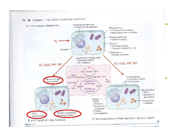
Oxidative stress n A condition in which cells are subjected to excessive levels of Reactive oxidizing species (Oxygen or nitrative species) & they are unable to counterbalance their deleterious effects with antioxidants. n It has been implicated in the ageing process & in many diseases (e. g. , atherosclerosis, cancer, neurodegenerative diseases, stroke)
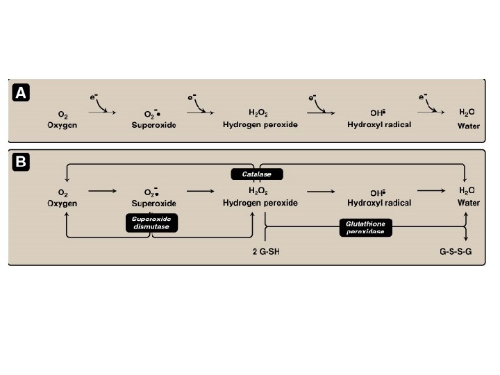
 & Reactive Nitrative Species (RNS) in Normal")
The Role of Reactive Oxygen Species (ROS) & Reactive Nitrative Species (RNS) in Normal Brain Physiology n They are mainly generated by microglia & astrocytes n They modulate synaptic transmission & non-synaptic communication between neurons & glia n During periods of increased neuronal activity, ROS & RNS diffuse to the myelin sheath of oligodendrocytes activating Protein kinase C (PKC) posttranslational modification of myelin basic protein (MBP) by phosphorylation n They regulate neuronal signaling in both central & peripheral nervous systems n They are required for essential processes as learning & memory formation

The brain and Oxidative stress n The brain is highly susceptible to ROSinduced damage because of: q q q High concentrations of peroxidisable lipids Low levels of protective antioxidants High oxygen consumption High levels of iron (acts as pro-oxidants under pathological conditions) The occurrence of reactions involving dopamine & Glutamate oxidase in the brain

Molecular & Vascular effects of ROS in ischemic stroke n Molecular effects: q q q q n DNA damage Lipid peroxidation of unsaturated fatty acids Protein denaturation Inactivation of enzymes Cell signaling effects (e. g. , release of Ca 2+ from intracellular stores) Cytoskeletal damage Chemotaxis Vascular effects: q q q Altered vascular tone and cerebral blood flow Increased platelet aggregability Increased endothelial cell permeability

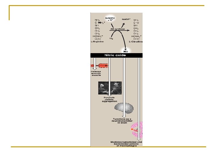
The role of NO in the pathophysiology of cerebral ischemia n Ischemia abnormal NO production n This may be both beneficial and detrimental, depending upon when and where NO is released n NO produced by endothelial NOS (e. NOS) improving vascular dilation and perfusion (i. e. beneficial). n In contrast, NO production by neuronal NOS (n. NOS) or by the inducible form of NOS (i. NOS) has detrimental (harmful) effects. n Increased i. NOS activity generally occurs in a delayed fashion after brain ischemia and trauma and is associated with inflammatory processes

Metabolic stress

Biochemical changes in The brain during ischemia Ischemia interruption or severe reduction of blood flow, O 2 & nutrients in cerebral arteries energy depletion (depletion of ATP & creatine phosphate) • Inhibition of ATP-dependent ion pumps • Membranes depolarization • Perturbance of transmembrane ion gradients Lactic acid in neurons acidosis promotes the prooxidant effect ↑ the rate of conversion of O 2. - to H 2 O 2 or to hydroxyperoxyl radical • Ca 2+ Influx (translocation from extracellular to intracellular spaces) activation of cellular proteases (Calpains) & lipases breakdown of cerebral tissue • Na+ influx • K+ efflux • K+-induced release of excitatory amino acids

Sources & consequences of increased cytosolic Calcium in cell injury

Neurochemical response

The neurochemical response to cerebral ischemia n Following cerebral ischemia, extracellular levels of various neurotransmitters are increased e. g. , q q Glutamate Glycine GABA Dopamine

The blood tests in patients with brain ischemia or hemorrhage n n n Complete blood count, including hemoglobin, hematocrit, white blood cell count, and platelet count Prothrombin time, international normalized ratio (INR), and activated partial thromboplastin time Thrombin time and/or ecarin clotting time if patient is known or suspected to be taking a direct thrombin inhibitor or a direct factor Xa inhibitor Blood lipids, including total, high-density lipoprotein (HDL), and low-density lipoprotein (LDL) cholesterol, and triglycerides. Cardiac enzymes and troponin http: //www. uptodate. com/contents/overview-of-the-evaluation-of-stroke

Biochemical basis of pharmacological intervention

Examples of Potential Biochemical Intervention in Cerebral Ischemia n Inhibitors of glutamate release n Ca 2+ channel blockers n Nitric oxide synthase inhibitors & free radical inhibition n Calpain inhibitors

To Summarize:

Ischemic cascade Lack of oxygen supply to ischemic neurones ATP depletion Malfunctioning of membrane ion system Depolarisation of neurones Influx of calcium Release of neurotransmitters, activation of proteases Further depolarisation of cells Further calcium influx

Cosequences of brain ischemia Energy failure / depolarisation / Oxidative stress Neurotransmitter release and receptor activation Lipolysis (DAG PKC ) Ca 2+ Protein phosphorylation Breakdown of cytoskeleton (FFAs ) Membrane damage Proteolysis Dysfunction of receptors and ion channels Inhibition of axonal transport, blebbing

Take Home Message Severe cerebral ischemic insults lead to a complex cascade of biochemical and molecular events, including: 1. Cell death 2. Oxidative stress 3. Metabolic stress and neurochemical changes

THANK YOU
- Slides: 32