
PASSIVE SPACE CONTROL Dr S E Jabbarifar Isfahan
PASSIVE SPACE CONTROL Dr S. E. Jabbarifar; Isfahan Dental School, Pediatric Dentistry Departement 2009
Prerequisite knowledge u. Understand that arch length is greatest at age four years u. Tooth position is maintained by balance of forces – shift vs. drift u. Greatest amount of space closure – within first 6 months of premature tooth loss u. Sequence & timing of exfoliation/eruption
Space control vs. space maintenance u. Space control u Dynamic u Careful ongoing supervision u. Space maintenance u Utilization of appliance to preserve existing space u Not always the rule!
Variables influencing space control u. Oral musculature & habits u. Time elapsed since extraction u. Dental age, eruption sequence & bony covering u. Available space u. Interdigitation u. Absence of anomalies
Considerations in premature 1 o tooth loss u. Preserve the arch length! u. Causes: u Anterior – primarily trauma, caries u Posterior – primarily caries u. If space lost: u Space maintenance u Space regaining u No treatment
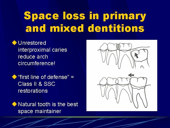
Space loss in primary and mixed dentitions u Unrestored interproximal caries reduce arch circumference! u “first line of defense” = Class II & SSC restorations u Natural tooth is the best space maintainer
Planning for space maintenance u. No medical contraindications u. Patient must be dentally fit u. Patient must be able to demonstrate good OH
Planning for space maintenance u. Parents must all understand costs involved u. Parents must understand importance of & be willing to attend regularly for appliance supervision/maintenance – teeth lost in primary dentition stage may cause delayed eruption of succedaneous teeth u. Periodic recementation may be required
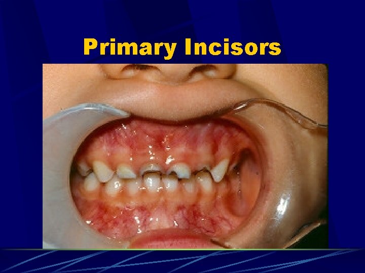
Primary Incisors
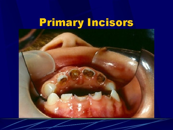
Primary Incisors
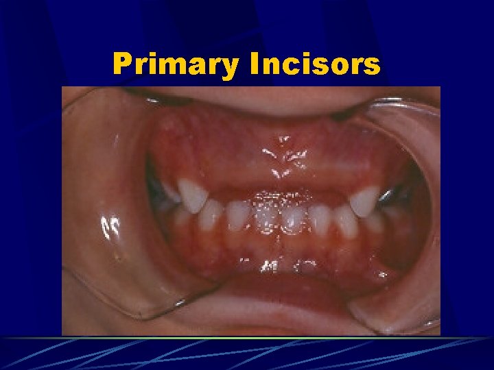
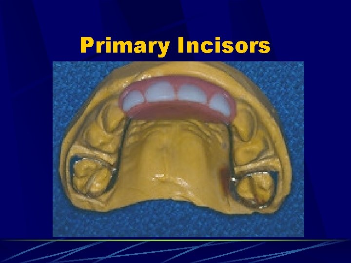
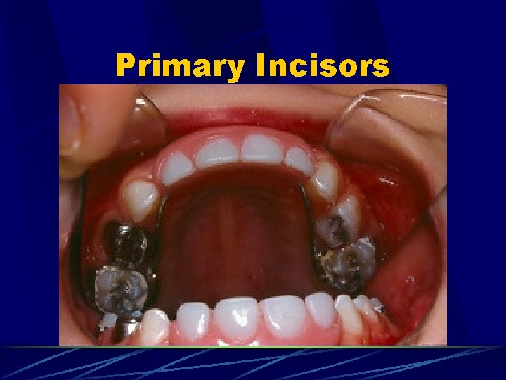
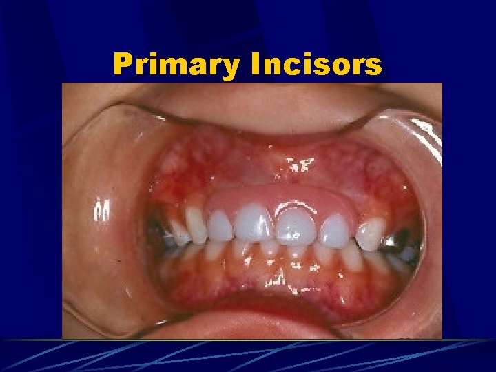
Primary Incisors u. Why replace primary incisors? u Primarily for esthetic reasons u Rarely see long-term effects on speech development and function u Once 1 o cuspids have erupted in occlusion the anterior arch length is established
Primary Incisors u. Problems with replacement: u Appliances u High are weak maintenance – close monitoring req’d u Frequent alterations as dentition changes u Appliance may enhance caries risk
Primary Incisors
Primary Incisors
Primary Incisors
Primary Incisors
Primary Canine u. Loss due to trauma or caries – rare u. Space maintainer: B&L vs. RPD u Must be removed to accommodate lateral u. No space maintainer: u Midline shift u Lingual collapse in mandible
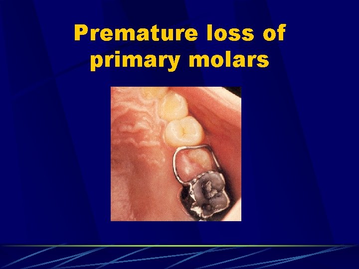
Premature loss of primary molars
Band-loop space maintainer u. Indications: u Unilateral loss of the 1 st primary molar before eruption of the 1 st permanent molar u Unilateral loss of the 1 st or 2 nd primary molar after eruption of the 1 st permanent molar u Bilateral loss of the 1 st primary molars before eruption of the permanent incisors and 1 st permanent molars u Bilateral loss of the 2 nd primary molars after eruption of the 1 st permanent molar
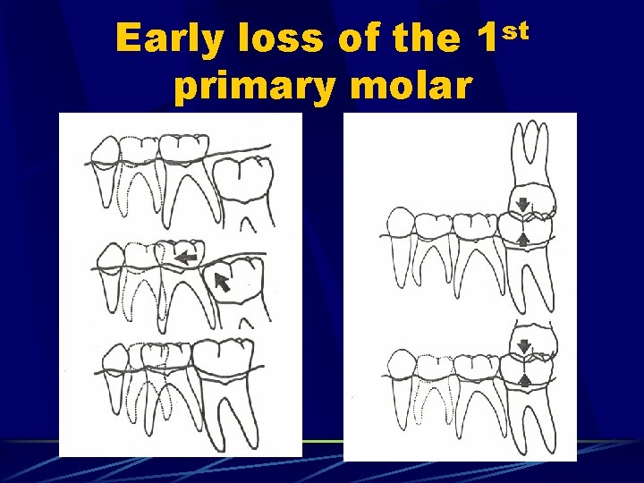
Early loss of the primary molar st 1
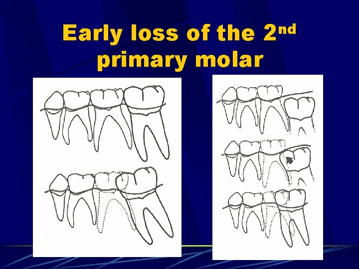
Early loss of the 2 nd primary molar
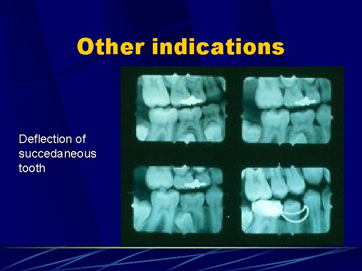
Other indications Deflection of succedaneous tooth
Band-loop space maintainer FABRICATION & DESIGN
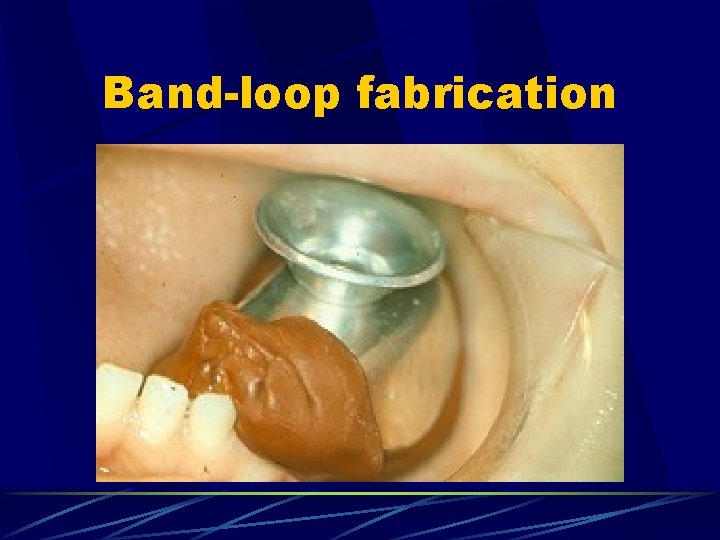
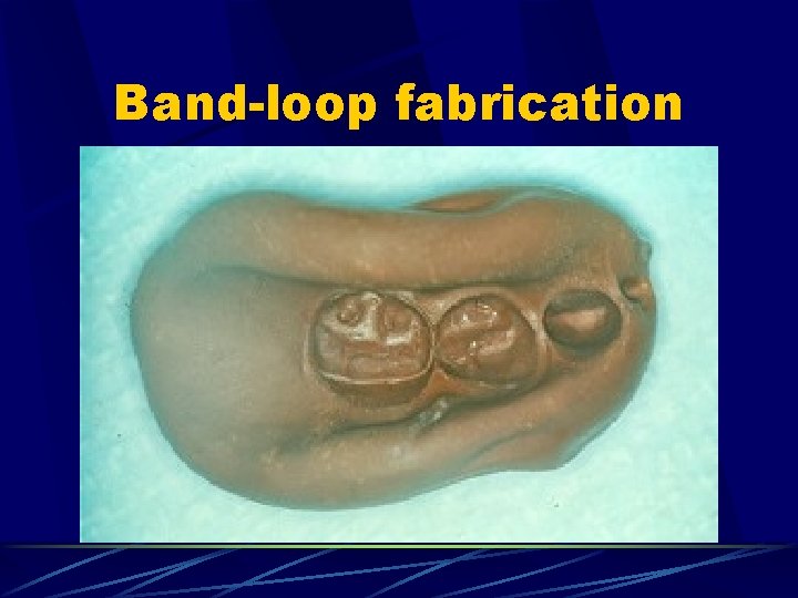
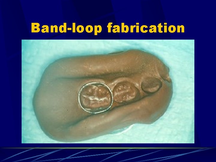
Band-loop fabrication u. Technique: u Properly fitting band on abutment tooth (pg. 389 – Pinkham) u Segmental impression (compound/alginate) u Remove band from tooth & secure in impression u Create working model
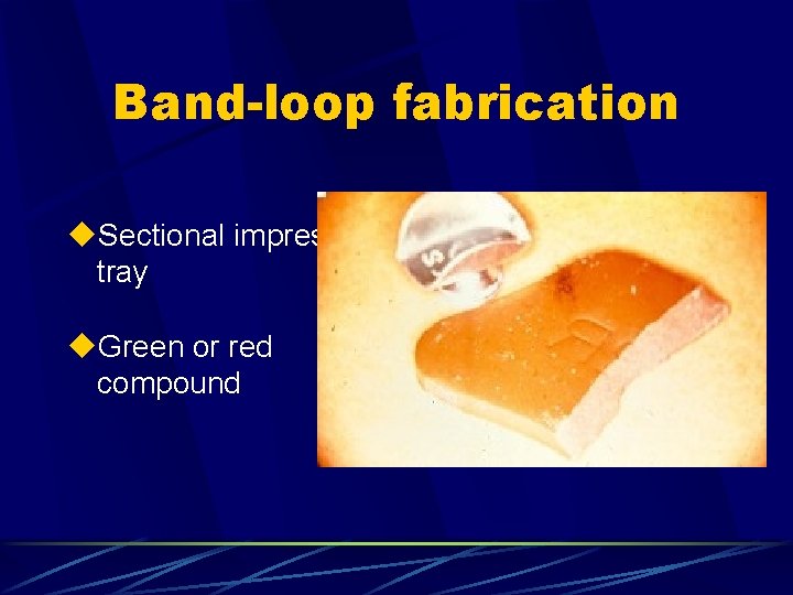
Band-loop fabrication u. Sectional impression tray u. Green or red compound
Band-loop fabrication
Band-loop fabrication
Band-loop fabrication
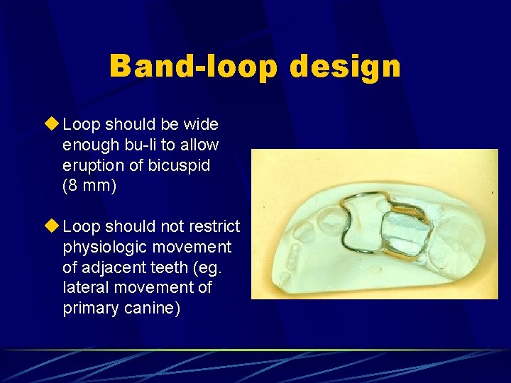
Band-loop design u Loop should be wide enough bu-li to allow eruption of bicuspid (8 mm) u Loop should not restrict physiologic movement of adjacent teeth (eg. lateral movement of primary canine)
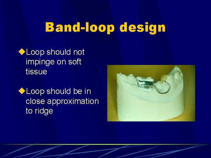
Band-loop design u. Loop should not impinge on soft tissue u. Loop should be in close approximation to ridge
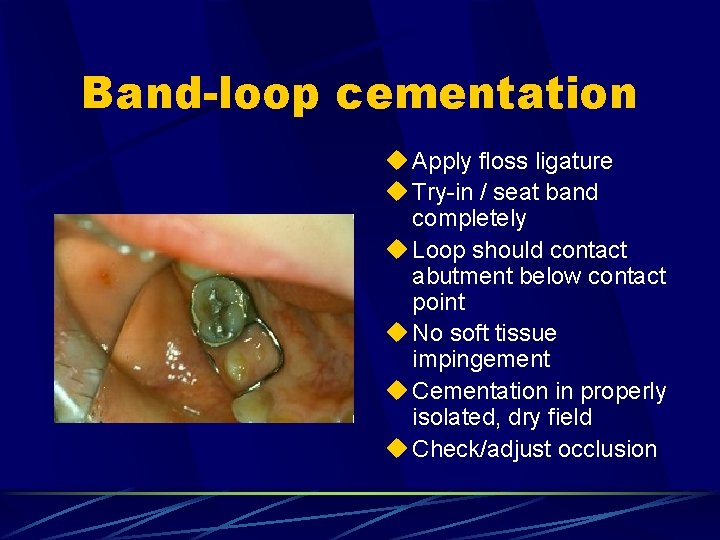
Band-loop cementation u Apply floss ligature u Try-in / seat band completely u Loop should contact abutment below contact point u No soft tissue impingement u Cementation in properly isolated, dry field u Check/adjust occlusion
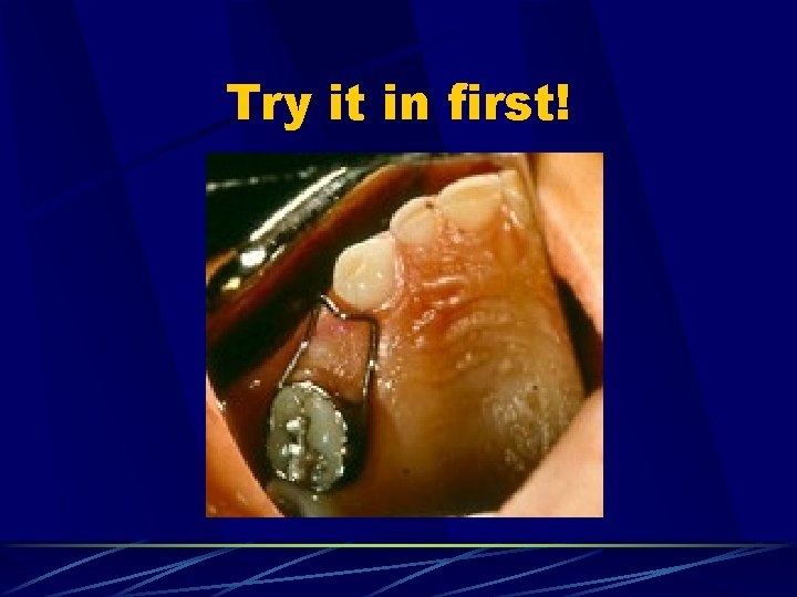
Try it in first!
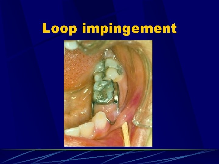
Loop impingement
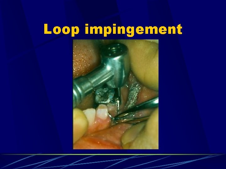
Loop impingement
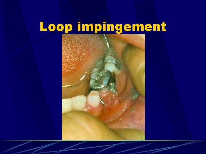
Loop impingement
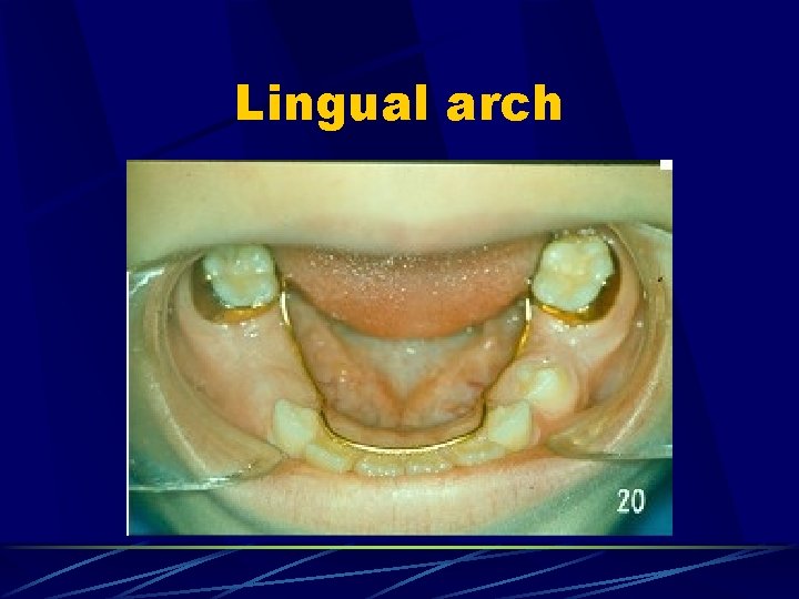
Lingual arch
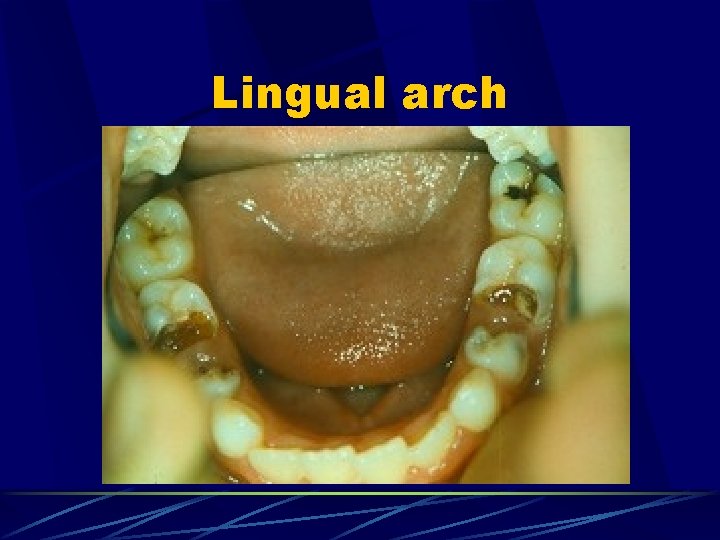
Lingual arch u. Indications: u Bilateral single or multiple tooth loss in mandible u Not recommended when primary incisors still present
Lingual arch
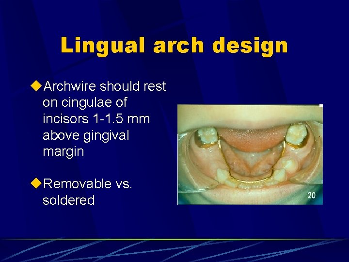
Lingual arch design u. Archwire should rest on cingulae of incisors 1 -1. 5 mm above gingival margin u. Removable vs. soldered
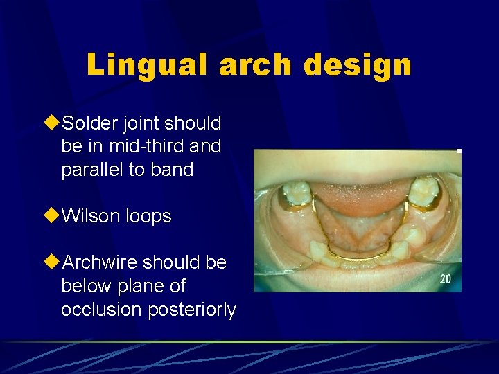
Lingual arch design u. Solder joint should be in mid-third and parallel to band u. Wilson loops u. Archwire should be below plane of occlusion posteriorly
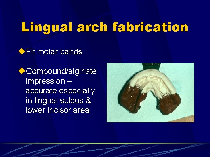
Lingual arch fabrication u. Fit molar bands u. Compound/alginate impression – accurate especially in lingual sulcus & lower incisor area
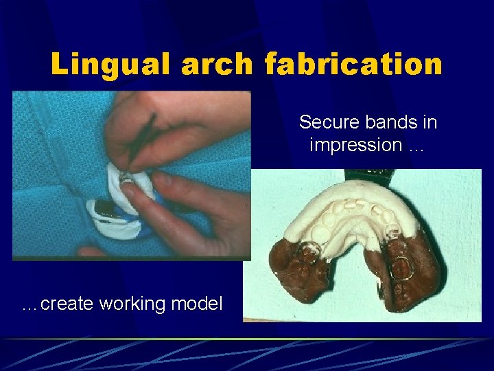
Lingual arch fabrication Secure bands in impression … …create working model
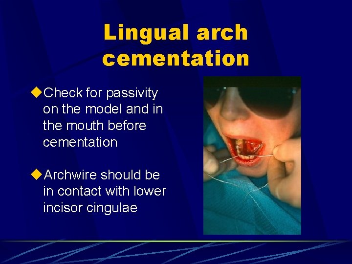
Lingual arch cementation u. Check for passivity on the model and in the mouth before cementation u. Archwire should be in contact with lower incisor cingulae
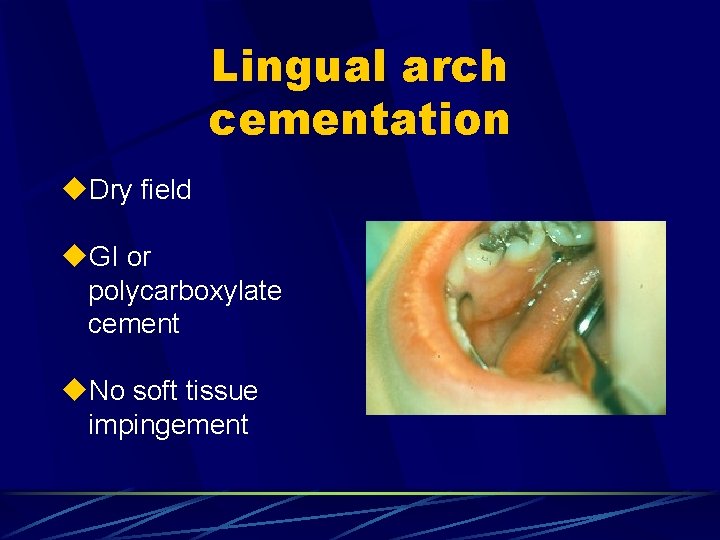
Lingual arch cementation u. Dry field u. GI or polycarboxylate cement u. No soft tissue impingement
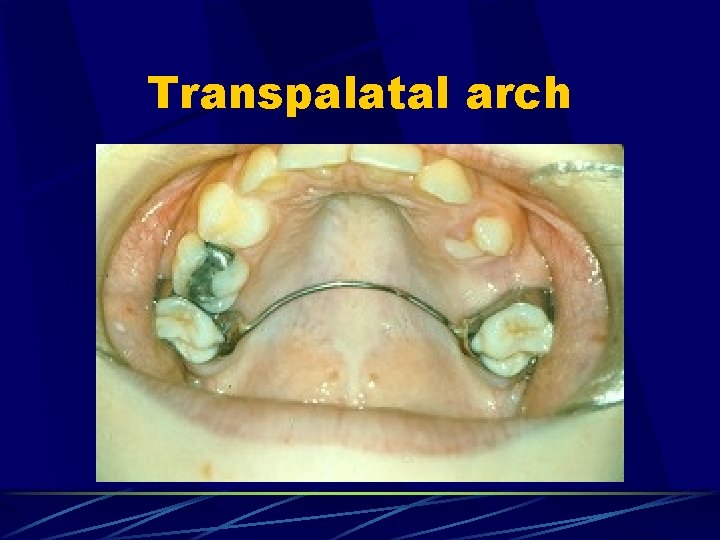
Transpalatal arch
Transpalatal arch u. Rarely recommended for bilateral tooth loss in maxilla u. Can prevent mesio-palatal rotation of palatal root of Mx 1 st permanent molar but allows mesial tipping of molars & space loss
Transpalatal arch u. May have an indication for use when one side of the arch is intact but several primary teeth are missing contralaterally u. Some designs incorporate omega loop: when active can prevent bodily movement of molars
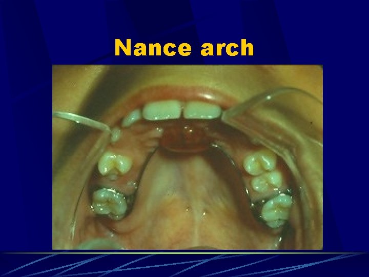
Nance arch
Nance arch u. Used commonly in maxilla for bilateral tooth loss u. Incorporates acrylic button in contact with palate to prevent molars from tipping u. Can be very unhygenic
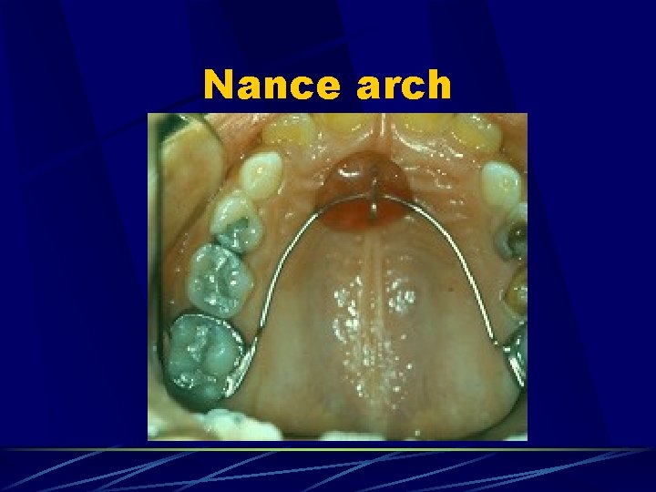
Nance arch
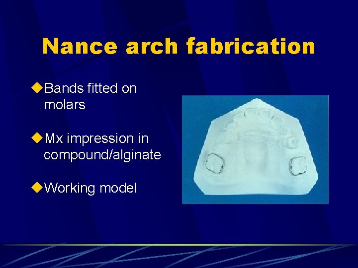
Nance arch fabrication u. Bands fitted on molars u. Mx impression in compound/alginate u. Working model
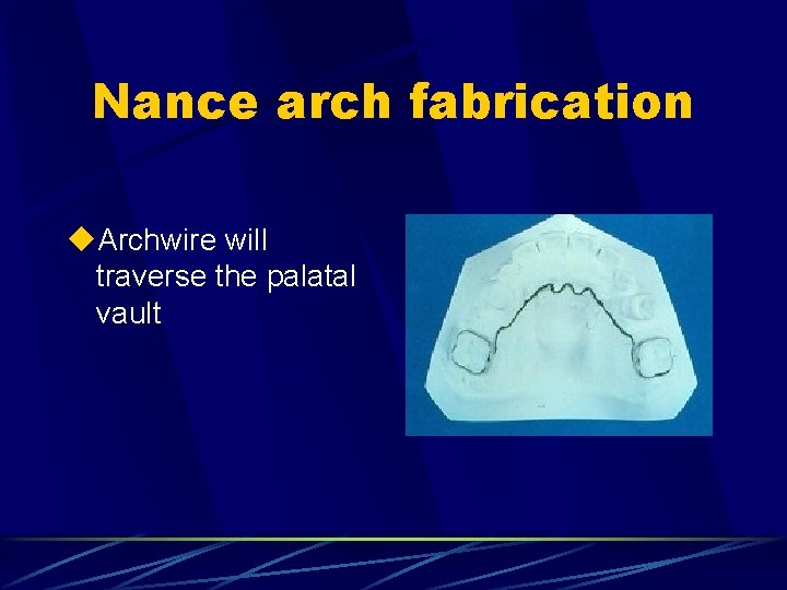
Nance arch fabrication u. Archwire will traverse the palatal vault
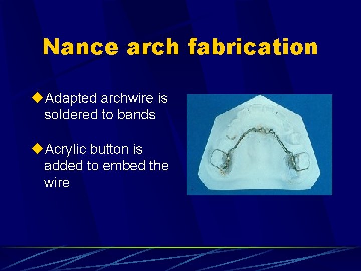
Nance arch fabrication u. Adapted archwire is soldered to bands u. Acrylic button is added to embed the wire
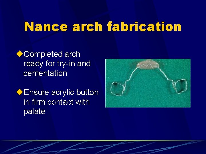
Nance arch fabrication u. Completed arch ready for try-in and cementation u. Ensure acrylic button in firm contact with palate
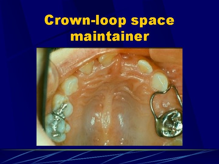
Crown-loop space maintainer
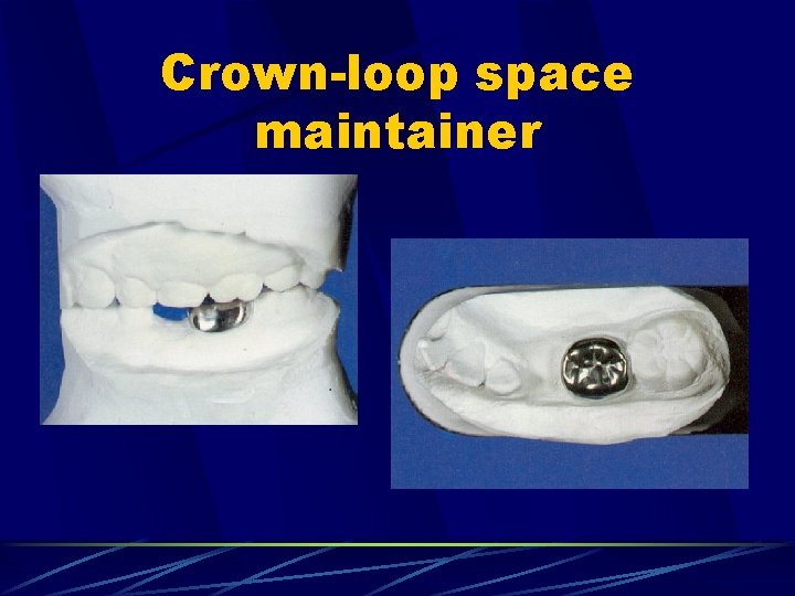
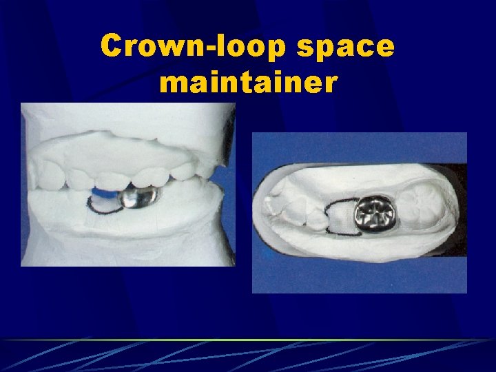
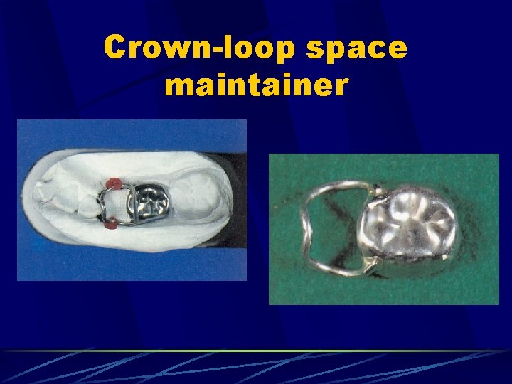
Crown-loop space maintainer u. Indications: u As for band-loop u Abutment tooth requires full coverage SSC due to multi-surface caries or pulp treatment
Crown-loop fabrication u. Abutment tooth prepared for SSC u. Properly contoured SSC seated, but not cemented u. Compound impression u. SSC placed into impression u. Working model u. Another SSC fitted and cemented with temporary cement
Crown-loop space maintainer
Crown-loop space maintainer
Crown-loop space maintainer
Crown-loop cementation u. Temporary SSC removed, under LA if necessary u. Try-in crown-loop to verify loop contours u. Cementation in dry field
Crown-loop space maintainer u. Disadvantages: u If solder joint fails, there is no way to repair the appliance without entire re-make u Cost is higher (extra SSC)
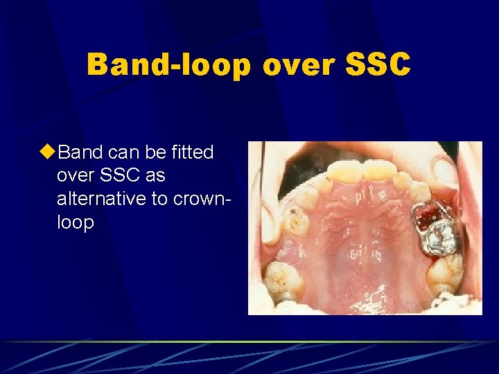
Band-loop over SSC u. Band can be fitted over SSC as alternative to crownloop
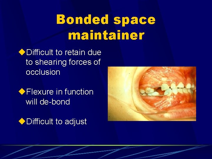
Bonded space maintainer u. Difficult to retain due to shearing forces of occlusion u. Flexure in function will de-bond u. Difficult to adjust
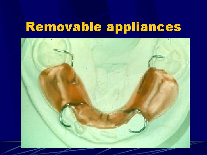
Removable appliances u. Indicated for mulitple primary tooth loss when no suitable abutment teeth exist u. Need to restore occlusal function over longer span u. Clasping difficult for primary teeth therefore retention a problem u. Compliance issues
Removable appliances
INTRA-ALVEOLAR SPACE MAINTENANCE D 362/QP 362 Division of Orthodontics and Paediatric Dentistry 2004 -2005 Karen M. Campbell, DDS
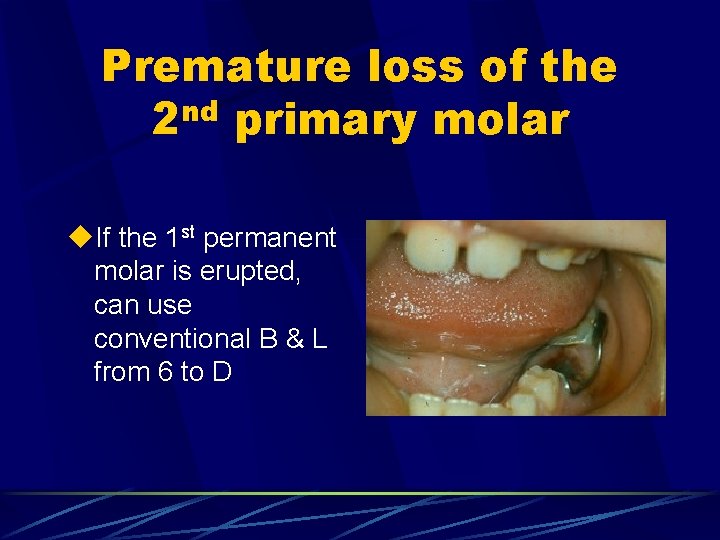
Premature loss of the 2 nd primary molar u. If the 1 st permanent molar is erupted, can use conventional B & L from 6 to D
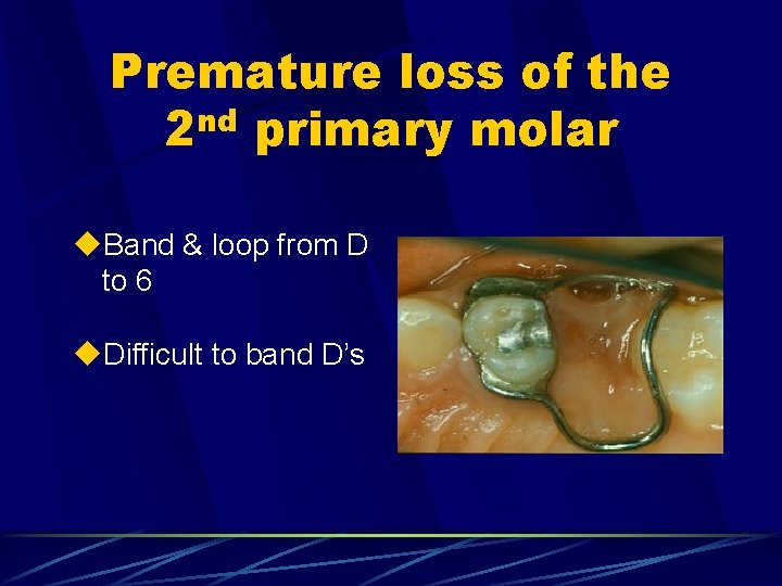
Premature loss of the 2 nd primary molar u. Band & loop from D to 6 u. Difficult to band D’s
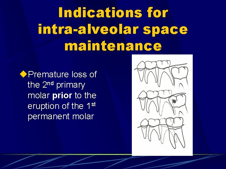
Indications for intra-alveolar space maintenance u. Premature loss of the 2 nd primary molar prior to the eruption of the 1 st permanent molar
Contraindications u. Medically compromised: u Cardiac patients requiring SBE prophylaxis u Immunosuppression u Chemotherapy/radiation therapy, pre-BMT u. Demonstrated lack of commitment to -up follow
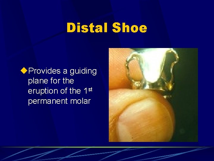
Distal Shoe u. Provides a guiding plane for the eruption of the 1 st permanent molar
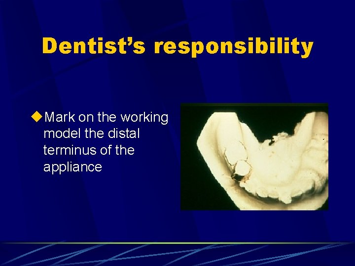
Dentist’s responsibility u. Mark on the working model the distal terminus of the appliance
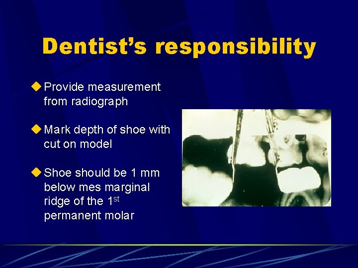
Dentist’s responsibility u Provide measurement from radiograph u Mark depth of shoe with cut on model u Shoe should be 1 mm below mes marginal ridge of the 1 st permanent molar
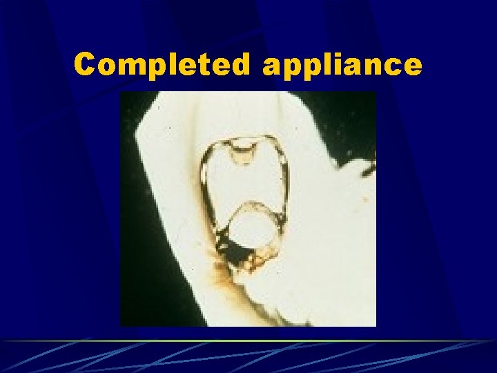
Completed appliance
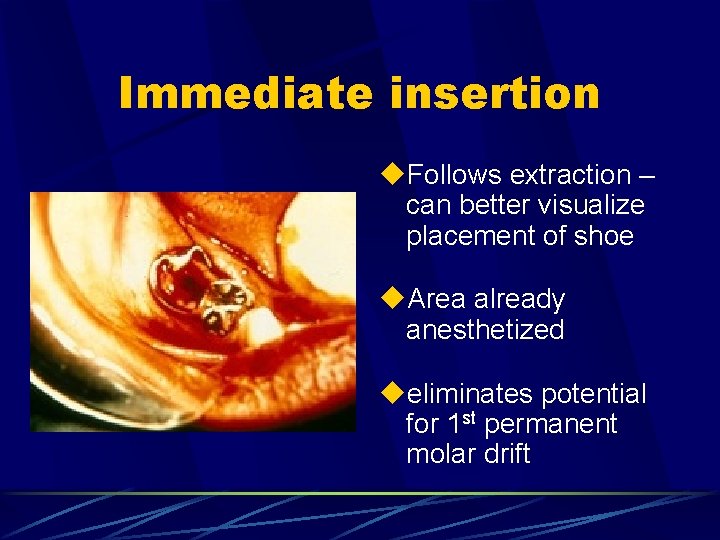
Immediate insertion u. Follows extraction – can better visualize placement of shoe u. Area already anesthetized ueliminates potential for 1 st permanent molar drift
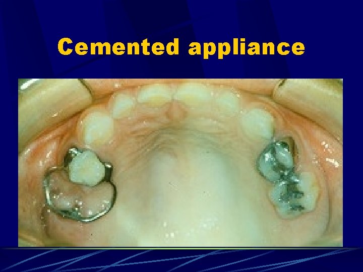
Cemented appliance
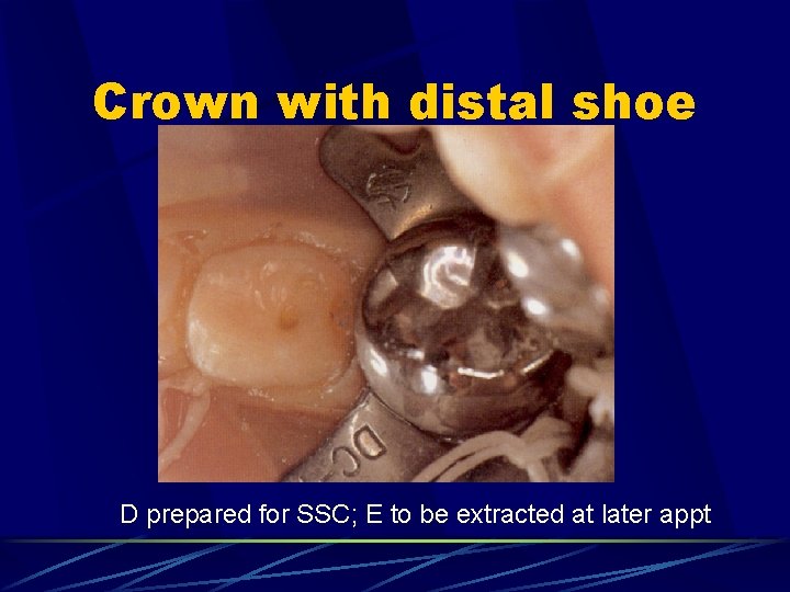
Crown with distal shoe D prepared for SSC; E to be extracted at later appt
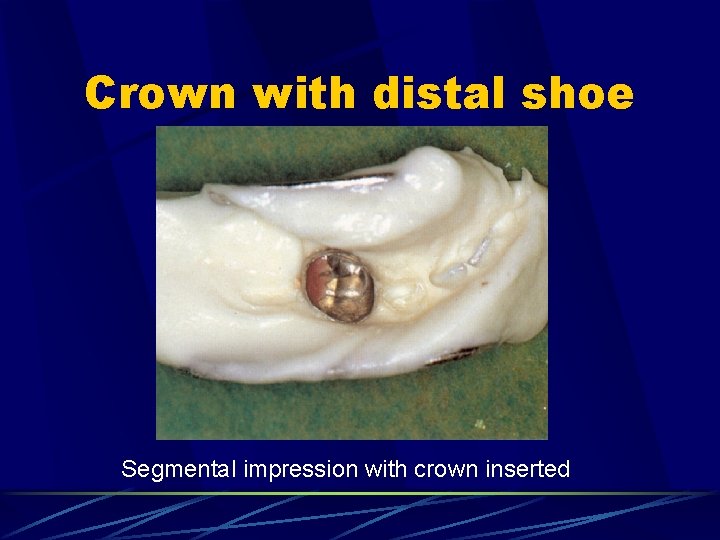
Crown with distal shoe Segmental impression with crown inserted
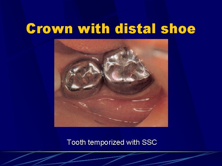
Crown with distal shoe Tooth temporized with SSC
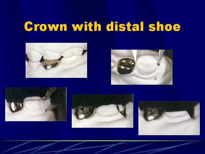
Crown with distal shoe
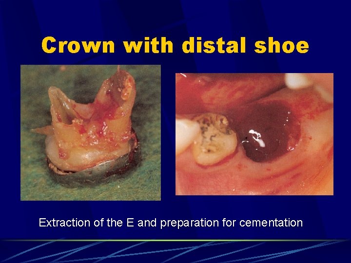
Crown with distal shoe Extraction of the E and preparation for cementation
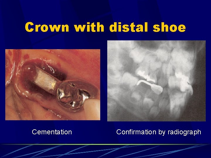
Crown with distal shoe Cementation Confirmation by radiograph
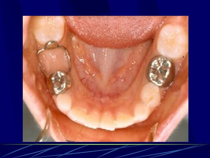
Following eruption of the 1 st permanent molar u. Distal shoe no longer appropriate – 1 st permanent molar may tip mesially above shoe u. Parents must be aware of need for second appliance from the beginning u. Conventional B & L or lingual arch may be required
Drawbacks of the appliance u. Can only replace a single tooth due to its cantilever design u. Inherent lack of strength u. Cannot restore occlusal function u. D’s are very difficult to fit bands u. Epithelium perforated in area of distal shoe
- Slides: 86