Partial Pressure Daltons law The air we breath

Partial Pressure & Dalton’s law: The air we breath is not a single gas but a mixture of gases. N 2 molecules are the most abundant about 78. 6% of atmospheric gas molecules. O 2 molecules are the second most abundant about 20. 9% of atmospheric content. Most of the remaining 0. 5% consists of H 2 O molecules, with Co 2 about 0. 04% of total atmosphere. Atmospheric pressure is 760 mm Hg, each of the gases contributes to the total pressure in proportion to its percentage, this relationship is known as (Dalton’s law).

Partial Pressure of a gas is the pressure contributed by a single gas within a mixture of gases. All the partial pressures added together equal the total pressure of a gas mixture. ***PN 2 + PO 2 + PH 2 O + PCO 2 = 760 mm Hg atmospheric pressure. So the partial pressure of oxygen (PO 2) = 20. 9 percent of the 760 mm Hg or 159 mm Hg. Diffusion between liquids & gases & Henry’s law: -differences in pressure move gas molecules from one place to another. -Pressure differences also affect the movement of gas molecules into and out of solutions. The amount of a particular gas in solution is directly proportional to the partial pressure of that gas. This is known as (Henry’s law). * when a gas under pressure contacts a liquid, the pressure tends to force gas molecules into solution.

- if partial pressure goes up, more gas molecules will go into solution. - if partial pressure goes down, gas molecules will come out of solution. (e. g. for Hanry’s law is the soda can, when opened pressure falls & gas molecules begin coming out of solution). - at equilibrium, gas molecules are diffusing out of the liquid as quickly as they are entering, (& the total number of gas molecules remains constant).

Diffusion at the respiratory membrane: Gas exchange at the respiratory membrane is efficient for the fallowing five reasons: 1. The differences in partial pressure across the respiratory membrane is great and the rate of gas diffusion is fast. 2. The distances involved in gas exchange are small: the capillary & alveolar basement membranes reduces the distance to an average of 0. 5µm. 3. The gases are lipid soluble: both O 2 & Co 2 diffuse readily through the surfactant layer, the alveolar & endothelial cell membranes. 4. The total surface area is large: the alveolar surface area at inspiration is about 140 m². 5. Blood flow & air flow coordinated: the arrangement improves the efficiency of both pulmonary ventilation & pulmonary circulation.

Partial Pressure in the Alveolar Air & Alveolar Capillaries: Blood arriving in the pulmonary arteries has a lower PO 2 & a higher PCO 2 than alveolar air. Diffusion between the alveolar air & pulmonary capillaries thus elevates the PO 2 of the blood & lowering its PCO 2. ( by the time the blood enters the pulmonary venules, it has reached equilibrium with the alveolar air. PO 2 = 100 mm Hg & PCO 2=40 mm Hg).

Partial Pressure in the Systemic Circuit: The oxygenated blood leaves the alveolar capillaries & returns to the heart, to be discharged in to the systemic circuit. ((the oxygenated blood from the alveolar capillaries mix with blood that flowed the conducting passageways capillaries, & blood leaving the conducting passageways carries relatively little oxygen. So PO 2 in pulmonary veins therefore drops to about 95 mm. Hg, & this is the PO 2 in blood that enters the systemic circuit)). Intestinal fluid has a PO 2 of 40 mm Hg & PCO 2 of 45 mm Hg so O 2 diffuses out of the capillaries & Co 2 diffuses in. until capillaries partial pressures are the same as the adjacent tissue.
,")
**This Figure shows the Partial Pressure in the Alveolar Air & Alveolar Capillaries (a), & Partial Pressure in the Systemic Circuit (b):

#Oxygen Transport: The blood leaving the alveolar capillaries carries about 20 ml of O 2 per 100 ml. and only 0. 3 ml (1. 5%) of O 2 molecules consist in solution, the rest are bound to Hemoglobin (Hb) molecules. (a Hb molecule consists of four globular proteins each contain a heme unit so a Hb molecule can bind to four molecules of O 2 forming (Oxyhemoglobin)). This is a reversible reaction Hb + O 2 = Hb. O 2 ((There approximately 280 million molecules of Hb in each RBC & because a Hb molecule contain 4 heme units, each RBC can carry more than a billion molecule of O 2)).

*Hb Saturation: Hb saturation is the percentage of heme units containing bound O 2. (Hb is a protein, so any changes in shape can affect O 2 binding). -if all the Hb molecule in blood are fully loaded there is 100% saturation. -if each Hb molecule carries two O 2 molecules there is 50% saturation. Hb & PO 2 : - (O 2 -Hb saturation ) • Binding of O 2 to Hb is a reversible reaction • It’s the relation between saturation of Hb & Partial Pressure of O 2 (PO 2). -if PO 2 increase (↑es) the reaction shift to the right, more O 2 bound to Hb. -if PO 2 decrease(↓es) the reaction shift to the left, more O 2 is released by Hb.

Hb & p. H : - active tissue generate acids that lower p. H of interstitial fluid. ( at a tissue PO 2 of 40 mm. Hg, when Co 2 diffuse into the blood, it rapidly diffuse into RBCs. Co 2 + H 2 O H 2 CO 3 H ˉ + HCO 3ˉ -when PCO 2 ↑es, the reaction proceeds from left to right & rate of H 2 CO 3 formation ↑es & H ˉ generated diffuse out of the RBCs & p. H of plasma drops. -When PCO 2 ↓es the reaction proceeds from right to left & H ˉ diffuses into RBCs & p. H of plasma rises.

Hb & temperature: - temperature changes also affect the slop of the Hb saturation curve. -as temperature ↑es, Hb releases more O 2. -as temperature ↓es Hb holds O 2 more tightly. An active skeletal muscle generate heat, the heat warms the blood that flows through the muscle & as blood warms the Hb molecules release more O 2 that can be used by the active muscle fibers.

# Carbon Dioxide Transport: The CO 2 is generated by aerobic metabolism in peripheral tissue after entering the blood stream. *CO 2 molecules may be: 1. converted to H 2 CO 3 (carbonic acid). 2. bound to the protein of Hb molecule in RBCs. 3. dissolve in the plasma. All three are completely reversible reactions = Carbonic Anhydrase CO 2 + H 2 O H 2 CO 3 H ˉ + HCO 3ˉ

Control of Respiration: 1 - local regulation: this inclued: *a. Changes in lung Perfusion = as blood flows towards the alveolar capillaries, its directed towards lobules in which the PO 2 is high. This shift in circulation tends to eliminate temporary differences in O 2 & CO 2 content of alveoli. *b. Changes in alveolar ventilation = smooth muscles in walls of bronchioles are sensitive to the PCO 2 of air they contain. - when the PCO 2 ↑es the bronchioles increase in diameter (bronchodilation). - when the PCO 2↓es the bronchioles constrict(bronchoconstriction). Air flow is therefore directed to lobules in which the PCO 2 is High.

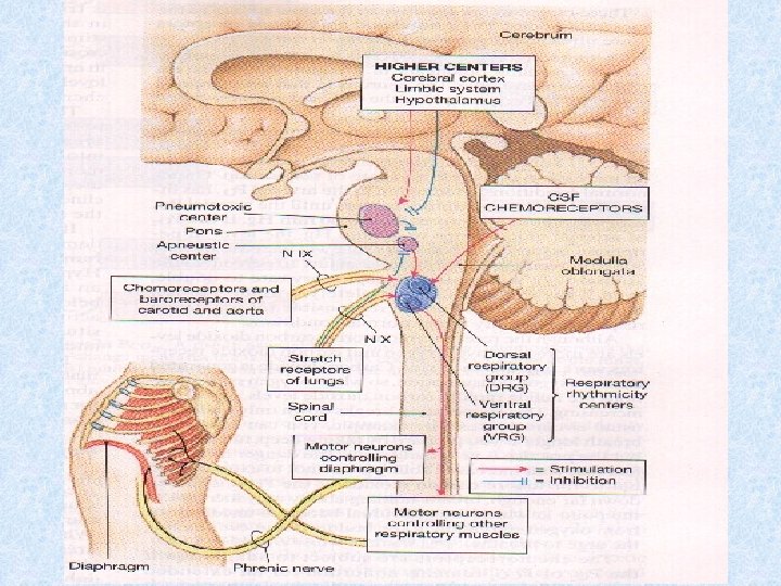
2 - the respiratory centers of the brain: Respiratory control has both involuntary & voluntary components. *the brain’s involuntary centers regulate the activity of respiratory muscles & controls the respiratory minute volume by adjusting the frequency & depth of pulmonary ventilation. **the voluntary control of respiration reflects activity in the cerebral cortex that affects either → output of respiratory centers in the medulla oblongata & pons. or → motor neurons in the spinal cord that control respiratory muscles. --the respiratory centers are three pairs of nuclei, in the medulla (the respiratory rhythmicity centers) & pons (the apneustic & pneumotaxic centers). --the motor neurons in spinal cord are generally controlled by (respiratory reflexes).
: These are paired centers, each")
# Respiratory Centers in Medulla Oblongata (Respiratory Rhythmicity center): These are paired centers, each center can be subdivided into a (dorsal respiratory group) DRG & a (ventral respiratory group) VRG. The DRG’s inspiratory center. . contain neurons that control lower motor neurons innervating the external intercostals muscles & diaphragm. (this group function in every respiratory cycle “whether quiet or forced”). The VRG functions only in forced respiration & it includes neurons that innervate lower motor neurons controlling accessory respiratory muscles involved in active exhalation (an expiratory center) & maximal inhalation (an inspiratory center).
: The apneustic centers &")
# Respiratory Centers of Pons (the Apneustic & Pneumotaxic centers): The apneustic centers & pnemotaxic centers of the pons are paired nuclei that adjust the output of the respiratory rhythmicity centers. ( their activities adjust the respiratory rate & depth of respiration in response to sensory stimuli or input from other centers in the brain). = Each apneustic center provides continuous stimulation to the DRG

# Respiratory Reflexes: Activities of the respiratory centers are modified by sensory information from the fallowing: 1. chemo receptors sensitive to the PCO 2, p. H &/or PO 2 of the blood or central spinal fluid (CSF). 2. changes in blood pressure in the aorta or carotid sinuses. 3. stretch receptors that respond to changes in the volume of the lung. 4. irritating physical or chemical stimuli in the nasal cavity, larynx or bronchial tree. 5. other sensations: pain, changes in body temperature.
- Slides: 18