PART 4 THE TISSUE LEVEL OF ORGANIZATION Unit


 inside the P. M. attaching")

 Not for linking cells but")
 form connexons (tiny fluid-filled tunnels")


 Two main types 1. Exocrine –")


 Epithelial membrane • • • Membranes")
 Epithelial membrane • • Membranes Areolar")
 Epithelial membrane Covers entire")


 generate force with ATP. Functions �")


 supportive functions Physical")


- Slides: 73
PART 4 THE TISSUE LEVEL OF ORGANIZATION Unit 1 Organization of the Human Body
Introduction As you already know, the human body is composed of trillions of cells. But, these cells don’t work alone – they work in groups called tissues. Furthermore, tissues are combined to form organs. Therefore, to understand a complex organ like a heart or a brain, you need to understand their individual components – tissues. Tissue level of Organization describes the organization of cells into tissues. The origin, structure, function, and location of each of the principal tissues types are examined. Throughout, the relationship between structure and function is emphasized. Attention is also given to extracellular materials, exocrine glands, and mucous, serous, and synovial membranes.
A Primary Tissue Types Definition of tissue Basic tissue types
Primary Tissue Types Tissue � Similar cells performing a similar function � May be hard, semisolid, even liquid Bone, fat, blood � Structure and properties of a tissue is influenced by factors such as: Extracellular material surrounding tissue cells Connections between cells composing the tissue
Study of Tissues Histology � Study of tissue Pathology � Study of diseased tissue � Patho = disease
Primary Tissue Types 1. Epithelium � 2. Connective tissue � 3. Support and protection Bind organs together, store energy reserves as fat, immunity. Ex. Blood Muscle � 4. Covering, lining, glandular Sheets when covering surfaces and lining cavities Forms glands Contraction and producing movement Nervous tissue � Receives and generates nerve impulses
Primary Tissue Types
Cell Junctions How cells are held together to form tissues – contact points between the P. M. of tissue cells. Most epithelial, some muscle and some nerve cells are held together very tightly. Five types of cell junctions (fig. 4. 2): 1. 2. 3. 4. 5. Tight Adherens Desmosomes Hemidesmosomes Gap
Tight Junctions Weblike strands of transmembrane proteins fusing cells together via their membranes. Prevents substances from passing between the cells. Prevents content leakage into blood and surrounding tissue. Ex. Epithelial cells of the stomach, intestine, and urinary bladder
Adherens Junctions Contain a dense layer of proteins (plaque) inside the P. M. attaching to membrane proteins and cytoskeletal microfilaments. Cadherins join cells. In epithelial cells – adherens junctions form zones called adhesion belts � They encircle the cell like a belt � Prevent contractile cells from separating
Desmosomes Desmo = band Plaque and cadherins attach cells Plaque doesn’t attach to microfilaments here, but to intermediate filaments of cytoskeleton made of keratin. Common in epidermis and cardiac muscles Prevents separation under tension and during contraction
Hemidesmosomes Look like half a desmosome (hemi = half) Not for linking cells but attaching cells to basement membrane Integrins instead of cadherins Integrins attach to different things: � Inside P. M. to intermediate filaments of keratin � Outside P. M. to protein laminin of basement membrane
Gap Junctions Allow for cell communication. Connexions (membrane proteins) form connexons (tiny fluid-filled tunnels for the diffusion of ions and small molecules) connecting cells. P. M. have small intercellular gaps (not tight) Transfer of nutrients and wastes in cornea and lens of eye, allows nerve and muscle impulses to happen quickly � Crucial for proper fnc of the NS, heart muscle, uterus and GI tract
Summary
Epithelium vs. Connective Tissue Number of cells vs. extracellular matrix Vascularization Epithelium – forms surface layers, free apical layer
B Epithelial Tissue Physical characteristics Functions Main types Glands
Epithelium Physical Characteristics � Lots of cells Little extracellular matrix Many cell junctions � Apical (apex � Basal surface – superficial) surface (base – deep) � Avascular � Well-innervated � Mitotically active
Figure 4. 2 Surfaces of epithelial cells and the structure and location of the basement membrane
Epithelium Functions � Provide a barrier � Control permeability � Provide sensation � Produce secretions
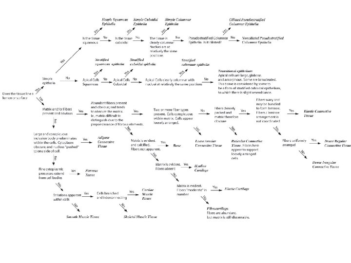
Epithelium Classification System
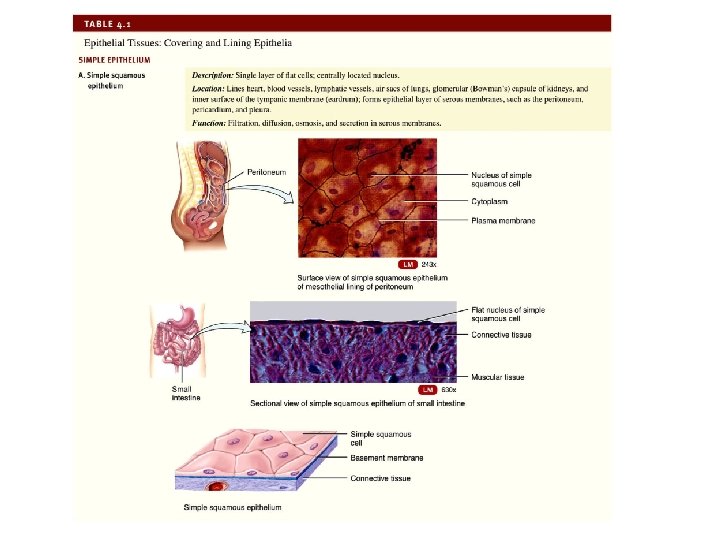
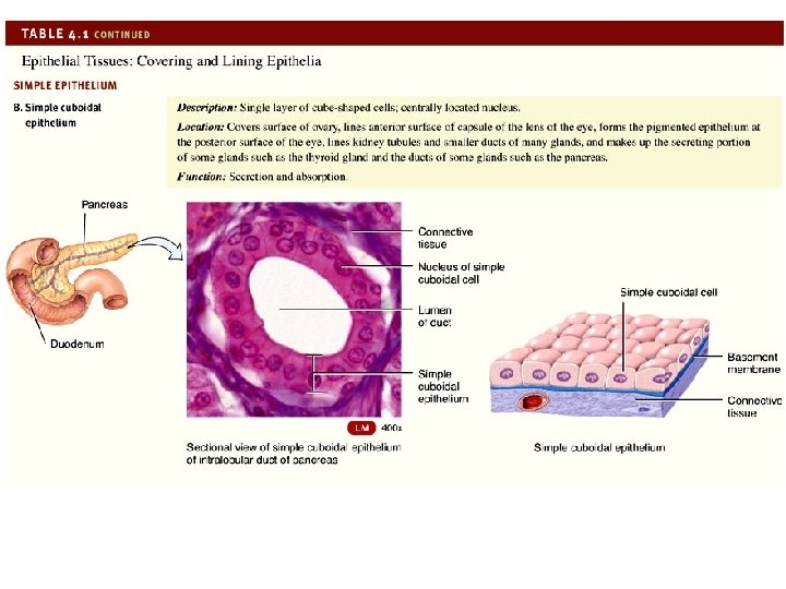
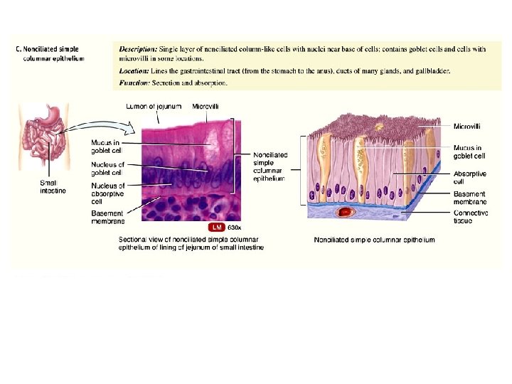
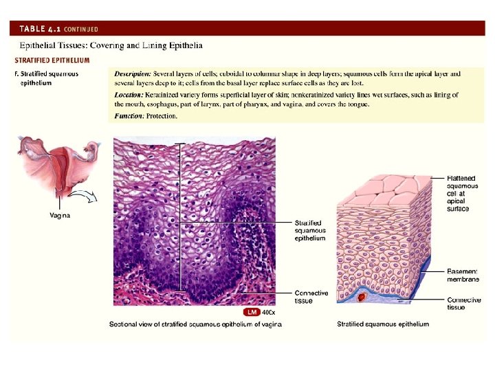
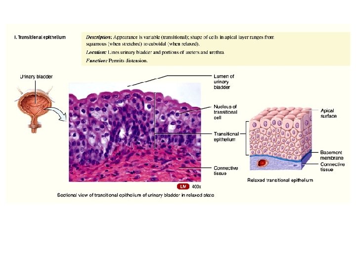
Epithelium Main Types � We will examine the main types of epithelium in the lab � See Exercise 6 in your lab manual � See Table 4. 1 in your text �A very brief overview:
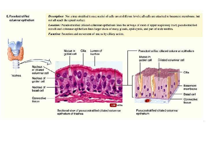
Mucociliary Elevator Ciliated pseudostratified columnar epithelium – covered with cilia instead of microvilli Bronchi, bronchioles, paranasal sinuses Mucus-producing goblet cells Cilia continuously beat Major barrier against infection � Microorganisms get caught in mucus and moved up by the elevator and expelled. Smoking paralyzes the cilia – how does this affect susceptibility? If you were an invader, how might you prevent yourself from being swept up by the elevator?
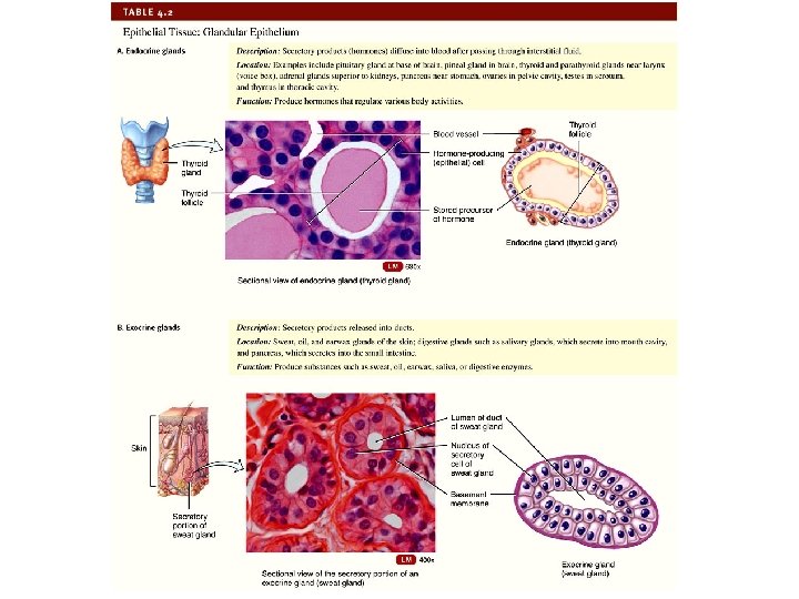
Glandular Epithelium Specialized epithelium with specialized secretions (glands) Two main types 1. Exocrine – ducts Secrete products (salvia, sweat, digestive fluids) into ducts that empty onto the surface of the epithelium (skin) or in the lumen. 2. Endocrine – no ducts Secrete products (hormones) directly into the bloodstream via diffusion out of interstitial fluid.
Figure 4. 6 summarizes exocrine glands
C Connective Tissue Physical characteristics Functions Main types
Connective Tissue Physical Characteristics � Lots of ECM Between widely spaced cells. Protein fibers & ground substance. � Always covered � Well-vascularized Exceptions – cartilage and tendons � Well-innervated Exception � Mitotically Exception - cartilage active - cartilage
Functions of Connective Tissue Binds, supports and strengthens other tissues Insulates and protects organs Compartmentalizes structures � Skeletal muscle – framework Major transport system � Blood Primary stored energy reserves � Adipose tissue Main immune response
Connective Tissue Cells CT came from embryonic cells called mesenchymal cells. CT contains immature cells � Mitotically active � ends in –blast (to bud or sprout) � Secrete ECM “Blast” cells mature into –cytes � Reduced ability to divide � Monitors and maintains ECM
Connective Tissue Common Cell Types � Fibroblasts � Fibrocytes � Adipocytes � Chondroblasts � Chondrocytes � Osteoblasts � Osteocytes
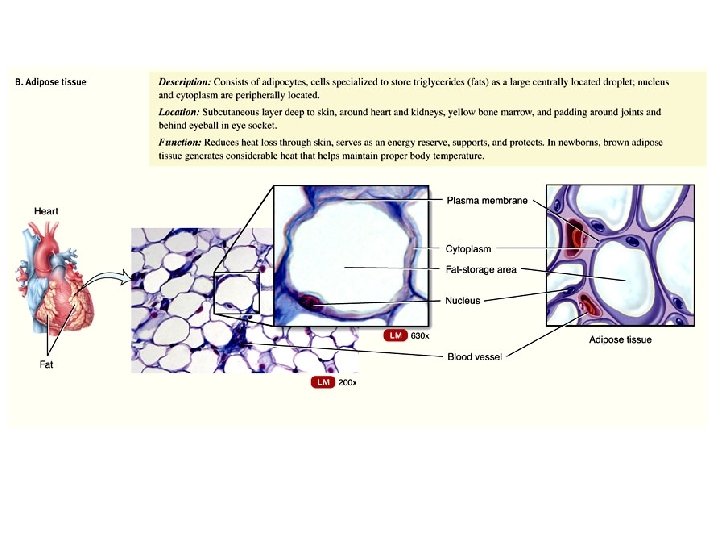
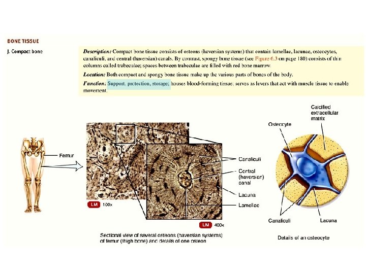
Connective Tissue – Common Cell Types Fibroblasts Chondroblasts • Large, flat, with branches • Usually most numerous type • Secrete fibers and parts of ground substance of ECM • Secrete ECM in cartilage Fibrocytes • Maintain ECM in areolar, reticular, dense regular, and dense irregular CTs Osteoblasts • Secrete ECM in bone Adipocytes • Adipose cells • Store triglycerides • Deep to skin and around heart and kidneys Osteocytes • Cells of mature bone • Maintain ECM in bone Chondrocytes • Cells of mature cartilage • Occur in spaces (lacunae) in ECM
Connective Tissue Common Fiber Types in ECM � Collagen fibers – “ropes” � Reticular fibers – “baskets” � Elastic fibers – “elastics”
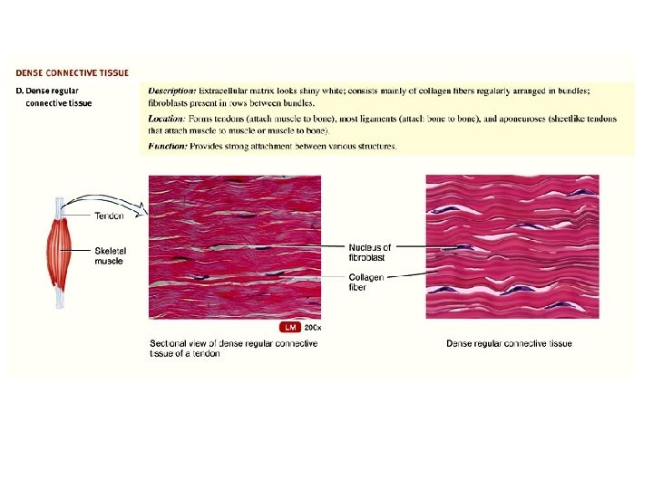
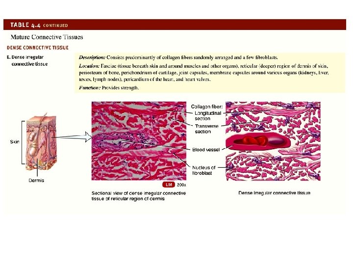
Common Fiber Types in ECM Collagen fibers – “ropes” � Most common type � Colla = glue � Collagen = most abundant protein in body (approx 25%) � Very strong, flexible, resist tension � Properties can vary depending on tissue type Location: � Bone, cartilage, tendons, ligaments
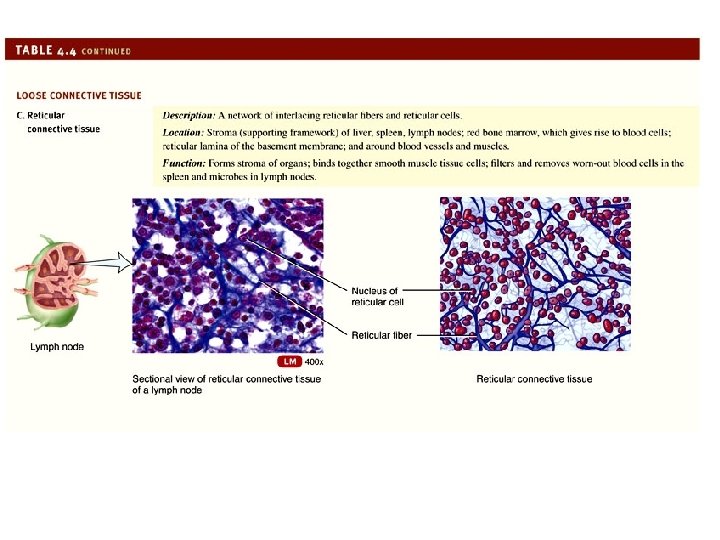
Common Fiber Types in ECM Reticular fibers – “baskets” � Reticul = net � Collagen in fine bundles Location: � Walls of blood vessels � Network around cells in tissues: Areolar connective tissue, adipose tissue, nerve fibers, smooth muscle � Supporting Spleen, framework for: lymph nodes, basement membrane
Common Fiber Types in ECM Elastic fibers – “elastics” � Form a fibrous network � Strength and stability from elastin and fibrillin proteins � Stretch up to 150% without breaking � Have elasticity Location � Skin, blood vessels, lung tissue
Common Fiber Types in ECM
Figure 4. 6 Representative cells and fibers present in connective tissues
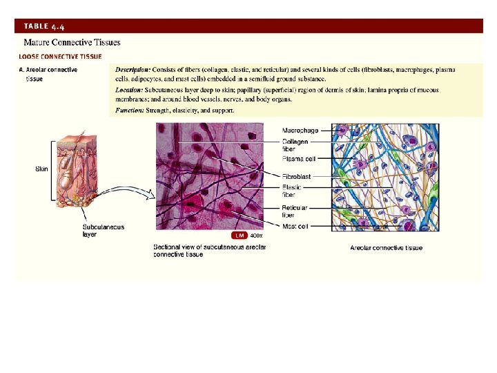
Connective Tissue Main Types � We will examine the main types of connective tissue in the lab � See Exercise 6 in your lab manual � See Table 4. 4 in your text
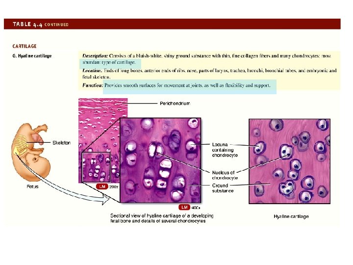
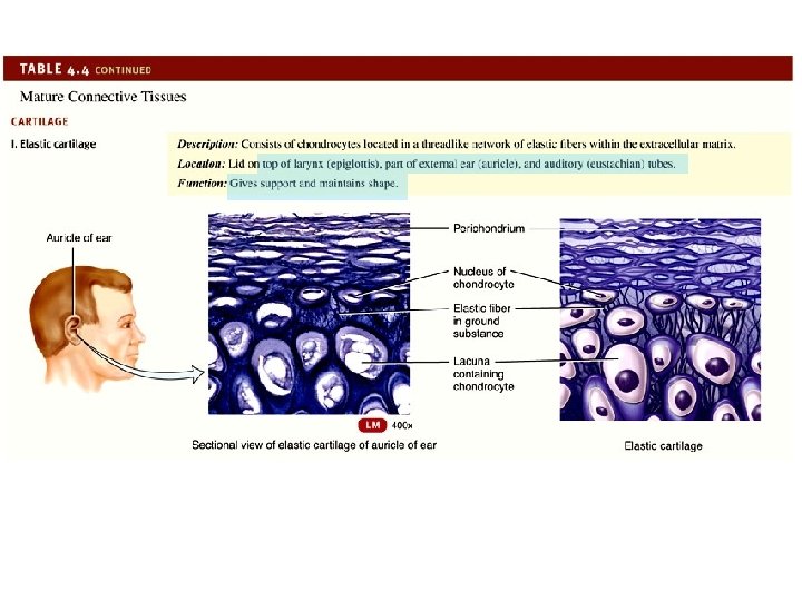
Cartilage Dense network of collagen and elastic fibers firmly embedded in ground substance Can endure more stress than loose or dense connective tissues due to collagen fibers and resilience to stretching Few cells – lots of ECM NO nerves or blood vessels � secretes a substance that inhibits blood vessel growth (antiangiogenesis factor) � heals poorly following injuries because of the lack of blood supply
Supports and joins structures together Strongest type of cartilage
D Membranes Definition of membrane Main types
Membranes Membrane � Flat sheets of pliable tissue that cover or line a body part Main Types 1. Mucous 2. Serous 3. Cutaneous 4. Synovial Epithelial membranes = epithelial layer on top of a connective layers Synovial membrane = lines joints, only connective tissue
Figure 4. 7 a • Mucous Membranes (mucosae) Epithelial membrane • • • Membranes Connective tissue = areolar CT called the lamina propria (one’s own) Epithelial tissue = Protective barrier Lines a body cavity that opens directly to exterior • Digestive, respiratory, reproductive and urinary tracts.
Figure 4. 7 b • Serous Membranes (serosae) Epithelial membrane • • Membranes Areolar CT covered by mesothelium (simple squamous epithelium) Recall – serous membranes have 2 layers – visceral and parietal Lubricating (serous = watery) mesothelium secretes fluid Lines a body cavity (thoracic / abdominal) that does not open directly to exterior. • Pleura, pericardium and peritoneum
Figure 4. 7 c • • • Cutaneous Membrane (skin) Epithelial membrane Covers entire body surface Epidermis (superficial) • • Membranes Keratinized squamous epithelium – protective barrier Dermis (deep) • Dense irregular CT and areolar CT
Figure 4. 7 d • • • Membranes Synovial Membranes Syn = together ova = egg (egg white slimy) Lines, lubricates and nourishes cartilage covering of bones and free movable joints Lacks epithelium – only CT • • Discontinuous layer of cells called synoviocytes closer to synovial cavity Areolar and adipose CT (deeper)
E Muscle Tissue Physical Characteristics Functions Main Types
Muscle Tissue Physical Characteristics � Contractile � Electrically excitable � Well-vascularized � Well-innervated � Not mitotically active
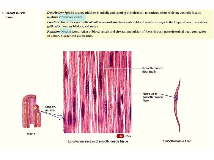
Muscle Tissue Myocytes or Muscle fibers (elongated cells) generate force with ATP. Functions � Produce movement � Maintain posture � Produce heat
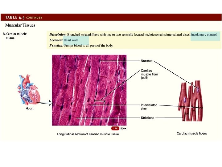
Muscle Tissue Main Types � We will examine the main types of muscle tissue in the lab Skeletal Cardiac Smooth � See Exercise 6 in your lab manual � See Table 4. 5 in your text
04_table_05 a
F Nervous Tissue Physical Characteristics Functions Main Types
Nervous Tissue Neurons – sensitive to stimuli Neuroglia – glia (glue) supportive functions Physical Characteristics � Electrically excitable Nerve action potentials Communicate to other neurons, muscles, glands � Well-vascularized � Not mitotically active
Nervous Tissue Functions � Responds to stimuli � Conducts electrical impulses
Nervous Tissue Main Types � We will examine the main types of nervous tissue in the lab � See Exercise 6 in your lab manual � See Table 4. 6 in your text
04_table_06