
PART 3 Chapter 7 YOUNG SMALL BREEED DOGS
PART 3 Chapter 7
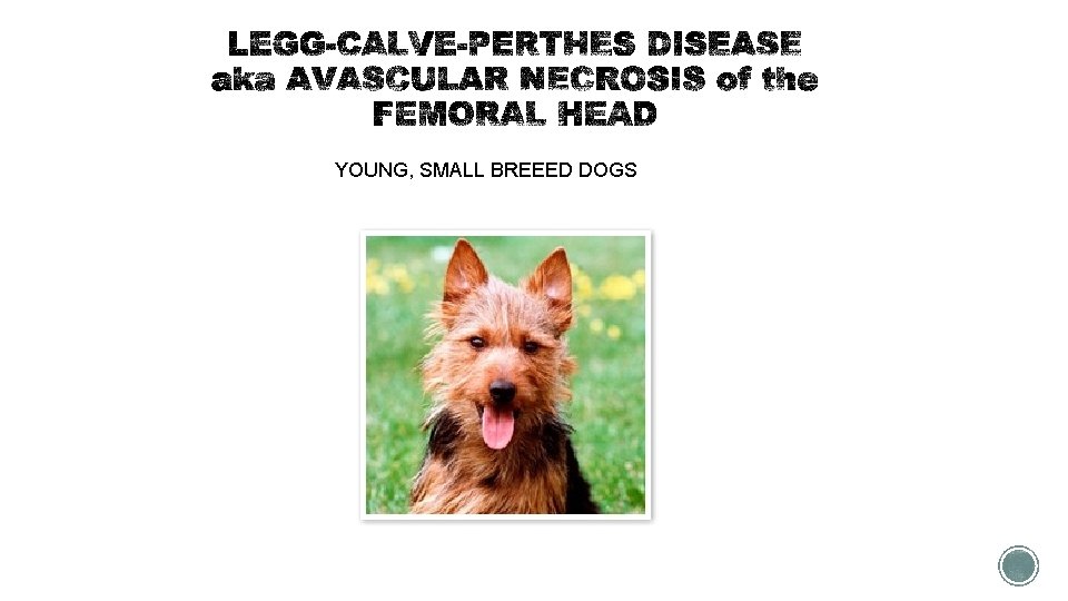
YOUNG, SMALL BREEED DOGS
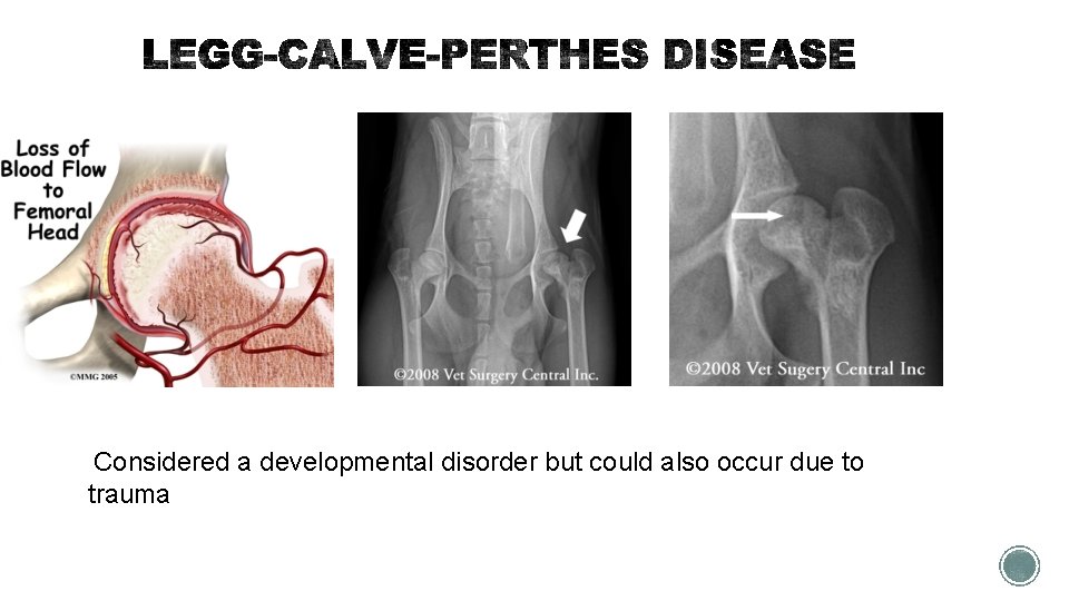
Considered a developmental disorder but could also occur due to trauma
§ Clinical signs § Pain in the hip § Atrophy of the muscles of the affected limb § Gradual onset of lameness § Chewing at the hip and flank area § Usually young dogs 5 -8 months § Treatment consist of surgery!
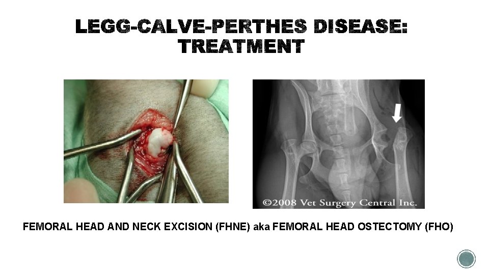
FEMORAL HEAD AND NECK EXCISION (FHNE) aka FEMORAL HEAD OSTECTOMY (FHO)
§ CLIENT INFO § Post-operative treatment requires early and active use of the limb as soon as 2 weeks after surgery § Exercise and passive range-of-motion exercises should be done daily § Animals may have both hips involved – surgeon is likely to separate procedures by 8 -10 weeks § https: //www. youtube. com/watch? v=u. Bz 3 YLl_QYo § Therapy for patients: hydrotherapy
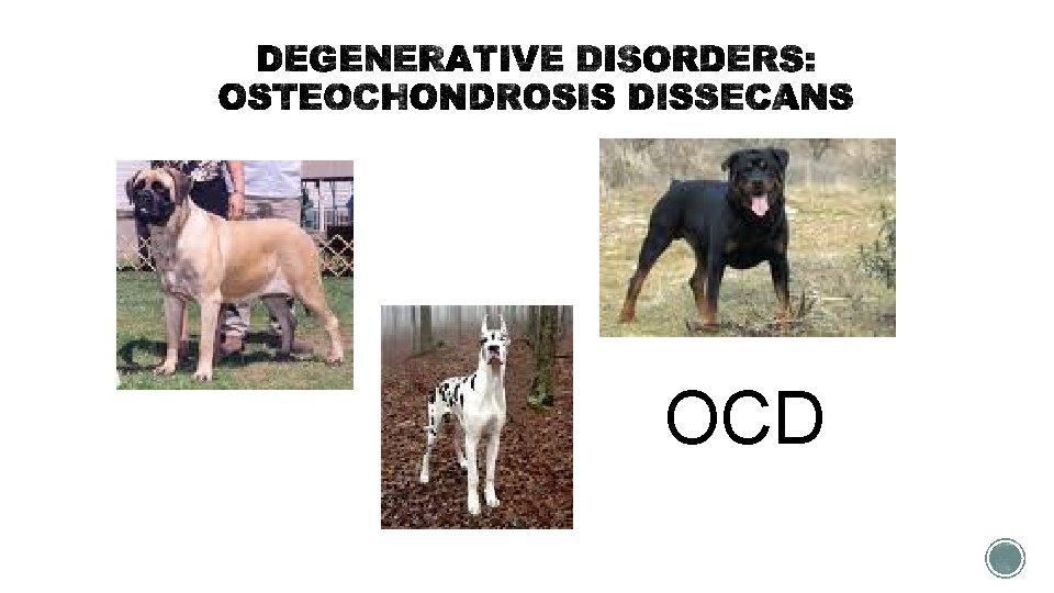
OCD
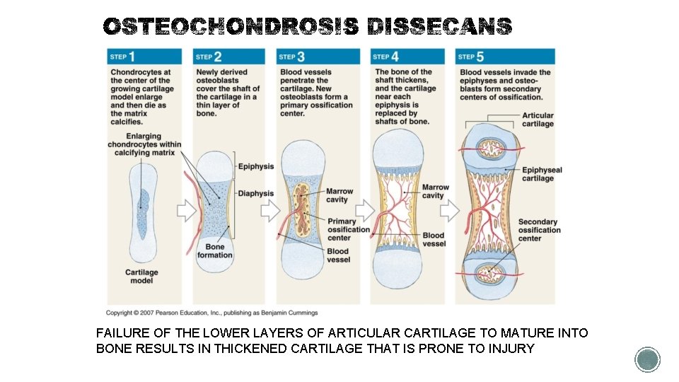
FAILURE OF THE LOWER LAYERS OF ARTICULAR CARTILAGE TO MATURE INTO BONE RESULTS IN THICKENED CARTILAGE THAT IS PRONE TO INJURY
§ Failure of the articular cartilage to become cemented to underlying bone, together with constant trauma during exercise, results in formation of a non-healing flap or joint mouse § This disorder can occur in several joints § Shoulder § Stifle § Hock § Elbow § CLINICAL SIGNS § Lameness in young, large breed dogs
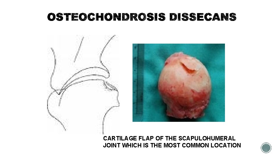
CARTILAGE FLAP OF THE SCAPULOHUMERAL JOINT WHICH IS THE MOST COMMON LOCATION
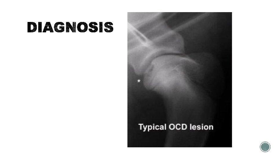
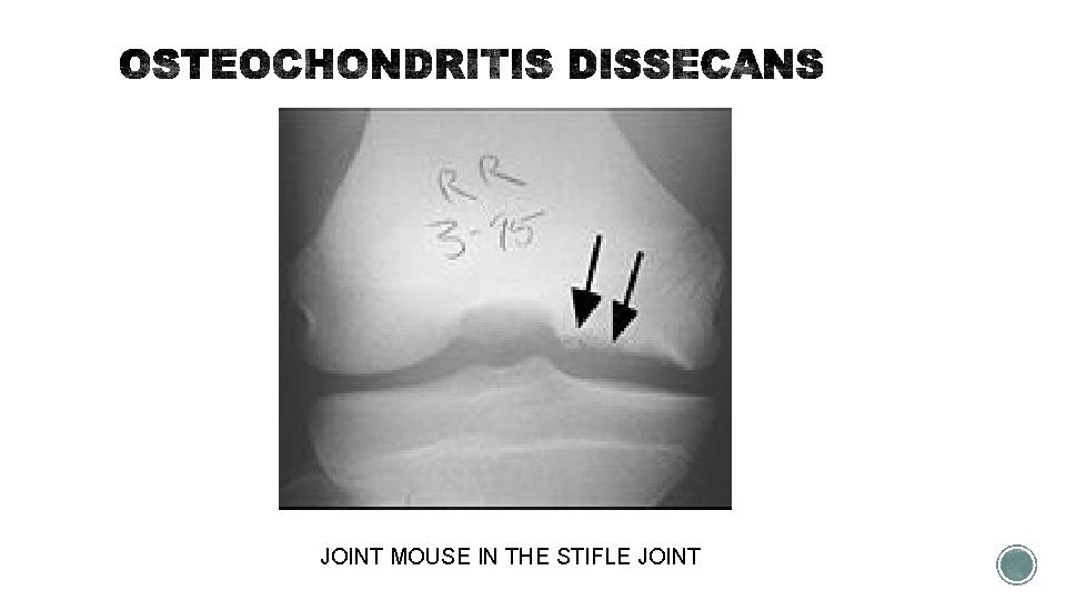
JOINT MOUSE IN THE STIFLE JOINT
§ TREATMENT § SURGICAL REMOVAL OF THE FLAP OR JOINT MOUSE via ARTHROSCOPY § ALMOST IMMEDIATE RETURN TO NORMAL FUNCTION § https: //youtu. be/KNEGny. M 39 uc § The surgery to remove the flap or joint mouse
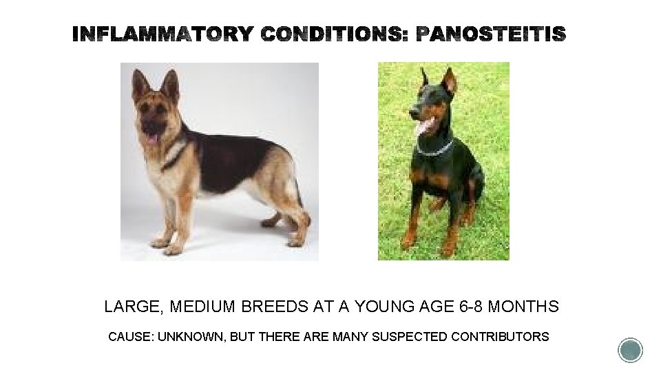
LARGE, MEDIUM BREEDS AT A YOUNG AGE 6 -8 MONTHS CAUSE: UNKNOWN, BUT THERE ARE MANY SUSPECTED CONTRIBUTORS
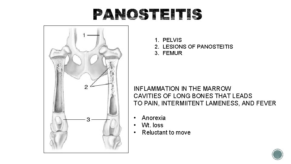
1. PELVIS 2. LESIONS OF PANOSTEITIS 3. FEMUR INFLAMMATION IN THE MARROW CAVITIES OF LONG BONES THAT LEADS TO PAIN, INTERMIITENT LAMENESS, AND FEVER • Anorexia • Wt. loss • Reluctant to move
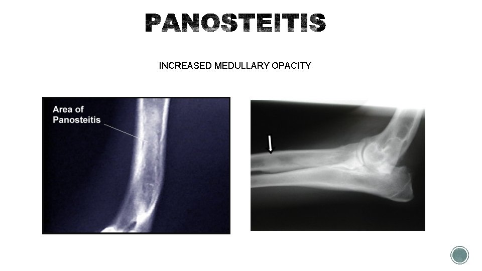
INCREASED MEDULLARY OPACITY
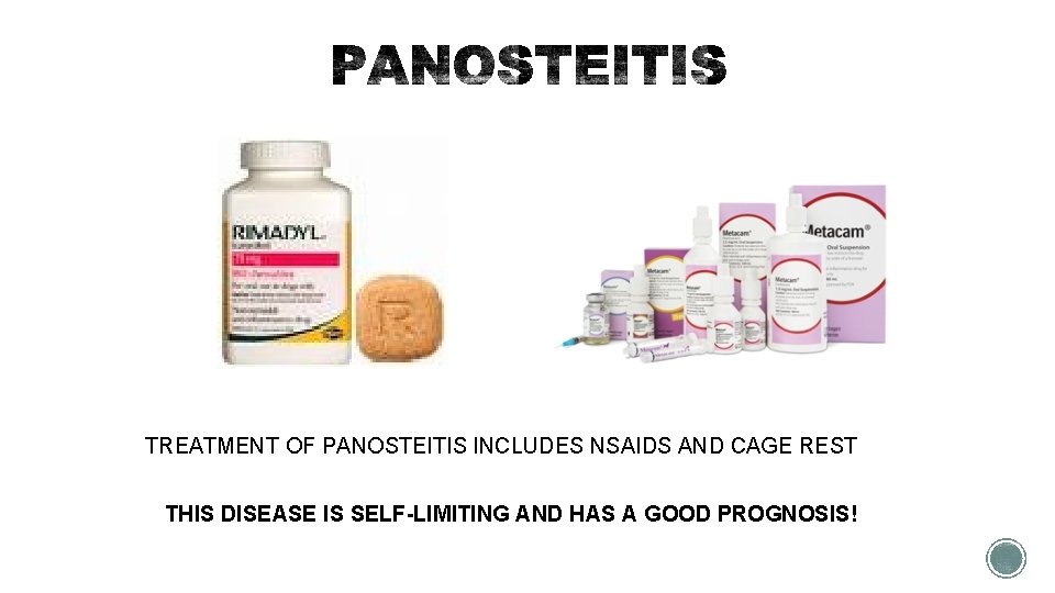
TREATMENT OF PANOSTEITIS INCLUDES NSAIDS AND CAGE REST THIS DISEASE IS SELF-LIMITING AND HAS A GOOD PROGNOSIS!
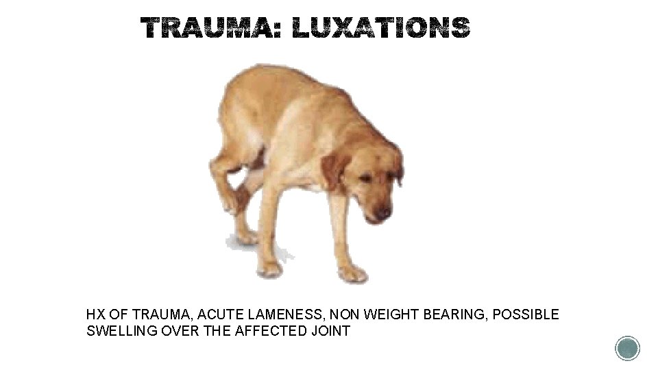
HX OF TRAUMA, ACUTE LAMENESS, NON WEIGHT BEARING, POSSIBLE SWELLING OVER THE AFFECTED JOINT
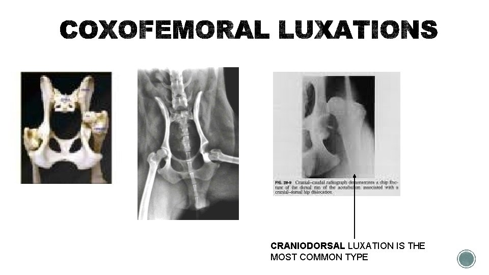
CRANIODORSAL LUXATION IS THE MOST COMMON TYPE
§ TREATMENT § Closed reduction: femoral head is replaced back into the acetabulum under anesthesia § Open reduction: femoral head is surgically replaced into the acetabulum and the soft tissue structures are used to secure the reduction.
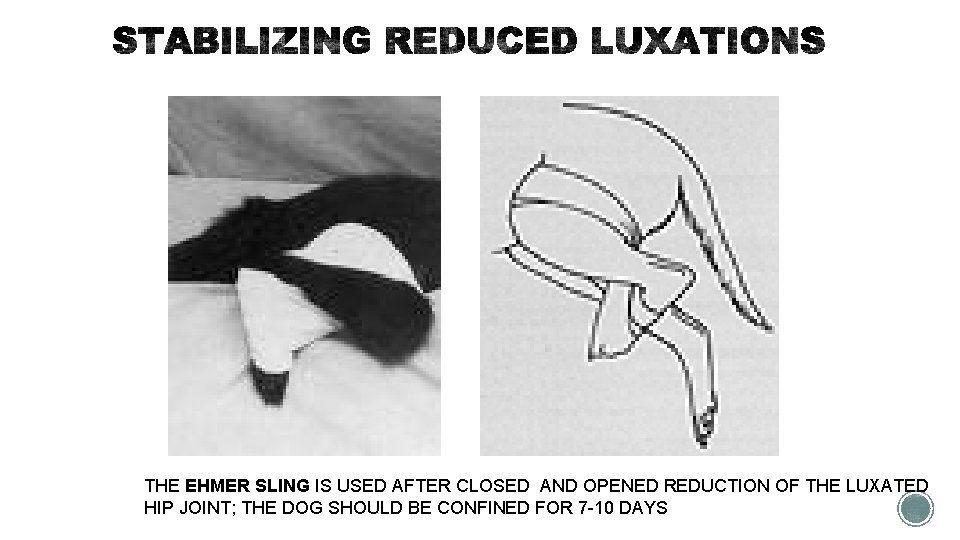
THE EHMER SLING IS USED AFTER CLOSED AND OPENED REDUCTION OF THE LUXATED HIP JOINT; THE DOG SHOULD BE CONFINED FOR 7 -10 DAYS
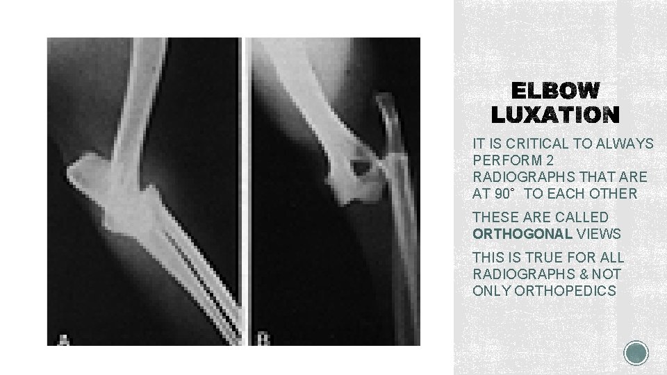
IT IS CRITICAL TO ALWAYS PERFORM 2 RADIOGRAPHS THAT ARE AT 90∘TO EACH OTHER THESE ARE CALLED ORTHOGONAL VIEWS THIS IS TRUE FOR ALL RADIOGRAPHS & NOT ONLY ORTHOPEDICS
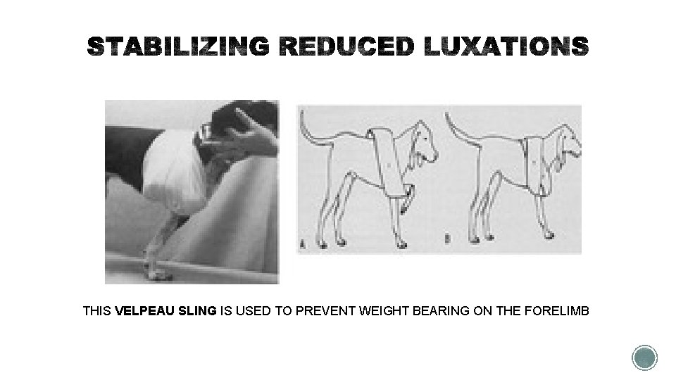
THIS VELPEAU SLING IS USED TO PREVENT WEIGHT BEARING ON THE FORELIMB
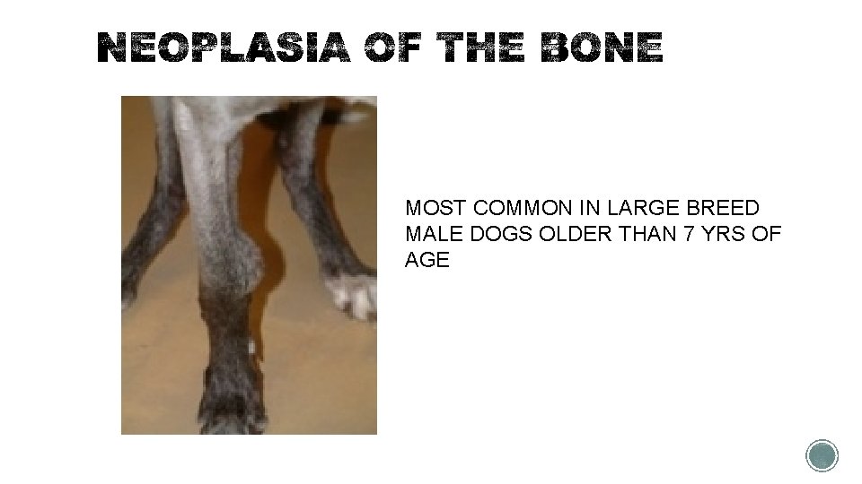
MOST COMMON IN LARGE BREED MALE DOGS OLDER THAN 7 YRS OF AGE
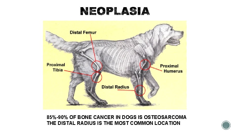
85%-90% OF BONE CANCER IN DOGS IS OSTEOSARCOMA THE DISTAL RADIUS IS THE MOST COMMON LOCATION
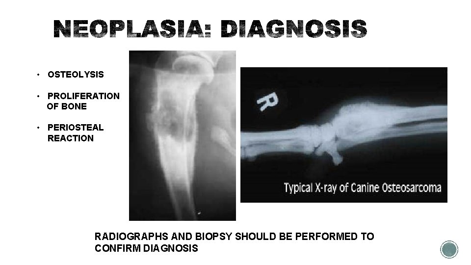
• OSTEOLYSIS • PROLIFERATION OF BONE • PERIOSTEAL REACTION RADIOGRAPHS AND BIOPSY SHOULD BE PERFORMED TO CONFIRM DIAGNOSIS
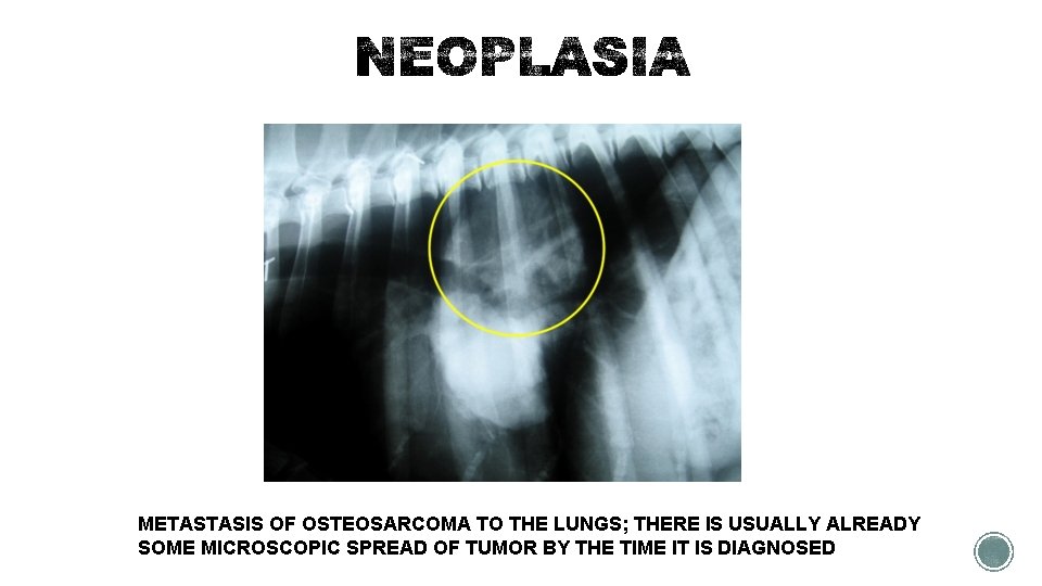
METASTASIS OF OSTEOSARCOMA TO THE LUNGS; THERE IS USUALLY ALREADY SOME MICROSCOPIC SPREAD OF TUMOR BY THE TIME IT IS DIAGNOSED
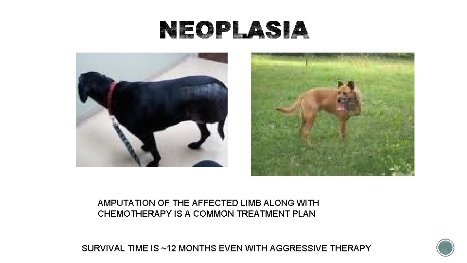
AMPUTATION OF THE AFFECTED LIMB ALONG WITH CHEMOTHERAPY IS A COMMON TREATMENT PLAN SURVIVAL TIME IS ~12 MONTHS EVEN WITH AGGRESSIVE THERAPY
§ MYOPATHIES are diseases that affect the muscle § Inflammatory (bacterial, protozoal) § Immune-mediated § Acquired (secondary to other diseases) § Diagnosis is usually made by muscle biopsy and treatment directed toward the primary cause
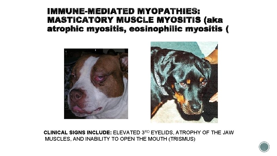
CLINICAL SIGNS INCLUDE: ELEVATED 3 RD EYELIDS, ATROPHY OF THE JAW MUSCLES, AND INABILITY TO OPEN THE MOUTH (TRISMUS)
§ The muscles of mastication contain a special type of fiber (2 M) that has antigenic properties possibly shared with bacteria. § The treatment of choice is glucocorticoids (steroids).
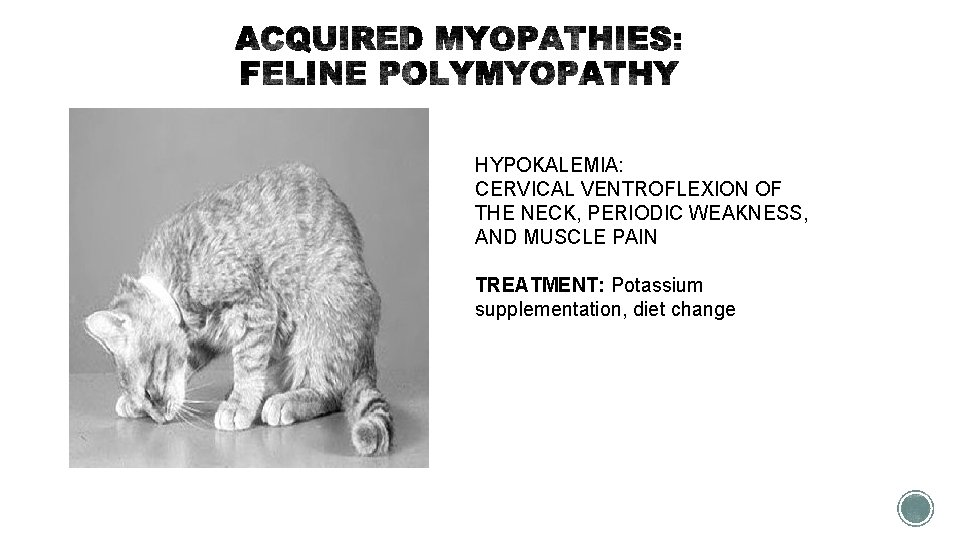
HYPOKALEMIA: CERVICAL VENTROFLEXION OF THE NECK, PERIODIC WEAKNESS, AND MUSCLE PAIN TREATMENT: Potassium supplementation, diet change
- Slides: 33