PAROTID REGION Parotid gland G para near otis

 Is the largest salivary gland it")
 is found below the level")


,")
: post belly of digastric, Cervical br of facial n and")


= *superficial temporal (sup surface)")
 Formed of small ducts coalesce at the anterosuperior aspect of")


 Emerges from the")

- Slides: 26
PAROTID REGION
Parotid gland G. para, near + otis, ear) Is the largest salivary gland it is composed mainly of serous acini. It secretes about 2030% of total saliva. Large, lobulated and irrgular Wt about 15 g
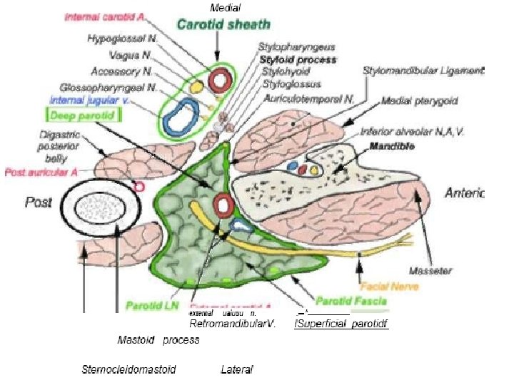
PG- Position (Nearly 80% of the parotid gland (PG) is found below the level of the external auditory canal, in deep hollow between the mandible and the SCM. The remaining 20% extends medially through the stylomandibular tunnel, which is formed ventrally by the posterior edge of the ramus dorsally by the anterior border of the SCM & posterior digastric muscle deeply and dorsally by the stylomandibular ligament.
PG- Extensions Project to mastoid process Down the anterior aspect of the SCM for a short distance Around the posterior border of the mandible. Superiorly to the to inferior margin of the zygomatic arch Superficial to the posterior aspect of the masseter mm Anterior extension for the facial process of the parotid duct. * Maybe separated from the remainder as the Accessory parotid gland.
Parotid Capsule Gland encapsulated by a fascial layer that is continuous with deep cervical fascia. superficial layer is dense and closely united with gland (parotidomasseteric fascia). the fascia forms the stylomandibular ligament, separate parotid from mandibular gland
PG- external features 3 -sided pyramid Apex: directed downward Four surfaces: superior- base, superficial(lateral), anteromedial(anterior) and posteromedial(deep). Three borders: anterior, posterior and medial.
PG- Relations Apex(lower pole): post belly of digastric, Cervical br of facial n and 2 divisions of retromadibular vein. Superior surface(upper pole): cartilagenous of ECM, post part TMJ, suptemporal vessels, and auriculotemporal n
PG- Relations- surfaces and borders
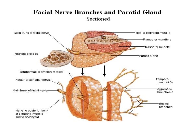
CN VII branches roughly divide the PG into superficial and deep lobes while coursing anteriorly from the stylomastoid foramen to the muscles of facial expression.
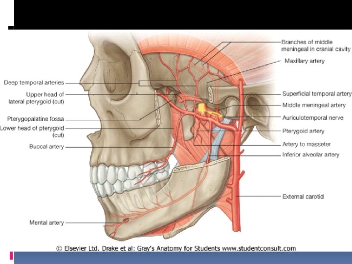
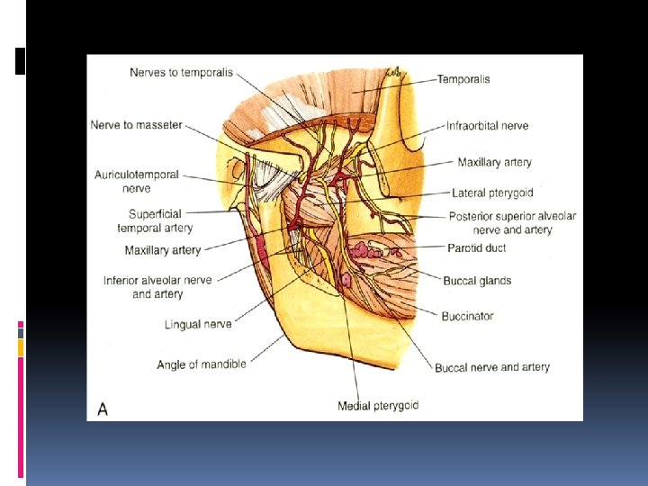
Structures embedded in the gland A. External Carotid (postmedial surface)= *superficial temporal (sup surface) *maxillary art. (anteromed. Surface) *transverse facial art. (ant. border) B. Retromandibular vein Formed within gland by maxillary V. + superficial temporal divides in lower part to its divisions other veins may present: *common facial and *external jugular vein • Branches of facial and auriculotemporal
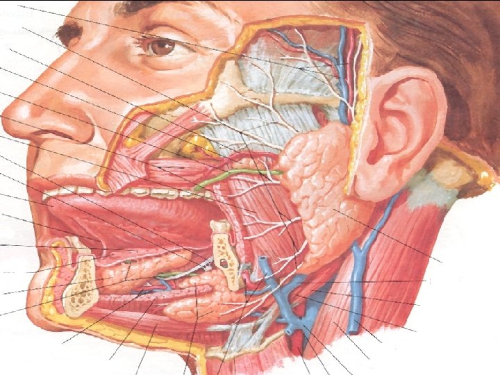
Parotid Duct (Stensen’s duct) Formed of small ducts coalesce at the anterosuperior aspect of the PG Arises from middle of anterior border lies superficial to the masseter muscle 1. 5 cm inferior to Zygomatic arch Is inferior to the transverse facial artery Buccal branchs of facial nerve travel with the duct It is thick walled: 1 -3 mm in diameter+4 -6 cm in length At the anterior edge of the masseter muscle it pierces medially through the buccal pad of fat, buccopharyngeal meebrane and Buccinator at 2 nd Molar The oblique course of the duct between the mucous membrane and the buccinator act as a valve.
Parotid Blood supply and Lymphatics Blood supply from: Arterial= ECA and branches. Venous= EJV Lymphatic drainage is to the superficial and deep cervical nodes Preauricular lymph nodes (LN) in the superficial fascia drain the temporal scalp, upper face, anterior pinna LN within the gland drain the parotid gland, nasopharynx, palate, middle ear and external auditory meatus
Innervation 1. Sympathetic : superior cervical ganglion- superficial temporal artery (Serous watery saliva, amylase decrease) 2. Parasympathetic secretomotor fibers reach the gland by a circuitous route: presynaptic neurons lie in the inferior salivatory nucleus of Glossopharyngeal nerve ( CNIX) thru lesser superficial petrosal nerve Postsynaptic neurons secretomotor fibers leave the otic ganglion and distributed by the Auriculotemporal nerve to the parotid gland. Mucoid viscosity saliva, amylase increase 3. Sensory innervation to gland from Auriculotemporal and to capsule from greater auricular nerve
Facial nerve Stylomastoid foramen Superficial lobe and deep lobe 3 Motor branches immediately: stylohoid muscle, posterior auricular muscle, digastric muscle posterior belly Pes Anserinus ((intraparotid plexus of CN 7) – 1. 3 cm from the stylomastoid foramen Temporofacial division, Cervicofacial division 5 terminal branches
Facial nerve identification 1. Antigrade dissection Tympanomastoid suture Tragal point Digastric muscle posterior belly Styloid process SCM muscle 2. Retrograde dissection 3. Mastoidectomy
Clinical application Frey’s syndrome: It develops after penetrating wounds of the gland. When the pat. eats beads of perspiration appear on the skin covering the gland. This is duo to damage of GEATER AURICUAL N and AURICULOTEMPORAL N during healing parasympathetic secretomotor fibres from the AURICULOTEMPORAL N grow out and join the GEATER AURICUAL N fibres. Eventually, they reach the sweat glands in the facial skin. * By this means, a stimulus intended for saliva production produces sweat secretion instead Tumors of Parotid gland: Usually arise at the superficial lobe without involvement of the facial nerve. Iatrogenic cause as in surgical injury of facial nerve results to facial paralysis.
Clinical application Abscess or cyst of the gland may result in pressure on the Facial nerve. The weakest part of investing fascia is between Styloid process and spine of sphenoid, therefore, infections breakout parotid fascia drain into the lateral pharyngeal space, which is in direct communication with the retropharyngeal space. That may track inferior along carotid sheath between visceral and prevertebral fascia.
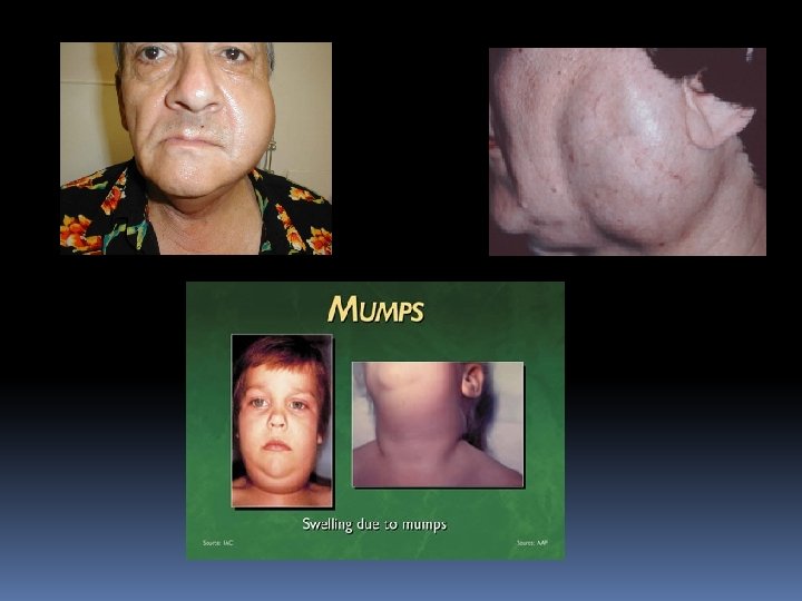
Clinical application Parotid Gland Infection: The gland may be acutely inflamed by retrograde bacterial infection from the mouth via the parotid duct or itself is infected by the bloodstream as in MUMPS. Which is due to PARAMYXOVIRUS. It is an infectious disease. Acute Parotitis: In both cases the gland is swollen, it is painful because of the capsule Sjogren’s syndrome: Xerostomia (dry mouth), keratoconjunctivitis sicca (dry eyes), rheumatoid arthritis, hypergammaglobulinemia Autoimmune disorder that affects not only salivary glands and lacrimal glands of Mikulicz’s disease, but also minor salivary glands and occasionally lymph nodes, lung, kidney, bone marrow, skeletal muscle, skin, liver
Nerves related to PG Great Auricular Nerve (C 2, C 3) Emerges from the posterior border of the SCM at Erb’s point. �It crosses the mid-portion of the SCM about 6. 5 cm beneath the EAM. Passes parallel and superior to the external jugular vein to supply the ear and pre-auricular region. Auriculotemporal Nerve Branch of V 3 Traverses the upper part of the parotid gland emerges from the superior surface with the superficial temporal vessels. It carries sensory fibers from the trigeminal and postganglionic parasympathetic (secretory)fibers.
Parotid Bed V: internal jugular vein A: external and internal carotid arteries N: glossopharyngeal N vagus N spinal accesory N hypoglossal N S: styloid process styloglossus mm stylohyloid mm