
Parathyroid Hormone Calcitonin Calcium and Phosphate Metabolism Prof

Parathyroid Hormone, Calcitonin, Calcium and Phosphate Metabolism Prof. dr. Zoran Valić Department of Physiology University of Split School of Medicine

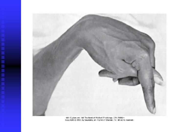
Calcium and Phosphate Regulation in the ECF and Plasma Ca+2 in extracellular fluid (ECF) – 2, 4 mmol/L q normally precisely regulated (few %) q key role of Ca+2 in many physiologic processes: muscle contraction, blood clotting, transmission of nerve impulses q hypercalcemia – depression; / hypocalcemia – excitetion (tetany) q 0. 1% Ca+2 in ECF, 1% in cells, rest is stored in bones q
q 85% of body's phosphate is stored in bones q 14 -15% in cells, 1% in ECF q concentration is not nearly as well regulated as calcium q same factors that regulate calcium

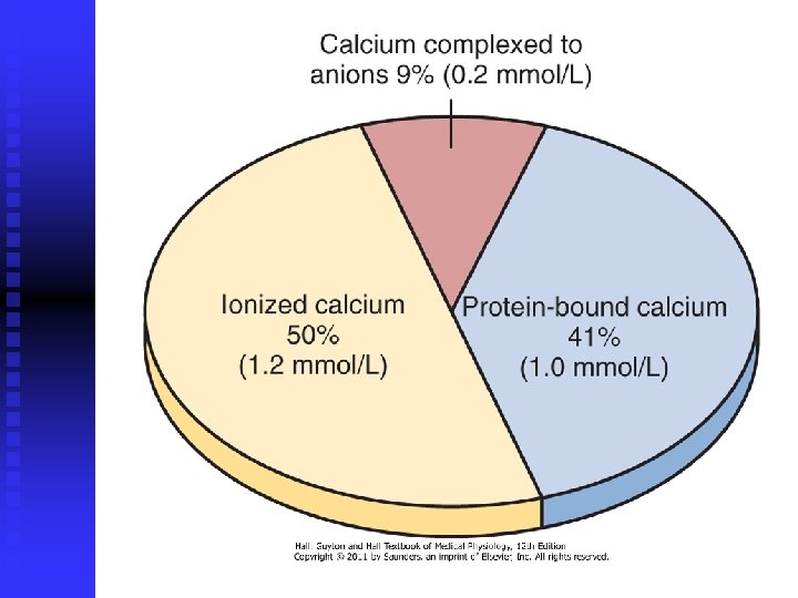
Calcium in the Plasma and Interstitial Fluid (IF) q present in three forms : 41% - combined with plasma proteins b) 9% - combined with anionic substances (citrate and phosphate) c) 50% - ionized a) q ionic calcium is the form that is important for most functions of calcium in the body
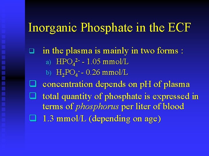
Inorganic Phosphate in the ECF q in the plasma is mainly in two forms : HPO 42 - - 1. 05 mmol/L b) H 2 PO 4 - - 0. 26 mmol/L a) q concentration depends on p. H of plasma q total quantity of phosphate is expressed in terms of phosphorus per liter of blood q 1. 3 mmol/L (depending on age)
Nonbone Physiologic Effects of Altered Calcium and Phosphate Concentrations q hypocalcemia causes nervous system excitement ( permeability to Na+) q tetany occurs when calcium decreases to 1. 5 mmol/L, and death when it is 1 mmol/L q hypocalcemia : dilatation of heart, changes in enzyme activities, increased membrane permeability of some cells, impaired blood clotting – nonphysiological decrease
Hypercalcemia q nervous system becomes depressed q lack of appetite and constipation q decreases the QT interval of the heart q depressive effects begin to appear when calcium rises above 3. 0 mmol/L, they can become marked above 3. 8 mmol/L
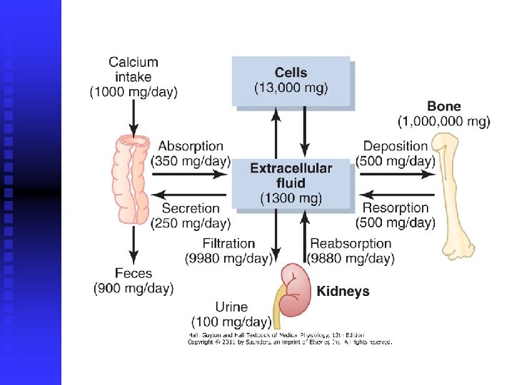
Absorption and Excretion of Calcium and Phosphate q daily intake is about 1000 mg/day each for calcium and phosphorus (25 mmol) – 1 L of milk q 90% of daily intake of calcium is excreted in feces q almost all the dietary phosphate is absorbed into the blood from the gut and later excreted in the urine
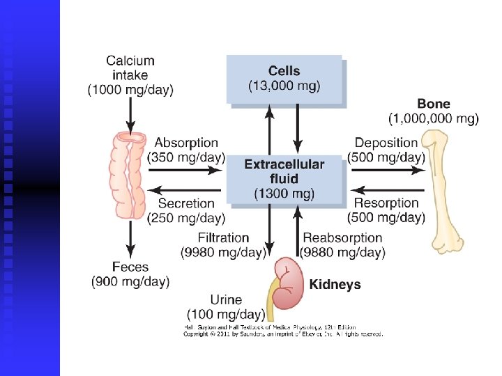
q kidney filers calcium (ionized and combined with anions) q renal tubules reabsorb 99 percent of the filtered calcium q renal phosphate excretion is controlled by an overflow mechanism (critical value of about 1 mmol/L – no phosphate is lost in the urine)
Bone and Its Relation to ECF Calcium and Phosphate q q q bone = organic matrix + calcium salts compact bone: 30% matrix, 70% salts organic matrix : a) b) q 90 -95 % collagen fibers (tensile strength) 5 -10 % ground substance (ECF + proteoglycans, chondroitin sulfate and hyaluronic acid) bone salts: calcium and phosphate

Bone Salts q Ca 10(PO 4)6(OH)2 q hydroxyapatite – shaped like long, flat plate q relative ratio of Ca/P 1. 3 -2. 0 q ions Mg 2+, Na+, K+ & HCO 3 q many ions of many types of ions (transuranic elements and heavy metals) q osteogenic sarcoma (bone cancer)
Tensile and Compressional Strength of Bone q compact bone is composed of repeating periodic segments of collagen q hydroxyapatite crystals bound tightly to it – prevents "shear" in the bone q collagen fibers – tensile strength q calcium salts – compressional strength
Precipitation and Absorption of Calcium and Phosphate in Bone q concentrations of calcium and phosphate ions in ECF are considerably greater than those required to cause precipitation of hydroxyapatite q pyrophosphate – inhibitor which prevent precipitation
Bone Calcification osteoblasts produce osteoid (collagen molecules + ground substance) q calcium salts begin to precipitate on the surfaces of the collagen fibers, forming minute nidi q mixture of amorphous compounds (noncrystalline) salts of Ca & P, converted into the hydroxyapatite crystals over a period of weeks or months q few percent may remain permanently in the amorphous form (important role) q mechanism is not fully understood q
Precipitation of Calcium in Nonosseous Tissues q arteriosclerosis q degenerating tissues q old blood clots
Calcium Exchange Between Bone and ECF q fast correction of plasma Ca concentration q exchangeable calcium – in equilibrium with the calcium ions in the ECF – rapid buffering mechanism q in all tissue cells (liver and gastrointestinal tract) & in the bone (amorphous calcium salts)
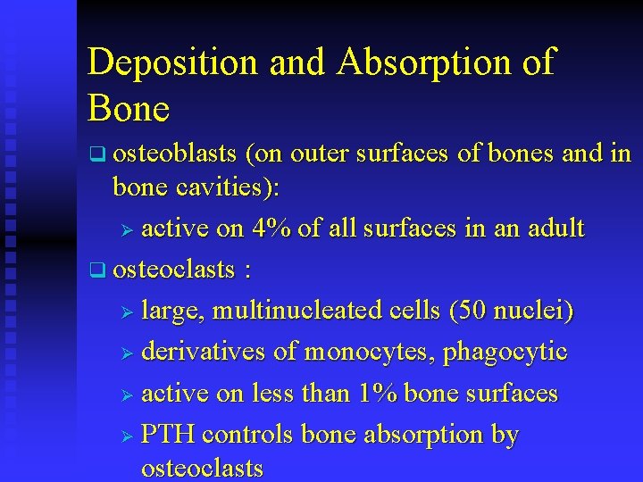
Deposition and Absorption of Bone q osteoblasts (on outer surfaces of bones and in bone cavities): Ø active on 4% of all surfaces in an adult q osteoclasts : Ø large, multinucleated cells (50 nuclei) Ø derivatives of monocytes, phagocytic Ø active on less than 1% bone surfaces Ø PTH controls bone absorption by osteoclasts
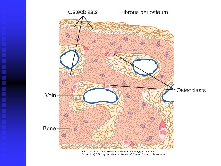
Absorption of Bone q q osteoclasts send out villus-like projections toward the bone (ruffled border) & secrete: a) proteolytic enzymes – organic matrix b) acids (citric, lactic acid) – bone salts PTH stimulates osteoclast activity and bone resorption, but through indirect mechanism
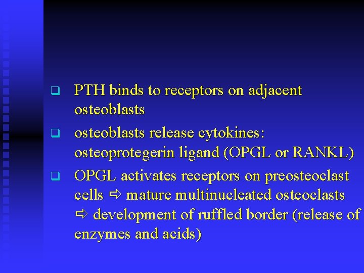
q q q PTH binds to receptors on adjacent osteoblasts release cytokines: osteoprotegerin ligand (OPGL or RANKL) OPGL activates receptors on preosteoclast cells mature multinucleated osteoclasts development of ruffled border (release of enzymes and acids)
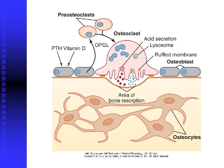
q q q osteoblasts also produce osteoprotegerin (OPG, also called OCIF) OPG inhibits bone resorption OPG acts as a "decoy" receptor binding to OPGL and preventing its actions vitamin D and PTH inhibit OPG production and stimulate OPGL formation estrogen stimulates OPG production used for treatment
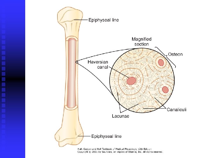
q q bone deposition and absorption are normally in equilibrium – total mass of bone remains constant. concentrated mass of osteoclasts tunnel in diameter 0, 2 -1 mm and several mm long tunnel is invaded by osteoblasts new bone begins to develop
q q new bone being laid down in successive layers of concentric circles (lamellae) deposition of new bone ceases when the bone begins to encroach on the blood vessels supplying the area haversian canal, is all that remains of the original cavity osteon
Value of Continual Bone Remodeling q bone strength in proportion to bone stress q shape of the bone can be rearranged q replacement of old organic matrix q bone is deposited in proportion to the compressional load (athletes & cast) q repair of fracture activates osteoblasts q callus, rings
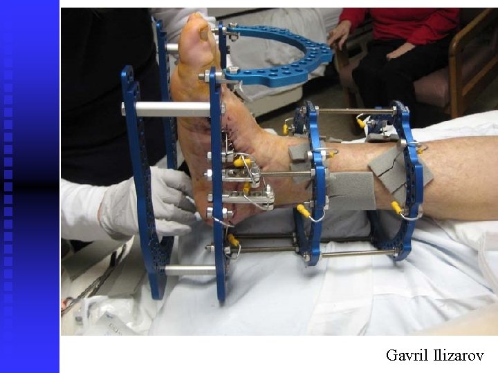
Gavril Ilizarov
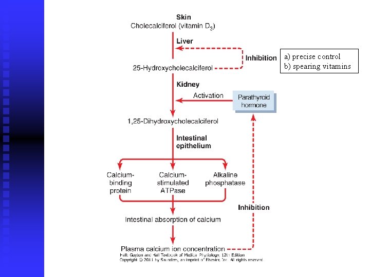
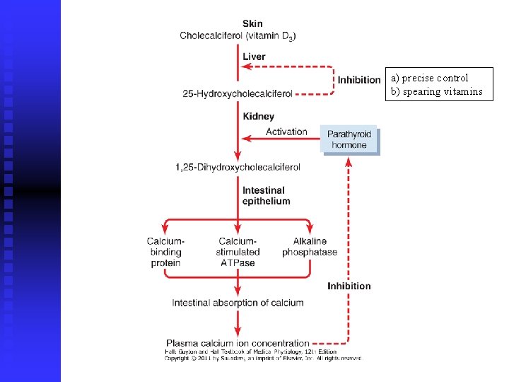
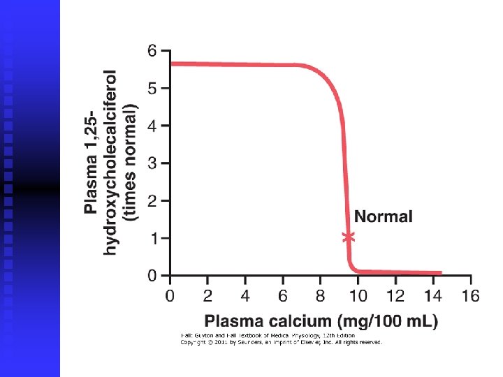
Vitamin D q potent effect to increase calcium absorption from the intestinal tract q vitamin D itself is not the active substance q must first be converted into: 1, 25 -dihydroxycholecalciferol q 7 -dehydrocholesterol + ultraviolet rays (in the skin) – cholecalciferol (vitamin D)
a) precise control b) spearing vitamins
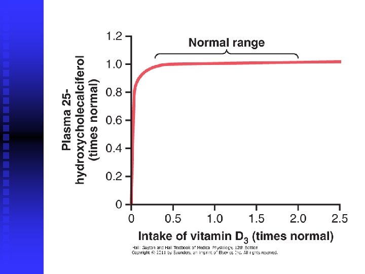
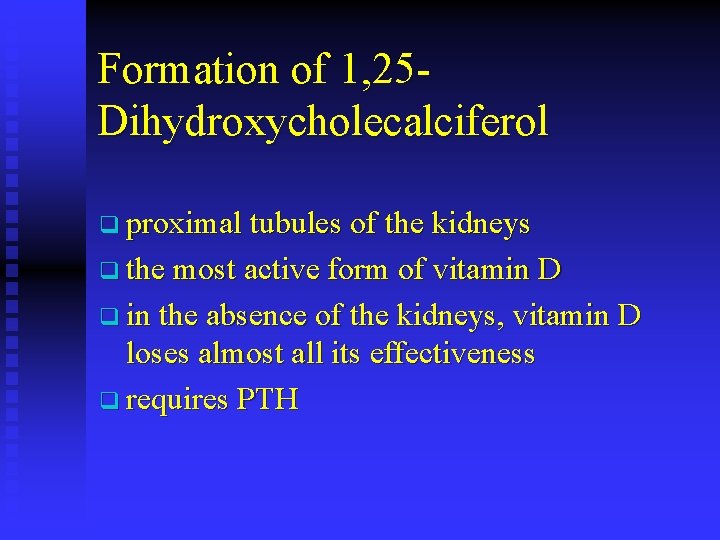
Formation of 1, 25 Dihydroxycholecalciferol q proximal tubules of the kidneys q the most active form of vitamin D q in the absence of the kidneys, vitamin D loses almost all its effectiveness q requires PTH
a) precise control b) spearing vitamins
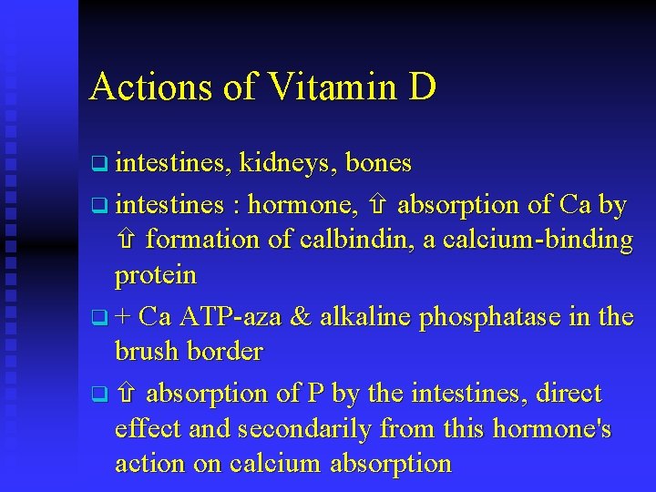
Actions of Vitamin D q intestines, kidneys, bones q intestines : hormone, absorption of Ca by formation of calbindin, a calcium-binding protein q + Ca ATP-aza & alkaline phosphatase in the brush border q absorption of P by the intestines, direct effect and secondarily from this hormone's action on calcium absorption
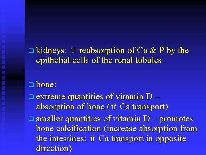
q kidneys: reabsorption of Ca & P by the epithelial cells of the renal tubules q bone: q extreme quantities of vitamin D – absorption of bone ( Ca transport) q smaller quantities of vitamin D – promotes bone calcification (increase absorption from the intestines; Ca transport in opposite direction)
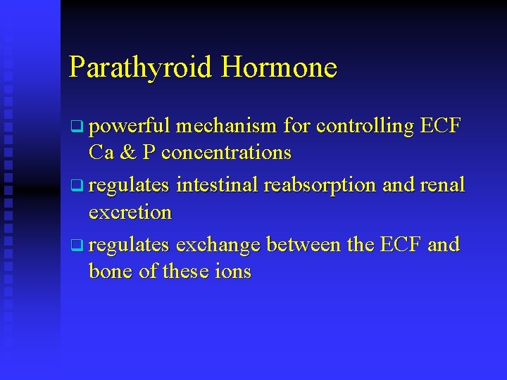
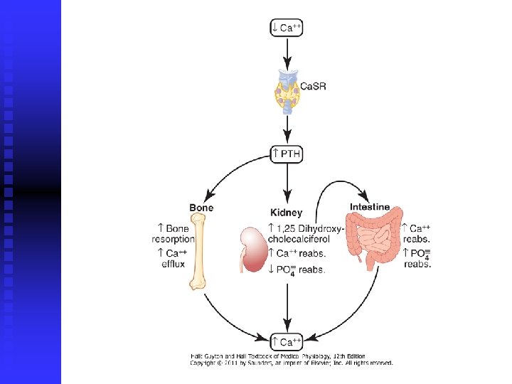
Parathyroid Hormone q powerful mechanism for controlling ECF Ca & P concentrations q regulates intestinal reabsorption and renal excretion q regulates exchange between the ECF and bone of these ions
q q increased secretion causes rapid absorption of calcium salts from the bones – hypercalcemia hypofunction of the parathyroid glands – hypocalcemia (tetany)
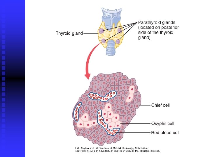
q q q normally – 4 parathyroid glands in humans located immediately behind thyroid gland 6 x 3 x 2 mm, appearance of dark brown fat danger of removal during thyroid operations (2(3) out of 4 – OK) chief cells (majority) and oxyphil cells (function unknown, absent in many animals and in young humans)
Chemistry of PTH q q PTH has been isolated in a pure form preprohormone (110 AA) prohormone (90 AA) PTH itself (84 AA) smaller compounds have also been isolated that exhibit full PTH activity kidneys rapidly remove big PTH within minutes but fail to remove many of the fragments for hours
Effect of PTH on Ca & P Concentrations in ECF
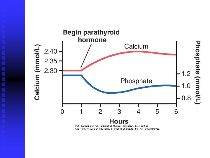
Effect of PTH on Ca & P Concentrations in Plasma q q q phosphate concentration falls more rapidly than the calcium rises both reach a plateau in about 4 hours increase Ca & P absorption from bone decrease excretion of Ca by kidneys increase renal P excretion
PTH Increases Ca & P Absorption from Bone q rapid phase: activation of the already existing osteocita (begins in minutes) q much slower phase: proliferation of the osteoclasts (requiring several days or even weeks)
Rapid Phase – Osteolysis q osteocytic membrane system – separates the bone itself from ECF q bone fluid – small amount between osteocytic membrane and bone q pumping of Ca ions from bone fluid into ECF – osteocytic pump
Rapid Phase – Osteolysis q bone fluid calcium concentration falls even lower absorbed of Ca & P from the bone – osteolysis q PTH strongly activates calcium pump ( Ca permeability of the bone fluid side of the osteocytic membrane)
Slow Phase of Bone Absorption q q PTH activates osteoblasts and osteocytes which send secondary "signals" to the osteoclasts (OPGL) 1) immediate activation of the osteoclasts that are already formed 2) formation of new osteoclasts osteoclastic resorption of bone can lead to weakened bones and secondary stimulation of the osteoblasts
PTH Decreases Ca Excretion and Increases P Excretion by Kidneys q increased P excretion is based on diminish proximal tubular reabsorption of P ions q simultaneous increase in renal tubular reabsorption of Ca q PTH increases reabsorption of Mg & H, and diminish reabsorption of Na, K, AA
Control of PTH Secretion by Ca Ion Concentration q PTH increases reabsorption of Ca & P from the intestines q c. AMP mediates the effects of PTH q Ca concentration controls secretion of PTH q parathyroid glands become greatly enlarged in rickets (5 x) in pregnancy, and during lactation
Decrease in PTH Secretion q excess quantities of Ca in the diet q increased vitamin D in the diet q bone absorption caused by factors other than PTH (immobilization)
q changes in ECF Ca ion concentration are detected by a calcium-sensing receptor (Ca. SR) in parathyroid cell membranes q Ca. SR is a G protein-coupled receptor q stimulated by calcium ions, activates phospholipase C ( in IP 3 and DAG) q stimulation of Ca release from intracellular stores decreases PTH secretion
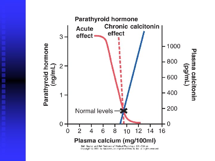
Calcitonin q synthesis and secretion of calcitonin occur in the parafollicular cells, or C cells of thyroid gland (0, 1% of the human thyroid gland, remnants of ultimobranchial glands) q effects opposite to those of PTH q 32 -amino acid peptide q ECF Ca ion concentration – primary stimulus for calcitonin secretion
Mechanism of Calcitonin Action q absorptive activities of the osteoclasts q osteolytic effect of osteocytic membrane q formation of new osteoclasts q minor effects on calcium handling in the kidney tubules and the intestines q calcitonin has bigger effect in children and certain diseases (Paget) than in adult human
Summary of Control of Ca Ions q first line of defense – buffer function of the exchangeable calcium in bones – amorphous compounds (Ca HPO 4) q + mitochondria of the liver and intestine q second line of defense – hormonal control
Pathophysiology q Hypoparathyroidism ( PTH secretion) osteocytic resorption of Ca + osteoclasts inactivity Ca tetany (laryngeal muscles) q extremely large quantities of vitamin D or giving PTH (expensive, short-acting, antibodies) q q Hyperparathyroidism (primary & secondary) ordinarily tumor (in women because pregnancy) q extreme osteoclastic activity Ca & P q fractures, decalcification, muscle weakness, q
q Rickets Ca (slightly) & P (greatly) in ECF, usually caused by lack of vitamin D q occur in spring months after depletion of stores q compensatory increase in PTH secretion causes extreme osteoclastic absorption of the bone q tetany occurs in later stages – death q treatment : vitamin D + Ca & P in the food q steatorrhea – osteomalacia (adult rickets) q renal rickets – failure of the damaged kidneys to form 1, 25 -dihydroxycholecalciferol q
q Osteoporosis most common of all bone diseases in adults q especially in old age q decreased bone matrix q osteoblastic activity is usually less than normal – osteoid deposition is depressed q lack of physical stress on the bones because of inactivity, malnutrition, lack of vitamin C, postmenopausal lack of estrogen secretion, old age ( h. GH), Cushing's syndrome ( glucocorticoids) q
q treatment: q antiresorptive drugs: bisphonates (Fosamax, Actonel & Boniva) q estrogen substitution therapy q SERMs (raloxifene, Evista) – osteoclasts q calcitonin q inhibitors of RANKL (monoclonal antibody, Denosumab) q q anabolic drugs: teriparatide (Forteo) – recombinant PTH q calcium salts (carbonates, citrates, lactates) q sodium-fluoride q
Physiology of the Teeth q teeth cut, grind, and mix the food eaten q forces: 250 -450 N & 650 -900 N q occlusion – fitting of upper set of teeth with the lower q major functional parts: enamel, dentin, cementum and pulp q another division: crown, neck and root
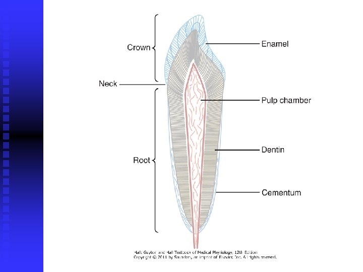
Enamel q outer surface of the tooth; ameloblasts q formed before eruption of the tooth q composed of large and dense crystals of hydroxyapatite with adsorbed Mg, Na, K + insoluble protein fibers (similar to keratin) q extremely hard and resistant to acids, enzymes, and other corrosive agents
Dentin q main body of tooth, strong, bony structure q hydroxyapatite crystals much denser than in bone, embedded in a strong meshwork of collagen fibers q does not contain: osteoblasts, osteocytes, osteoclasts, spaces for vessels or nerves q odontoblasts: formation and nurishment q calcium salts: compressional forces q collagen: toughness and tensional forces
Cementum q bony substance secreted by cells of the periodontal membrane – lines tooth socket q collagen fibers pass directly from jaw bone through periodontal membrane into cementum q increases in thickness and strength with age
Pulp q filling pulp (tooth) cavity q composed of connective tissue, nerve fibers, blood vessels, and lymphatics q odontoblasts – cells lining the surface of the pulp cavity q during the formative years lay down dentin q send projections into small dentinal tubules that penetrate all the way through the dentin
Dentition q humans and most other mammals develop two sets of teeth during a lifetime q deciduous or milk teeth (20) q q erupt between 7 th month and 2 nd year last until 6 th and 13 th year q permanent teeth (32)
Formation of the Teeth q invagination of the oral epithelium into the dental lamina and development of a toothproducing organ q epithelial cells above form ameloblasts (enamel) q epithelial cells below invaginate upward into the middle of the tooth to form the pulp cavity and the odontoblasts (dentin)
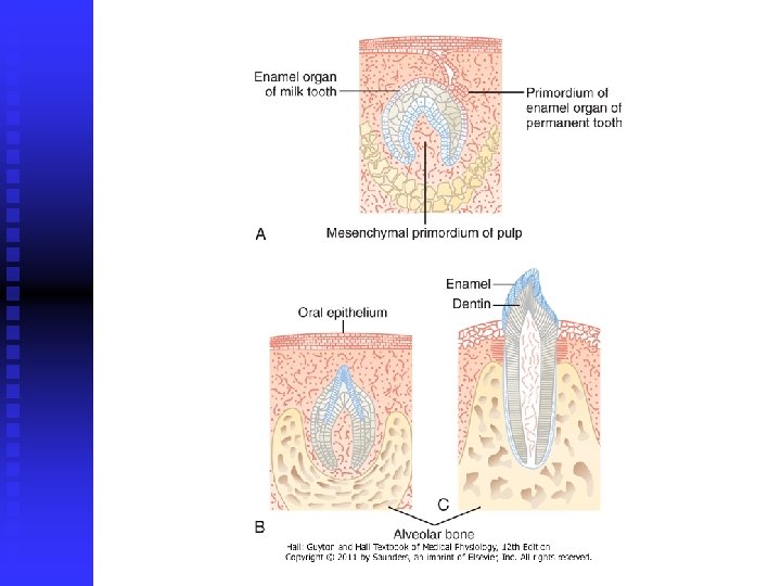
Eruption of Teeth q cause of "eruption" is unknown q most likely theory is that growth of the tooth root and the bone underneath the tooth progressively shoves the tooth forward
Development of Permanent Teeth q when each permanent tooth becomes fully formed, it pushes outward through the bone q erodes the root of the deciduous tooth and eventually causes it to loosen and fall out
Metabolic Factors q thyroid and growth hormones q availability of Ca & P in the diet q amount of vitamin D q rate of PTH secretion
Mineral Exchange in Teeth q salts composed of hydroxyapatite with adsorbed carbonates and various cations q new salts are constantly being deposited while old salts are being reabsorbed q deposition and reabsorbtion – in dentin and cementum, limited extent in enamel (saliva) q rate of exchange in dentin 3 x slower than in bone
Dental Abnormalities 1) 2) caries (erosion of the teeth) malocclusion (failure of the projections of the upper and lower teeth to interdigitate properly)
1) Caries q action of bacteria on the teeth (Streptococcus mutans) q deposit of plaque (film of saliva and food) q dependance on carbohydrates (form acids (lactic) and proteolytic enzymes) q acids – slow dissolvement of calcium salts q enamel of the tooth is primary and most important barrier to development of caries
q small amounts of fluorine develop enamel that is more resistant to caries q fluorine does not make the enamel harder q fluorine ions replace many of hydroxyl ions in hydroxyapatite crystals, which in turn makes enamel several times less soluble q fluorine may also be toxic to the bacteria q fluorine promote deposition of calcium phosphate to "heal" the enamel surface
2) Malocclusion q hereditary abnormality q teeth of one jaw grow to abnormal positions q teeth do not interdigitate properly and therefore cannot perform their normal grinding or cutting action adequately q pain in mandibular joint and deterioration of the teeth q treatment: continuous force on teeth

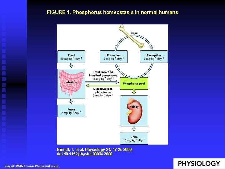
FIGURE 1. Phosphorus homeostasis in normal humans Berndt, T. et al. Physiology 24: 17 -25 2009; doi: 10. 1152/physiol. 00034. 2008 Copyright © 2009 American Physiological Society
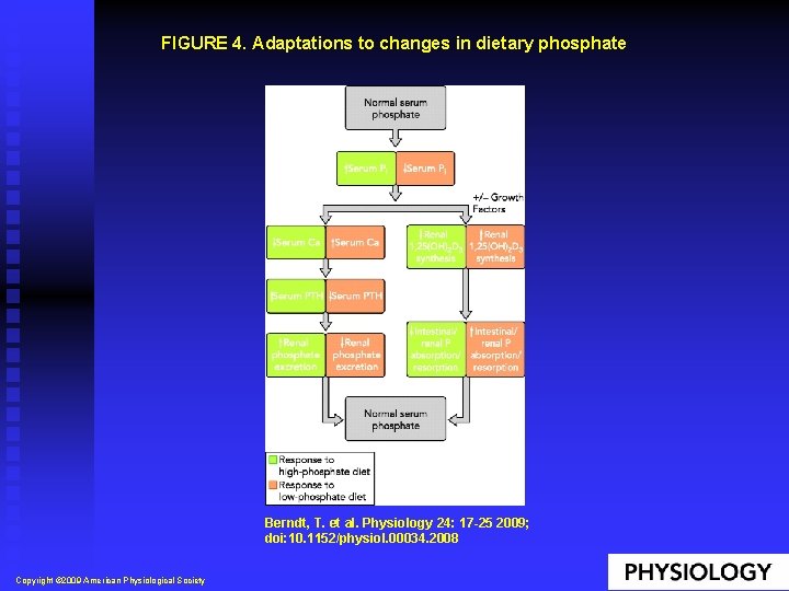
FIGURE 4. Adaptations to changes in dietary phosphate Berndt, T. et al. Physiology 24: 17 -25 2009; doi: 10. 1152/physiol. 00034. 2008 Copyright © 2009 American Physiological Society
- Slides: 84