Parasympathetic Nervous System Lecture Objectives Make a list

Parasympathetic Nervous System

Lecture Objectives • Make a list of the components of the system. • Make a list of cranial nerves having parasympathetic activity. • Describe the parasympathetic ganglia in the head and neck, their locations and target organs. • Describe the sacral parasympathetic outflow. • Make a list of its target organs.

Structure of the Parasympathetic Division • Craniosacral division: Preganglionic neurons originate from • Brainstem through cranial nerves III, VII, IX and X • Sacral spinal nerves S 2‐S 4 • Parasympathetic ganglia • terminal ganglia. • Presynaptic neuron usually synapses with 4‐ 5 postsynaptic neurons all of which supply a single visceral effector.

Autonomic Plexuses in the Thorax, Abdomen and Pelvis

Autonomic Plexuses A network of sympathetic and parasympathetic axons. • Cardiac plexus • heart. • Pulmonary plexus • the bronchial tree. • Esophageal Plexus • esophagus • Celiac plexus‐ largest. • Supplies the stomach, spleen, pancreas, liver, gallbladder, and adrenal medullae.

Autonomic Plexuses Continued. . • Superior mesenteric plexus • small intestine and proximal colon. • Inferior mesenteric plexus • distal colon and rectum. • Hypogastric plexus • urinary bladder and genital organs. • Renal plexus • kidneys and ureters.

Cranial Parasympathetic Outflow • Preganglionc neurons • III – Edinger‐Westphal nucleus – rostral midbrain • VII – superior salivatory nucleus – caudal pons • IX – inferior salivatory nucleus – rostral medulla • X – dorsal nucleus of vagus ‐‐ medulla • Vagus nerve carries nearly 80% of the total craniosacral flow. (thoracic and abdominal viscera)

Cranial Parasympathetic Outflow • Postganglionic neurons: • In Head and Neck Reside in four pairs of ganglia • Ciliary ganglia (III)‐ ciliary muscles (lens adaptation) & iris (constrictor) • Pterygopalatine ganglia (VII)‐ lacrimal gland • Submandibular ganglia (VII)‐ submandibular and sublingual glands • Otic ganglia (IX)‐ parotid gland In Thorax and Abdomen • • Terminal ganglia • Associated with the vagus nerve

Sacral Parasympathetic Outflow • Consists of S 2‐S 4. • Pelvic splanchnic nerves → postganglionic neurons (hypogastric plexus or walls of viscera) • Distal GIT (distal colon, sigmoid colon, rectum) • Urinary bladder (voiding) • Penis or clitoris (erection)
 • Inferior hypogastric plexus • Inferior")
Pelvic splanchnic nerves • Parasympathetic (S 2‐S 4) • Inferior hypogastric plexus • Inferior mesenteric plexus

Hypogastric Plexuses • Superior hypogastric plexuses • In front of promontory • Forms right & left hypogastric nerves • Inferior hypogastric plexuses • Hypogastric nerves + pelvic splanchnic nerves • Lateral to rectum, bladder & vagina

Parasympathetic Afferent Fibers • Follow the efferent pathway • Cell bodies • Cranial part ‐‐‐ sensory ganglia of cranial nerves • VII ‐‐‐‐ geniculate ganglion ‐‐‐‐ temporal bone • IX ‐‐‐‐ inferior (petrosal) ganglion ‐‐‐‐ jugular foramen • X ‐‐‐‐ inferior (nodose) ganglion ‐‐‐‐ jugular foramen • Sacral part ‐‐‐ dorsal root ganglia of sacral spinal nerves

Sympathetic Responses • Stress ↑ sympathetic system ↑ fight‐or‐flight response. • ↑ production of ATP. • Dilation of the pupils. • ↑ heart rate and blood pressure. • Dilation of the airways. • Constriction of blood vessels that supply the kidneys and gastrointestinal tract.

Sympathetic Responses continued. . • ↑ blood supply to the skeletal muscles, cardiac muscle, liver and adipose tissue • ↑ glycogenolysis ↑ blood glucose. • ↑ lipolysis.

Parasympathetic Responses • Rest‐and‐digest response. • Conserve and restore body energy. • ↑ digestive and urinary function. • ↓ body functions that support physical activity.

Integration and Control of Autonomic Functions • Direct innervation‐ brain stem and spinal cord. • Hypothalamus is the major control and integration center of the ANS. • It receives input from the limbic system.

Autonomic or Visceral Reflexes • Autonomic reflexes occur over autonomic reflex arcs. Components of that reflex arc: • • • sensory receptor sensory neuron integrating center pre & postganglionic motor neurons visceral effectors • Unconscious sensations and responses • changes in blood pressure, digestive functions etc • filling & emptying of bladder or defecation

Control of Autonomic NS • Not aware of autonomic responses because control center is in lower regions of the brain • Hypothalamus is major control center • input: emotions and visceral sensory information • smell, taste, temperature, osmolarity of blood, etc • output: to nuclei in brainstem and spinal cord • posterior & lateral portions control sympathetic NS • increase heart rate, inhibition GI tract, increase temperature • anterior & medial portions control parasympathetic NS • decrease in heart rate, lower blood pressure, increased GI tract secretion and mobility

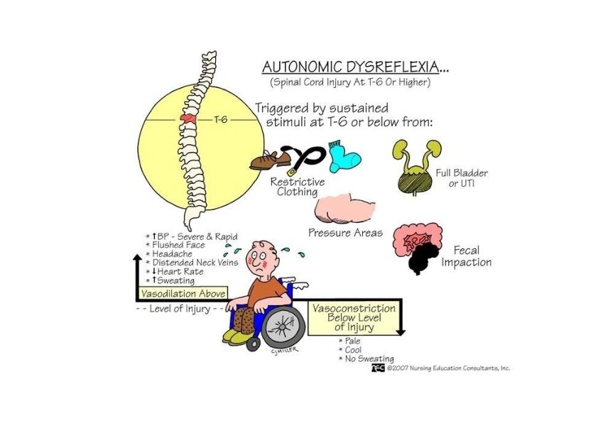
Autonomic Dysreflexia • Exaggerated response of sympathetic NS in cases of spinal cord injury above T 6 • Certain sensory impulses trigger mass stimulation of sympathetic nerves below the injury • Result • vasoconstriction which elevates blood pressure • parasympathetic NS tries to compensate by slowing heart rate & dilating blood vessels above the injury • pounding headaches, sweating warm skin above the injury and cool dry skin below • can cause seizures, strokes & heart attacks

Example of Spinal and Supraspinal Control of AN: Urinary Bladder Function • Urinary bladder function • Storage phase • Example of spinal reflex control on the AN • Voiding phase • Example of supraspinal control on the AN
 eliminates")
Effect of SCI on the Urinary Bladder Function • Spinal cord injury (SCI) eliminates the supraspinal control • Urinary bladder dysfunction

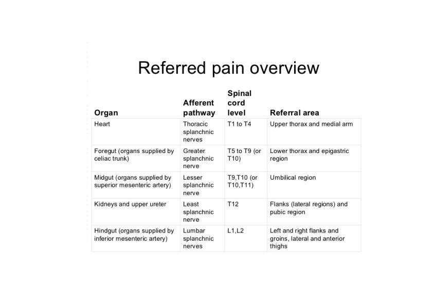
Visceral Pain Vague and poorly localized Referral pain depend on the spinal segment receiving the afferent
- Slides: 24