
PARASITIC PROTOZOA Sudipta Bhowmick MSc Ph D Assistant
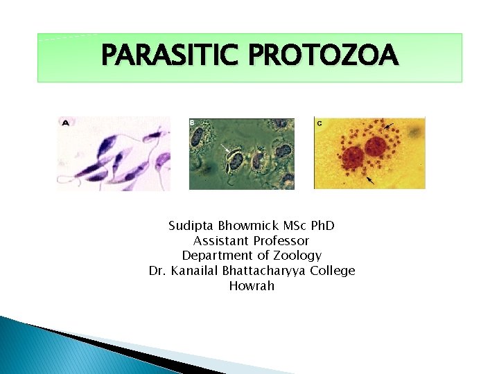
PARASITIC PROTOZOA Sudipta Bhowmick MSc Ph. D Assistant Professor Department of Zoology Dr. Kanailal Bhattacharyya College Howrah
Parasite & Parasitism Parasites are living organisms, which depend on a living host for their nourishment and survival. They multiply or undergo development in the host. Parasitism is a relationship in which one of the participants, the parasite, either harms its host or in some sense lives at the expense of the host.
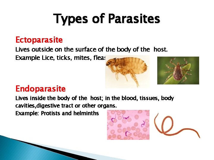
Types of Parasites Ectoparasite Lives outside on the surface of the body of the host. Example Lice, ticks, mites, fleas Endoparasite Lives inside the body of the host; in the blood, tissues, body cavities, digestive tract or other organs. Example: Protists and helminths
Types of Parasites Temporary Parasite Visits host for a short period. Example mosquitoes and bed bugs Permanent Parasites Leads a parasitic life throughout the whole period of its life. Example Intestinal helminth Facultative Parasite Lives a parasitic life when oppurtunity arises. Example Free living Amoeba Obligatory Parasites Cannot exist without a parasitic life. Example Trypanosoma. Wuchereria
Types of Parasites Occasional or Accidental Parasite Attacks an unusual host. Example Echinococcus granulosus infects man accidentally Aberrant or Wandering parasites: Infects a host where they cannot develop further are known as aberrant or wandering parasites, e. g. Toxocara canis (dog roundworm) infecting humans
Types of Host Definitive host: The host, in which the adult parasite lives and undergoes sexual reproduction is called the definitive host, e. g. mosquito acts as definitive host in malaria, man as definitive host e. g. filaria, roundworm, hookworm. Intermediate host: The host, in which the larval stage of the parasite lives or asexual multiplication takes place is called the intermediate host. In some parasites, 2 different intermediate hosts may be required to complete different larval stages. These are known as first and second intermediate hosts, respectively.
Types of Host Paratenic host (A Carrier or Transport Host) A host, in which larval stage of the parasite remains viable without further development is referred as a paratenic host. Such host transmits the infection to another host. Reservoir host: In an endemic area, a parasitic infection is continuously kept up by the presence of a host, which harbors the parasite and acts as an important source of infection to other susceptible hosts, e. g. dog is the reservoir host of hydatid disease. � Accidental host: The host, in which the parasite is not usually found, e. g. man is an accidental host for cystic echinococcosis.
Zoonosis The word zoonosis was introduced by Rudolf Virchow in 1880 to include the diseases shared in nature by man and animals. Later, in 1959, the World Health Organization (WHO) defined zoonosis as “those diseases and infections, which are naturally transmitted between vertebrate animals and man”. It is of following types: Protozoal zoonoses, e. g. toxoplasmosis, leishmaniasis, balantidiasis, and cryptosporodiasis Helminthic zoonoses, e. g. hydatid disease, taeniasis Anthropozoonoses: Infections transmitted to man from lower vertebrate animals, e. g. cystic Echinococcosis Zooanthroponoses: Infections transmitted from man to lower vertebrate animals, e. g. human tuberculosis to cattle.
Vectors Insect vectors: A vector is an agent, usually an arthropod that transmits an infection from man to man or from other animals to man, e. g. female Anopheles is the vector of malarial parasite. Biological vectors: The term biological vector refers to a vector, which not only assists in the transfer of parasites but the parasites undergo development or multiplication in their body as well. They are also called as true vectors. Example of true vectors are: Mosquito—Malaria, filariasis , Sandflies—Kala-azar, Tsetse flies— Sleeping sickness, Reduviid bugs—Chagas’ disease, Ticks—Babesiosis. Mechanical vectors: The term mechanical vector refers to a vector, which assists in the transfer of parasitic form between hosts but is not essential in the life cycle of the parasite. Example of Mechanical vectors is: �. Housefly—amoebiasis�.
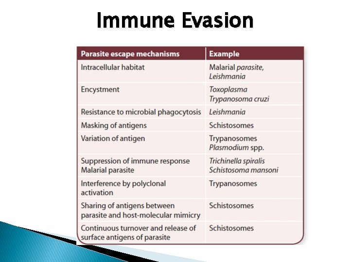
Immune Evasion
Protozoa General Features q Single-celled eukaryotic microorganisms belonging to kingdom protista are classified as Protozoa (Greek Protos: first; zoon: animal). q q q Unicellular ( consist of one cell ) this cell performs all phsiological function ( activities ) of life such as locomation , reproduction , respiration , ingestion , digestion and excretion. They vary in size and shape some are visible while others require high magnification ( by microscope ). The majority of protozoa are free living but some are parasitic. Both sexual and asexual reproduction occur in protozoa ( the majority of protozoa reproduce by asexual reproduction ). Locomotory organs psedopodia, flagella, cilia.

Giardia intestinalis Giardia, a microscopic parasite, is responsible for disease giardiasis. There are many species of Giardia intestinalis (synonymous Giardia lamblia) can infect human beings. Phylum: Sarcomastigorphora Habitat Duodenum and upper jejunum. It is the only protozoan parasite found in the lumen of the human small intestine.
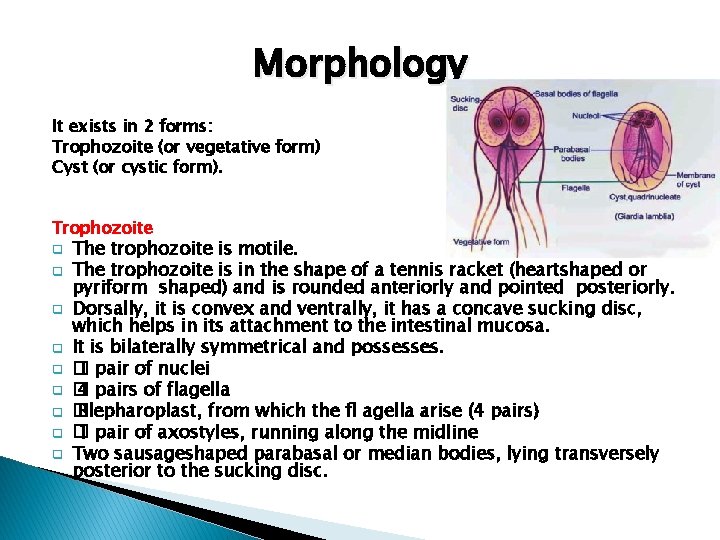
Morphology It exists in 2 forms: Trophozoite (or vegetative form) Cyst (or cystic form). Trophozoite q q q q q The trophozoite is motile. The trophozoite is in the shape of a tennis racket (heartshaped or pyriform shaped) and is rounded anteriorly and pointed posteriorly. Dorsally, it is convex and ventrally, it has a concave sucking disc, which helps in its attachment to the intestinal mucosa. It is bilaterally symmetrical and possesses. � 1 pair of nuclei � 4 pairs of flagella � Blepharoplast, from which the fl agella arise (4 pairs) � 1 pair of axostyles, running along the midline Two sausageshaped parabasal or median bodies, lying transversely posterior to the sucking disc.
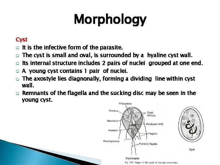
Morphology Cyst q It is the infective form of the parasite. q The cyst is small and oval, is surrounded by a hyaline cyst wall. q Its internal structure includes 2 pairs of nuclei grouped at one end. q A young cyst contains 1 pair of nuclei. q The axostyle lies diagnonally, forming a dividing line within cyst wall. q Remnants of the flagella and the sucking disc may be seen in the young cyst.
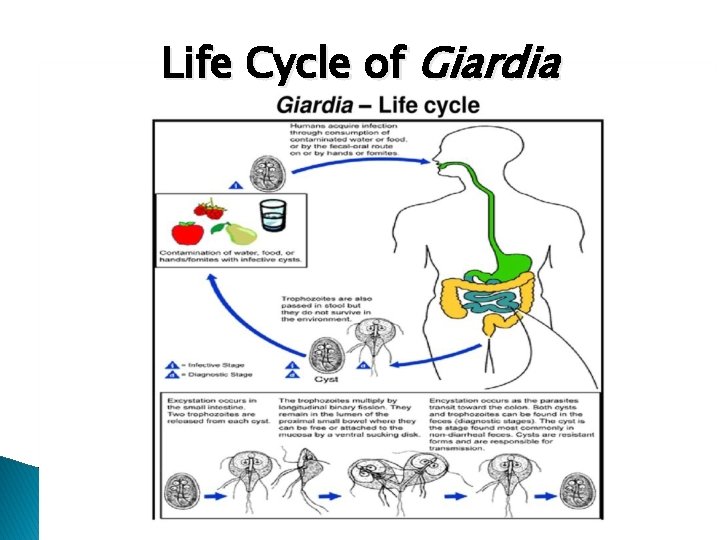
Life Cycle of Giardia
Life Cycle of Giardia passes its life cycle in 1 host. Infective form: Mature cyst. Mode of transmission: q Man acquires infection by ingestion of cysts in contaminated water and food. Other frequent mode of infection is from person-to-person in day-care centers or workplace. Potential zoonotic risks can berendered by infected dogs and cats which evidently serve as a source for contamination of the environment with the organism. q Within half an hour of ingestion, the cyst hatches out into two trophozoites, which multiply successively by binary fission and colonize in the duodenum q The trophozoites live in the duodenum and upper part of jejunum, feeding by pinocytosis. q During unfavorable conditions, encystment occurs usually in colony. q Cysts are passed in stool and remain viable in soil and water for several weeks. q There may be 200, 000 cysts passed per gram of feces. q Infective dose is 10– 100 cysts.
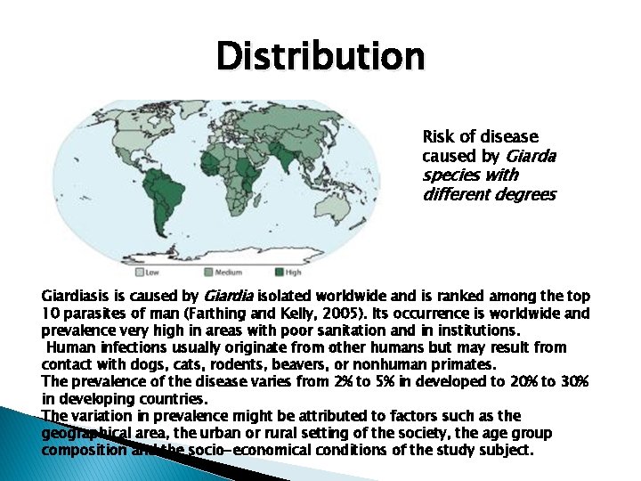
Distribution Risk of disease caused by Giarda species with different degrees Giardiasis is caused by Giardia isolated worldwide and is ranked among the top 10 parasites of man (Farthing and Kelly, 2005). Its occurrence is worldwide and prevalence very high in areas with poor sanitation and in institutions. Human infections usually originate from other humans but may result from contact with dogs, cats, rodents, beavers, or nonhuman primates. The prevalence of the disease varies from 2% to 5% in developed to 20% to 30% in developing countries. The variation in prevalence might be attributed to factors such as the geographical area, the urban or rural setting of the society, the age group composition and the socio-economical conditions of the study subject.
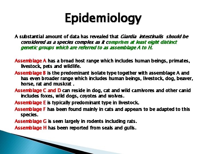
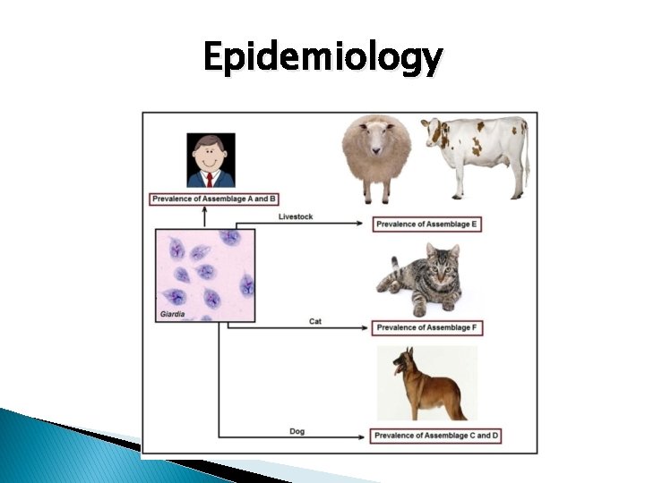
Epidemiology A substantial amount of data has revealed that Giardia intestinalis should be considered as a species complex as it comprises at least eight distinct genetic groups which are referred to as assemblage A to H. Assemblage A has a broad host range which includes human beings, primates, livestock, pets and wildlife. Assemblage B is the predominant isolate type together with assemblage A and has even broader range which includes human beings, livestock, dog, beaver, horse, rat and muskrat. Assemblage C and D can reside in dog, cat and wild carnivores and other canid includes foxes, wild dogs, coyotes and wolves. Assemblage E is typically predominant type in livestock. Assemblage F has been found mainly in cats and appears to be adapted to this species. Assemblage G is seen largely in rodents including rats. Assemblage H has been reported from seals and gulls.
Epidemiology
Pathogenesis q q q q Giardia is typically seen within the crypts of duodenal and jejunal mucosa. It does not invade the tissue, but remains tightly adhered to intestinal epithelium by means of the sucking disc. They may cause abnormalities of villous architecture by cell apoptosis and increased lymphatic infiltration of lamina propria. Variant specifi c surface proteins (VSSP) of giardia play an important role in virulence and infectivity of the parasite. Often they are asymptomatic, but in some cases, Giardia may lead to mucus diarrhea, fat malabsorption (steatorrhea), dull epigastric pain, and flatulence. The stool contains excess mucus and fat but no blood. Children may develop chronic diarrhea, malabsorption of fat, vitamin A, protein, sugars like xylose disaccharides, weight loss, and spruelike syndrome. Occassionally, Giardia may colonize the gall bladder, causing biliary colic and jaundice. Incubation period is variable, but is usually about 2 weeks.
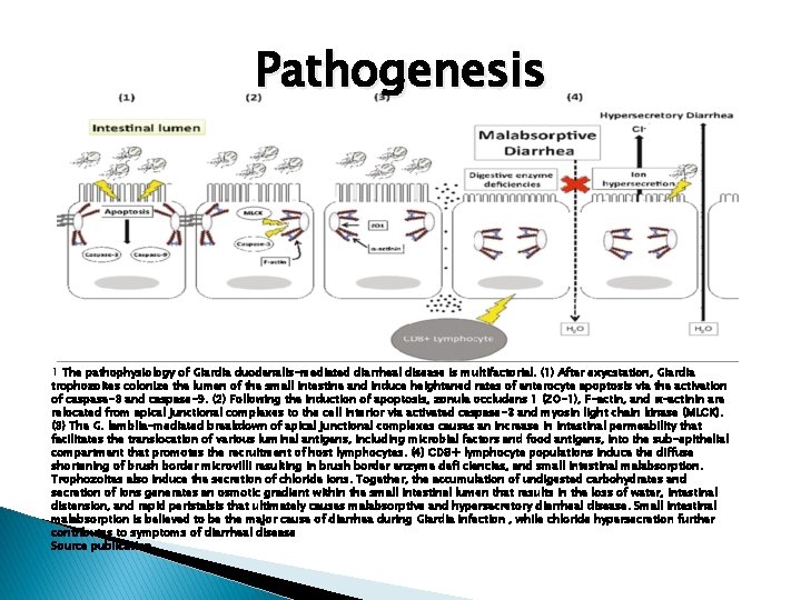
Pathogenesis 1 The pathophysiology of Giardia duodenalis-mediated diarrheal disease is multifactorial. (1) After exycstation, Giardia trophozoites colonize the lumen of the small intestine and induce heightened rates of enterocyte apoptosis via the activation of caspase-3 and caspase-9. (2) Following the induction of apoptosis, zonula occludens 1 (ZO-1), F-actin, and α-actinin are relocated from apical junctional complexes to the cell interior via activated caspase-3 and myosin light chain kinase (MLCK). (3) The G. lamblia-mediated breakdown of apical junctional complexes causes an increase in intestinal permeability that facilitates the translocation of various luminal antigens, including microbial factors and food antigens, into the sub-epithelial compartment that promotes the recruitment of host lymphocytes. (4) CD 8+ lymphocyte populations induce the diffuse shortening of brush border microvilli resulting in brush border enzyme defi ciencies, and small intestinal malabsorption. Trophozoites also induce the secretion of chloride ions. Together, the accumulation of undigested carbohydrates and secretion of ions generates an osmotic gradient within the small intestinal lumen that results in the loss of water, intestinal distension, and rapid peristalsis that ultimately causes malabsorptive and hypersecretory diarrheal disease. Small intestinal malabsorption is believed to be the major cause of diarrhea during Giardia infection , while chloride hypersecretion further contributes to symptoms of diarrheal disease Source publication
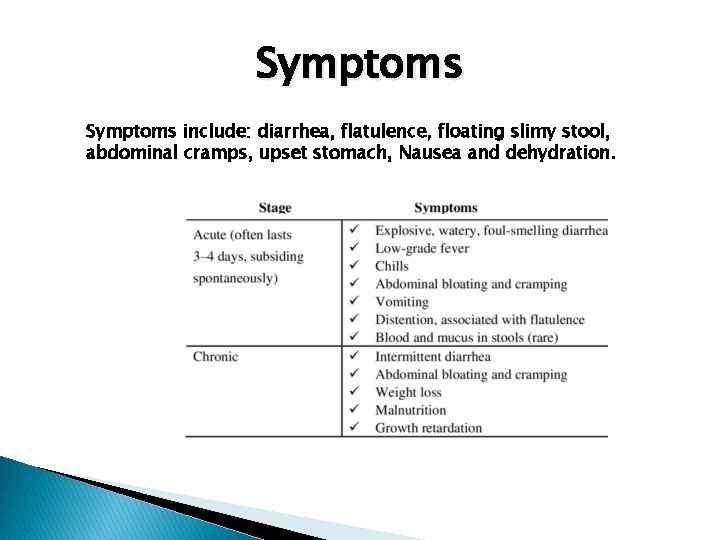
Symptoms include: diarrhea, flatulence, floating slimy stool, abdominal cramps, upset stomach, Nausea and dehydration.
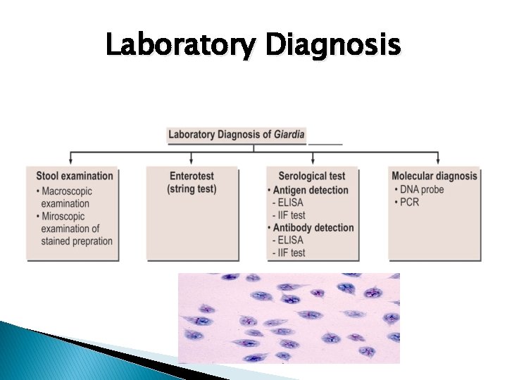
Laboratory Diagnosis
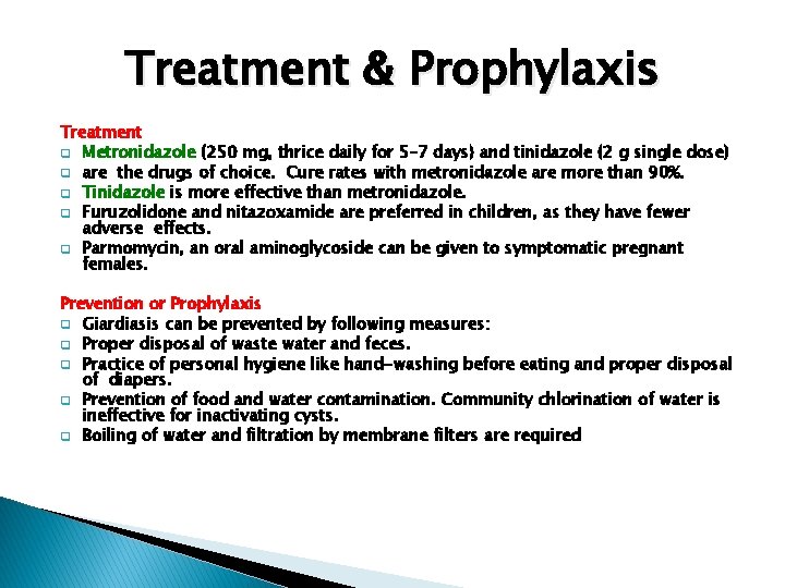
Treatment & Prophylaxis Treatment q Metronidazole (250 mg, thrice daily for 5– 7 days) and tinidazole (2 g single dose) q are the drugs of choice. Cure rates with metronidazole are more than 90%. q Tinidazole is more effective than metronidazole. q Furuzolidone and nitazoxamide are preferred in children, as they have fewer adverse effects. q Parmomycin, an oral aminoglycoside can be given to symptomatic pregnant females. Prevention or Prophylaxis q Giardiasis can be prevented by following measures: q Proper disposal of waste water and feces. q Practice of personal hygiene like hand-washing before eating and proper disposal of diapers. q Prevention of food and water contamination. Community chlorination of water is ineffective for inactivating cysts. q Boiling of water and filtration by membrane filters are required
- Slides: 24