Paracentral lobule Secondary visual areas 1819 Primary Visual

")
Paracentral lobule Secondary visual areas 18&19 Primary Visual area (area 17)

Cortical Centers of the medial surface 1 - Paracentral lobule; - It is continues with the motor and sensory areas in the lateral surface. - It is the center to the leg, foot and perineum of the opposite side. - It controls the micturition and defecation reflexes. 2 - primary Visual area (area 17); - It lies on the depth of calcarine sulcus - It receives visual sensation. 3 - secondary Visual (association) area (area 18, 19): - It lies in the occipital lobe surrounding the primary visual area. - Damage of this area causes visual agnosia (people can not identify the objects).

Sulci & Gyri of the inferior surface

gyrus rectus Olfactory Sulcus H- shaped orbital sulcus olfactory bulb, tract orbital gyri The inferior surface of the frontal lobe

Stem of lateral sulcus Rhinal sulcus L. Occipitotemporal gyrus Occipitotemporal sulcus M. Occipitotemporal gyrus Collateral sulcus The tentorial surface

A- On the orbital surface: 1 - Olfactory sulcus; on the orbital surface close and parallel to the medial orbital border. It contains olfactory bulb and tract. • Gyrus rectus: between medial orbital border and olfactory sulcus. 2 - Orbital sulcus: is H shaped sulcus lateral to the olfactory sulcus. 2 - Anterior, posterior, lateral and medial orbital gyri: on the orbital surface. B- On the tentorial surface: 1 - Stem of the lateral sulcus between the frontal and temporal lobes. 2 - Occipito-temporal sulcus: from occipital pole to temporal pole. 3 - Rhinal su. Icus: extends from the temporal pole. 4 - Collateral sulcus: begins close to the posterior end of the rhinal sulcus to the occipital pole. 3 - Medial and Lateral occipitotemporal gyrus: medial and lateral to occipitotemporal sulcus.

uncus rhinal sulcus parahippocampal gyrus hippocampal sulcus calcarine sulcus collateral sulcus lingual gyrus The tentorial surface
 Anterior part of parahippocampal gyrus")
Olfactory area uncus (area 34, 28) Anterior part of parahippocampal gyrus

On the tentorial surface: - Lingual gyrus between collateral sulcus and calcarine sulcus - Para hippocampal gyrus anterior to the lingual gyrus (Limbic system) - Uncus anterior to Para hippocampal gyrus, a hook-shaped convolution close to the temporal pole medial to the rhinal sulcus. Center of the olfactory

Limbic system

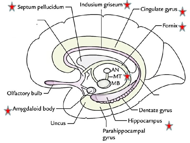
• Limbic system • The name applied to the number cortical and subcortical structures lying on the medial surface of the cerebral hemisphere in the form of an arch (limbus). ** Functions of the Limbic System 1 - Emotional and behavior reaction. 2 - Recent Memory. 3 - Responsible for olfactory related stimuli (a person may be vomit or salivate when smell a certain odor)

5 3 1 2 4 9 13 10 12 8 6 11 7
 System 1 - Cingulate gyrus. 2 - Indusium griseum")
Parts of Limbic (arch) System 1 - Cingulate gyrus. 2 - Indusium griseum (Thin grey matter which cover the superior surface of the corpus callosum). 3 - Septum pellucidum. 4 - Fornix 5 - Anterior nucleus of thalamus (AN). 6 - Mammillary body (MB) 7 - Mamillo-thalamic tract (MT). 8 - Amygdaloid body 9 - Stria terminalis (efferent fibres of Amygdaloid body to hypothalamus). 10 - Hippocampal formation (hippocampus, parahippocampual gyrus and Denatate gyrus in between, below the fimbria, it has a toothed surface). 11 - Fimbria (efferent pathway of hipocamppus, reaches posterior crus of fornix to end in mammillary body). 12 - Anterior perforated substance

HIPPOCAMPAL FORMATION

** Hippocampal formation Hippocampus ﺍﻟﺤﺼﻴﻦ - Hippocampus is a part of the cerebral cortex which is projected into the floor of the inferior horn of the lateral ventricle in the temporal lobe. - The hippocampus (named after its resemblance to the seahorse) - It contains two main parts: the hippocampus proper (called Cornu Ammonis, Its abbreviation CA is used in naming the hippocampal subfields: CA 1, CA 2, CA 3, and CA 4) and the dentate gyrus. . - The hippocampus is involved in the storage of long-term memory, which includes all past knowledge and experiences

- Hippocampus - Its anterior end broad and continues medially with the uncus. - Its posterior end is narrow and ends below the splenium of the corpus callosum. - It is continues with the parahippocampal gyrus. - On its ventricular surface is a thin film of white mater called alveolus (axons of hippocampal cells). - The fibers of alveolus thicken medially to form fimbria. - Fimbria, passes backward on the medial margin of the hippocampus above the dentate gyrus. It reaches the posterior crus of the fornix forming efferent pathway of the hipocamppus to end in the mammillary body.

How to estimate the vertebral levels of spinal segments The cervical Vertebra + 1 regions C 8 segment lies opposite C 7 v Upper thoracic Vertebra + 2 region (T 1 -T 6) T 6 segment lies opposite T 4 v Lower thoracic Vertebra + 3 region (T 6 -T 12) T 12 segment lies opposite T 9 v C 8 s T 6 s T 12 s C 7 V T 4 V T 9 V

• The 5 lumber lies opposite cord segments T 10, T 11 v L 1 -2 cord segments 10 th thoracic vertebra L 3 -5 cord segments 11 th thoracic vertebra T 10 V T 11 V T 12 V L 1 V • The sacral, lies opposite coccygeal segments T 12 th-L 1 v 13

45

Lamina of Rexed Grey matter of the spinal cord is classified by Rexed into 10 laminae; - Lamina I, at the apex of dorsal horn and receives rapid sharp pain. - Lamina II and III, They correspond to SGR and receive pain and temperature sensation. - Lamina IV and V, They correspond to Main sensory nucleus and receive crude touch sensation. - Lamina VI, it present only in the cervical and lumbo-sacral enlargements responsible for spinal reflexes. - Lamina VII, It corresponds the Clark's nucleus and lateral horn. - Lamina VIII, it includes the large motor cells of the medial group of the anterior horn which supply the trunk muscles. - Lamina IX, it includes the large motor cells of the central and lateral group of the ventral horn which supply the limb muscles. - Lamina X, (commissural lamina), lie around the central canal. 46
- Slides: 22