
Pancreatitis Koray Topgl MD Prof Department of General





- Slides: 48
Pancreatitis Koray Topgül, MD, Prof Department of General Surgery
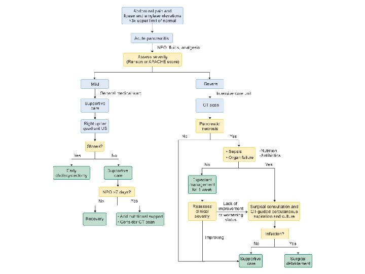
Acute Pancreatitis
Definition and Etiology An acute inflammation process of the pancreas with associated escape of the pancreatic enzyme into surrounding tissue. The primary etiologic factors are biliary tract disease & alcoholism May be a complication of viral or bacterial disease, peptic ulcer, trauma.
Pathophysiology Although different etiologies produce distinct inciting events, the final common pathway is premature activation of enzymes within the acinar cell. Ordinarily, pancreatic proenzymes become activated on release within the duodenum. Pancreatitis results when activation of pancreatic enzymes occurs early within the acinar cells, producing autodigestion of the pancreas and surrounding tissues. Exposure of trypsinogen to lysosomal enzymes such as cathepsin B has been shown as a mechanism for early trypsin activation
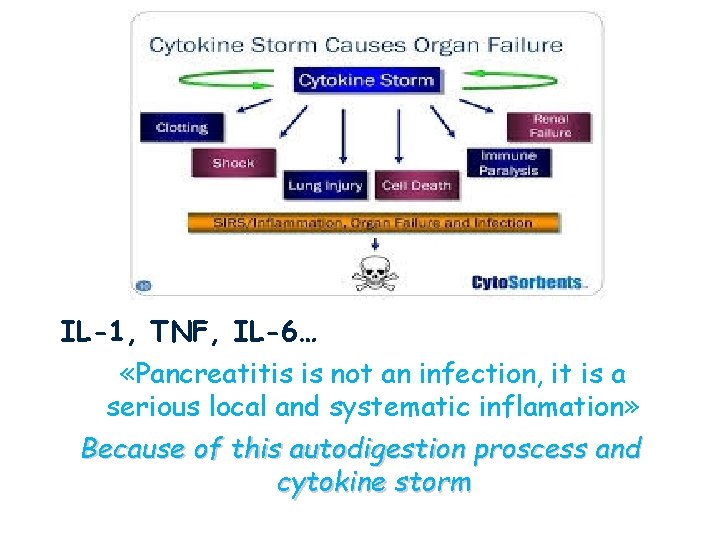
IL-1, TNF, IL-6… «Pancreatitis is not an infection, it is a serious local and systematic inflamation» Because of this autodigestion proscess and cytokine storm
Incidence & Risk Factors Major- Biliary stones, Alcohol use/abuse Minor- Age: 55 to 65 yrs. for biliary pancreatitis 45 - 55 yrs. For alcohol-related
*Female for biliary tract pancreatitis; Male-for alcohol-related pancreatitis. *Trauma, Infectious disease, drug toxicities, chronic diseases (inflammatory diseases).
Assessment Pain: Steady & severe in nature; located in the epigastric or umbilical region; may radiate to the back. Worsened by lying supine; may be lessened by flexed knee, curved-back position. Vomiting Varies in severity, but is usually protracted, worsened by ingestion of food or fluid. Does not relieve the pain. Usually accompanied by nausea.
Fever: Rarely exceeds 39 0 C. Abdominal Finding: Rigidity, tenderness, guarding, distended decreased or absent peristalsis and paralytic ileus. Fatty stools(steatorrhea) Laboratory Finding: Elevation of white count- 20, 00050, 000. Elevated serum lipase and amylase(5 to 40 times); glucose, bilirubin, alkaline phosphatase. Urine amylase elevated. Abnormal low serum Ca, Ca Na & Mg. -due to dehydration. Binding of Ca in areas of fat necrosis
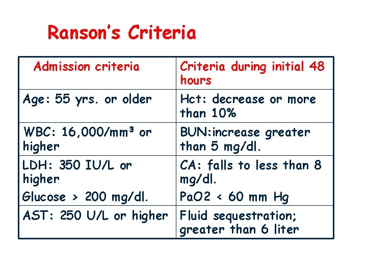
Ranson’s Criteria Admission criteria Criteria during initial 48 hours Age: 55 yrs. or older Hct: decrease or more than 10% WBC: 16, 000/mm 3 or higher BUN: increase greater than 5 mg/dl. LDH: 350 IU/L or higher Glucose > 200 mg/dl. CA: falls to less than 8 mg/dl. Pa. O 2 < 60 mm Hg AST: 250 U/L or higher Fluid sequestration; greater than 6 liter
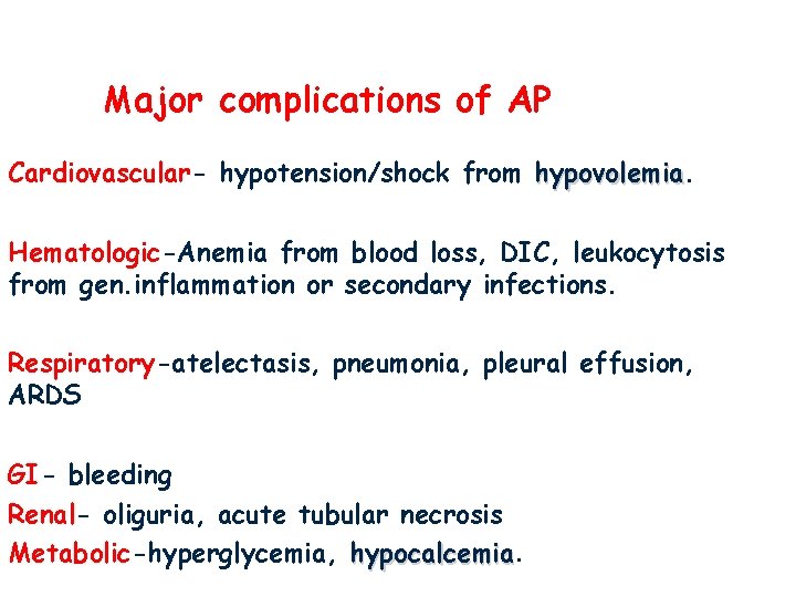
Major complications of AP Cardiovascular- hypotension/shock from hypovolemia Hematologic-Anemia from blood loss, DIC, leukocytosis from gen. inflammation or secondary infections. Respiratory-atelectasis, pneumonia, pleural effusion, ARDS GI- bleeding Renal- oliguria, acute tubular necrosis Metabolic-hyperglycemia, hypocalcemia
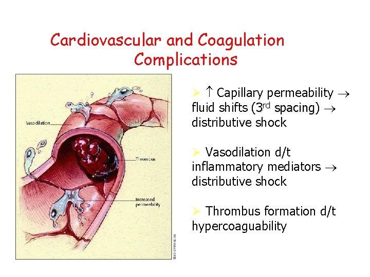
Cardiovascular and Coagulation Complications Capillary permeability fluid shifts (3 rd spacing) spacing distributive shock Vasodilation d/t inflammatory mediators distributive shock Thrombus formation d/t hypercoaguability DIC 13
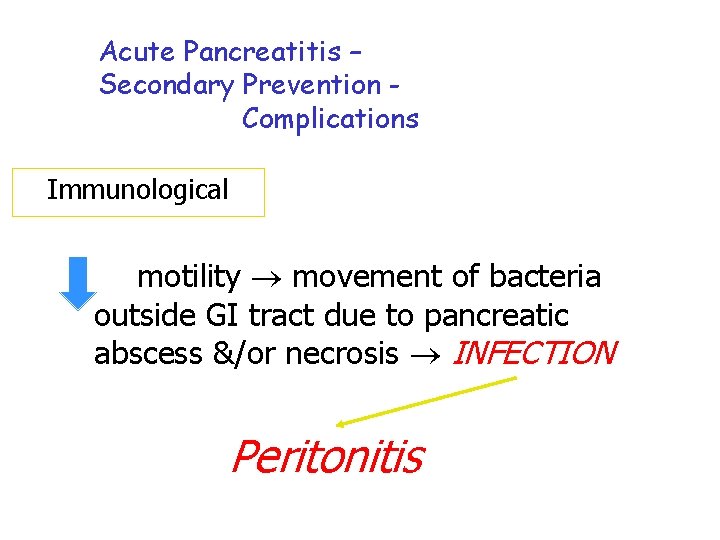
Acute Pancreatitis – Secondary Prevention Complications Immunological GI motility movement of bacteria outside GI tract due to pancreatic abscess &/or necrosis INFECTION Peritonitis 14
Acute Pancreatitis – Secondary Prevention Complications Renal Hypovolemia GFR, renal perfusion development of clots in renal circulation Acute tubular necrosis & Acute renal failure 15
Treatment of Acute Pancreatitis Alleviate pain & anxiety. Anxiety increases pancreatic secretions. Analgesicsthen morphine. Reduce pancreatic stimulus. NPO If there is ileus findings use NGT to remove gastric secretions. Na. HCO-reverse metabolic acidosis. Regular insulin to treat hyperglycemia.
Treat infection-antibiotics are not routine!!! Aggressive respiratory care- monitor ABG. Reduce body metabolism- bedrest, cool quiet environment. Avoid alcohol, coffee, heavy meals and spicy food.
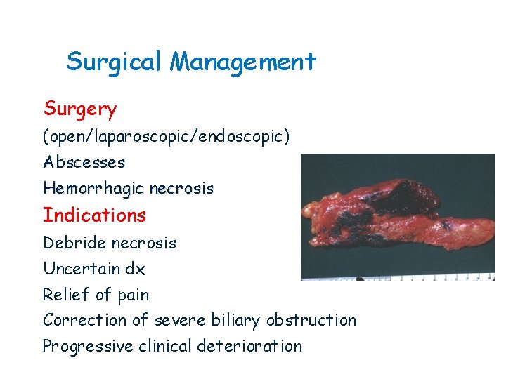
Surgical Management Surgery (open/laparoscopic/endoscopic) Abscesses Hemorrhagic necrosis Indications Debride necrosis Uncertain dx Relief of pain Correction of severe biliary obstruction Progressive clinical deterioration
CHRONIC PANCREATITIS .
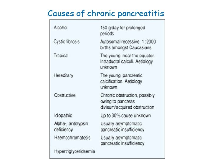
Causes of chronic pancreatitis
Definition Irreversible damage to the pancreas with histologic evidence of inflammation, fibrosis and destruction of exocrine (acinar) and endocrine (islets of Langerhans) tissue Etiologic classification – clinically useful Histologic – accessibiliy of tissue Imaging – late morphologic changes
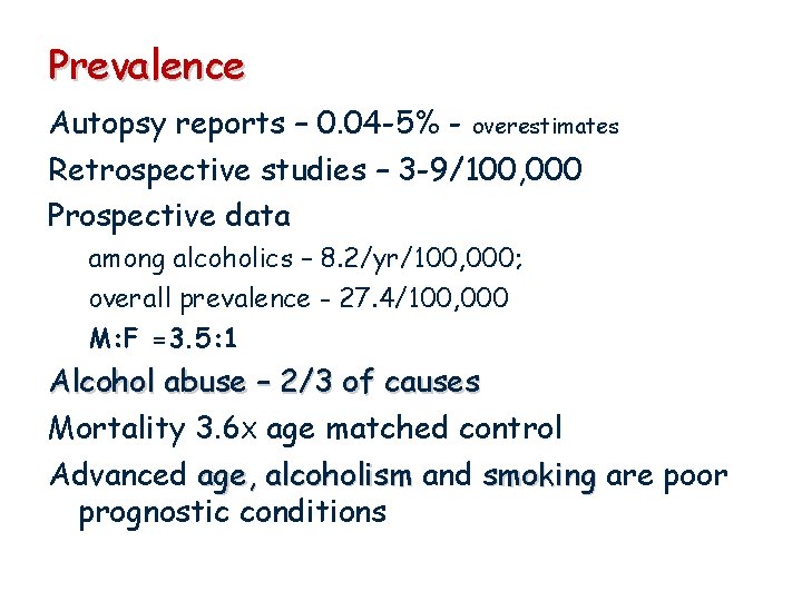
Prevalence Autopsy reports – 0. 04 -5% - overestimates Retrospective studies – 3 -9/100, 000 Prospective data among alcoholics – 8. 2/yr/100, 000; overall prevalence - 27. 4/100, 000 M: F =3. 5: 1 Alcohol abuse – 2/3 of causes Mortality 3. 6 X age matched control Advanced age, alcoholism and smoking are poor prognostic conditions
pathophysiology Incompletely understood Why 10% heavy alcoholics develop chronic pancreatitis and the rest not, or limited to asymptomatic pancreatic fibrosis Alcohol is the most studied
Ductal obstruction hypothesis Chronic alcohol use acinar and ductal cell protein rich pancreatic juice, low in volume and HCO 3 formation of protein precipitates – plug calcification of ppt – ductal stone formation ductule obstruction parenchymal damage Pancreatic ductal stone are seen in alcoholic, tropical, hereditary, idiopathic Histologic changes of CP may be seen with out ductal obstruction
Toxic metabolic hypothesis (alcohol) Direct injurious effect on acinar and ductal cells Increased membrane lipid peroxidation (oxidative stress), free radical production Increase acinar cell sensitivity to pathogenic stimuli Stimulate CCK production (duodenal I cells) – activation of proinflammatory transcription factors Activation of pancreatic stellate cells (alcohol, cytokines) – produce proteins of extracellular matrix
Necrosis fibrosis hypothesis Repeated episodes of acute pancreatitis with cellular necrosis or apoptosis, healing replaces necrotic tissue with fibrosis Evidence from natural history studies - more severe and frequent attacks More evidence from hereditary pancreatitis and animal models But some have evidence of chronic pancreatitis at time of first clinical acute attack
Clinical features Abdominal pain Acute pancreatic inflammation Increased intrapancreatic pressure Alterations in pancreatic nerves Steatorrhea – DM lipase secretion <10%
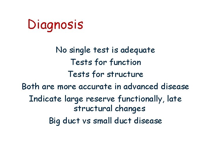
Diagnosis No single test is adequate Tests for function Tests for structure Both are more accurate in advanced disease Indicate large reserve functionally, late structural changes Big duct vs small duct disease
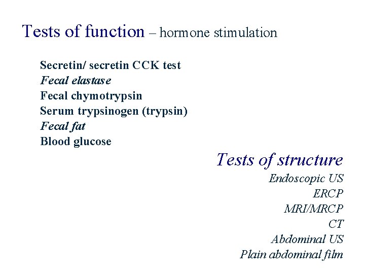
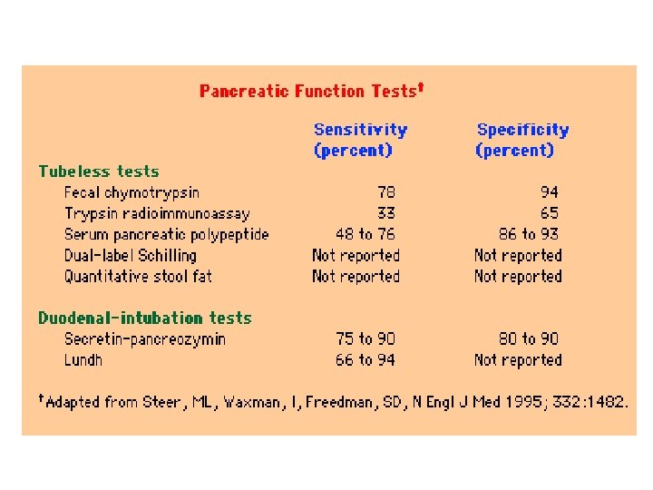
Tests of function – hormone stimulation Secretin/ secretin CCK test Fecal elastase Fecal chymotrypsin Serum trypsinogen (trypsin) Fecal fat Blood glucose Tests of structure Endoscopic US ERCP MRI/MRCP CT Abdominal US Plain abdominal film
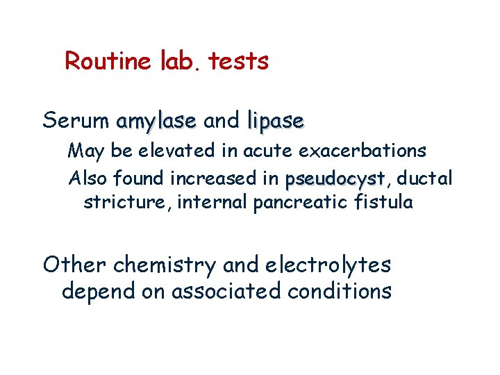
Routine lab. tests Serum amylase and lipase May be elevated in acute exacerbations Also found increased in pseudocyst, pseudocyst ductal stricture, internal pancreatic fistula Other chemistry and electrolytes depend on associated conditions
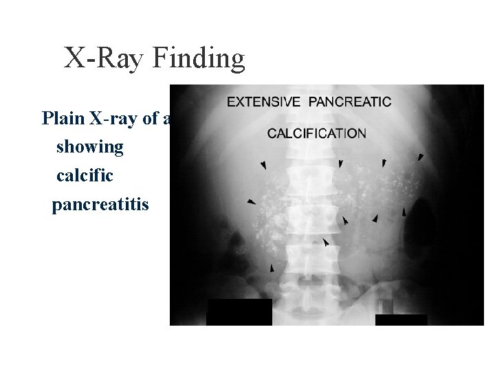
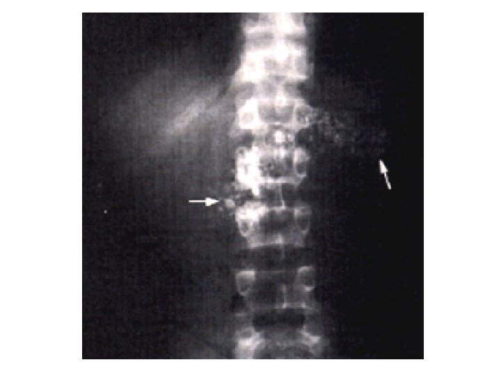
X-Ray Finding Plain X-ray of abdomen showing calcific pancreatitis
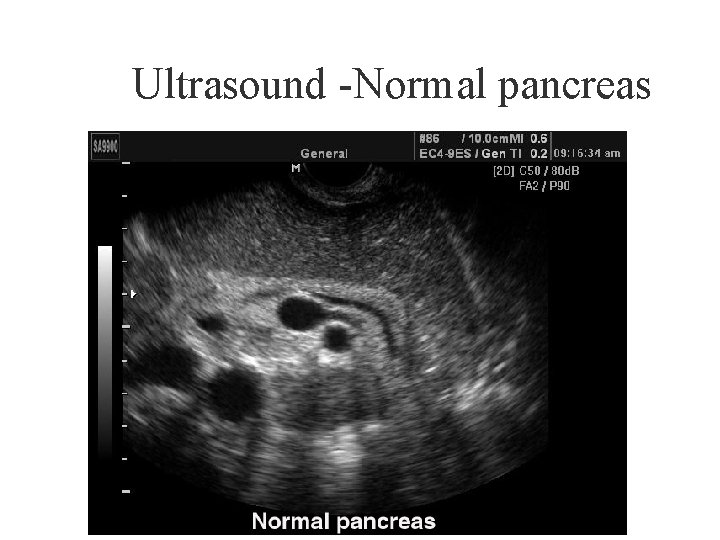
Ultrasound -Normal pancreas
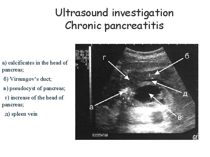
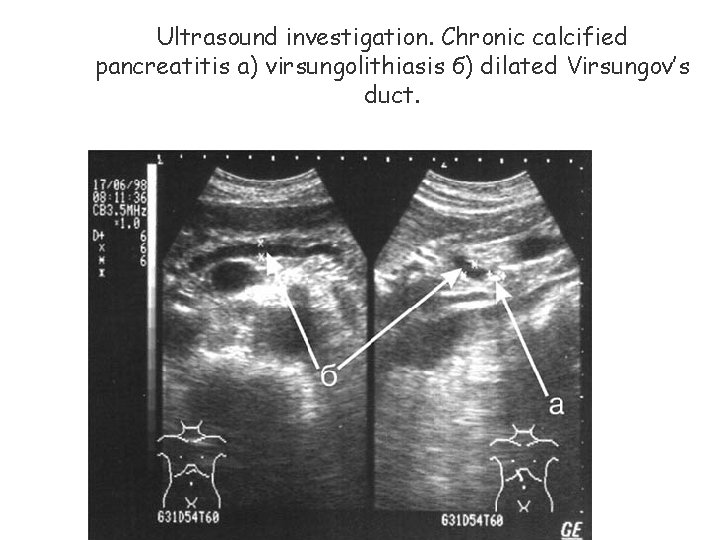
Ultrasound investigation Chronic pancreatitis а) calcificates in the head of pancreas; б) Virsungov’s duct; в) pseudocyst of pancreas; г) increase of the head of pancreas; д) spleen vein
Ultrasound investigation. Chronic calcified pancreatitis а) virsungolithiasis б) dilated Virsungov’s duct.
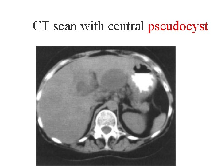
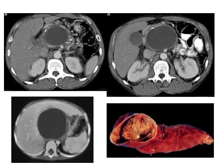
CT scan with central pseudocyst
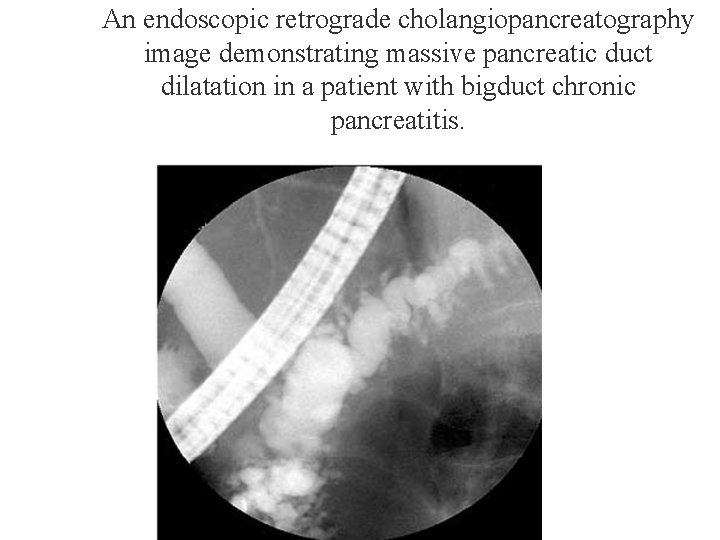
An endoscopic retrograde cholangiopancreatography image demonstrating massive pancreatic duct dilatation in a patient with bigduct chronic pancreatitis.
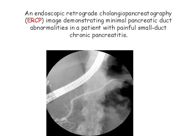
An endoscopic retrograde cholangiopancreatography (ERCP) image demonstrating minimal pancreatic duct abnormalities in a patient with painful small-duct chronic pancreatitis.
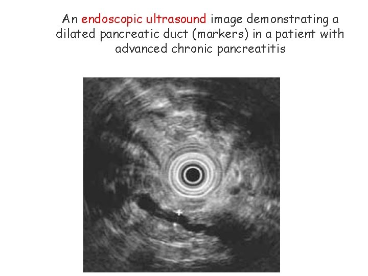
An endoscopic ultrasound image demonstrating a dilated pancreatic duct (markers) in a patient with advanced chronic pancreatitis
Classics of Chronic Pancreatitis Pancreatic calcification Steatorrhea Diabetes mellitus Found in less than a third of pts with CP abnormal secretin stimulations test when >60 % affected Serum trypsinogen < 20 ng/ml, fecal elastase < 100 mcg/mg stool - severe exocrine insuf.
Complications Malabsorption Steatorrhea, A, D, E and K vitamin defficiency DM – but end organ damages of DM and DKA are rare Non DM retinopathy (peripheral) due to Vit A and Zn defc. Pleural, peritoneal and pericardial effusions with high amylase
GI bleeding – PUD, gastritis, pseudocyst, varies (SV thrombosis) Cholestasis, icterus, cholangitis, biliary cirrhosis Fistula – internal or external Subcutaneous fat necrosis – tender red nodules on the shins Pseudocyst, Obstruction, Pancreatic carcinoma – 4% life time risk Narcotic addiction
Pseudocyst A pancreatic pseudocyst is a circumscribed collection of fluid rich in pancreatic enzymes, blood, and necrotic tissue, tissue typically located in the lesser sac of the abdomen. Pancreatic pseudocysts are usually complications of pancreatitis. Pancreatic pseudocysts account for approximately 75% of all pancreatic masses. The prefix pseudo- (Greek for "false") distinguishes them from true cysts, which are lined by epithelium; pseudocysts are lined with granulation tissue Treatment—internal (Cystogastrostomi/Cystojejunostomi) or external drainage
Treatment Aim - Pain control and mx of maldigestion Pain Avoid alcohol Low fat meals Antipain – narcotics (addiction) Surgical pain control Resection (local - - 95%) – pancreatic insufficiency Splanchinectomy, celiac ganglionectomy, nerve block Endoscopic tx Sphinctorotomy, dilatation of strictures, caliculi removal, duct stenting Cpx – acute pancreatitis, abscess, ductal damage, death Pancreatic enzymes
Tx of maldigestion Pancreatic enzyme replacement 2 -3 enteric coated or 8 conventional tablets with meals adjuvants with conventional tablets – H 2 blockers, PPI, Na bicarbonate,