Pancreatic Diseases Dr Abdulsalam Alsharabi Asst Professor Hepatobiliary

Pancreatic Diseases Dr. Abdulsalam Alsharabi Asst. Professor Hepatobiliary and Liver Transplant Consultant King Saud University and King Khalid University Hospital

Anatomy The pancreas has an extensive arterial system arising from multiple sources n The venous drainage parallels arterial anatomy n – The veins terminate in the portal vein Multiple lymph nodes drain the pancreas n Neural function is controlled by duel sympathetic and parasympathetic innervation n

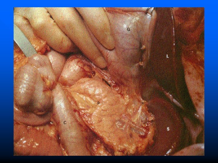
Anatomy Is a retroperitoneal structure found posterior to the stomach and lesser omentum n It has a distinctive yellow/tan/pink color and is multilobulated n The gland is divided into four portions n – The head – The neck – The body – The tail

Anatomy & Physiology II

Histology n n There are two distinct organ systems within the pancreas The endocrine portion of the pancreas is served by structures called the islet of Langerhans – The islet of Langerhans have several distinct cell types » Alpha cells-produce glucagon and constitute approximate 25% of the total islet cell number » Beta cells-the insulin producing cells (majority of the cells) » Delta cells-produce somatostatin (smallest number) n The exocrine portion of the pancreas is made up of acini and ductal systems – Acinar cells contain zymogen

Acute Pancreatitis n Includes a broad spectrum of pancreatic disease – Varies from mild parenchymal edema to severe hemorrhagic pancreatitis associated with gangrene and necrosis n The clinical presentation is quite variable – Mild abdominal discomfort to hypotension, metabolic derangements, sepsis, multi-organ failure and death

Etiology n n n Many causes exist 90% of cases are caused by biliary tract disease or excessive alcohol intake – The exact mechanism of alcohol-related injury is unknown Other frequent causes include – Hyperlipidemia, hypercalcemia, trauma, ischemia and some medications (acetaminophen, NSAIDS, thiazides and sulfonamides)

Clinical Presentation n The predominant clinical feature is abdominal pain – The pain normally begins in the mid epigastrium and may present in the right or left upper quadrants (nonlocalized abdominal pain may also occur) – Maximal pain occurs several hours into the illness – The pain has a penetrating quality that radiates to the back – In patients with alcohol associated pancreatitis the pain often begins 12 -48 hrs after inebriation – Nausea and vomiting frequently occur with the pain

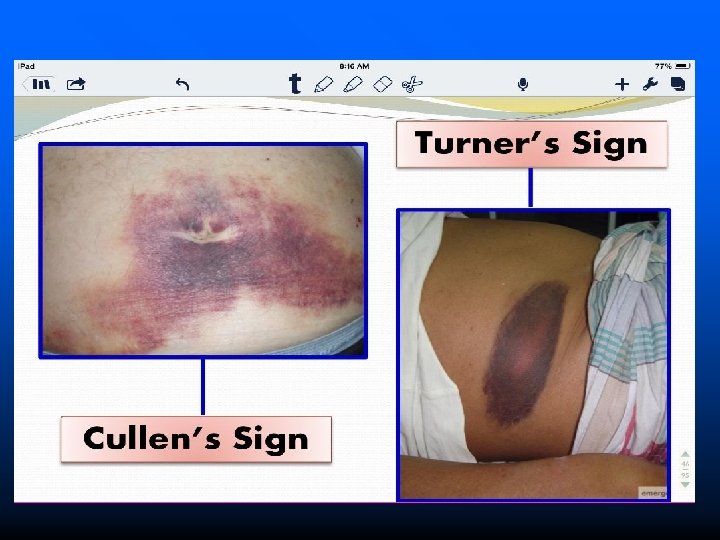
Physical Exam n n Fever, tachycardia, epigastric tenderness and abdominal distention are typical findings Severe pancreatitis associated with hemorrhage into the retroperitoneum may produce two distinctive physical signs-Turner’s sign and Cullen’s sign Jaundice is an uncommon finding at the initial presentation but may be seen with gallstone associated pancreatitis With severe pancreatitis – Major circulatory derangements such as hypotension, hypovolemia, hypoperfusion, and obtundation may occur

Diagnosis n Laboratory determinations – Serum amylase » Elevated within 24 hours of onset of symptoms » Amylase levels rapidly returned to normal » Persistent hyperamylasemia indicates the development of complications » The degree of amylase elevation is not a reliable predictor of the severity of disease » The magnitude is an independent predictor to differentiate gallstone associated pancreatitis from alcohol induced pancreatitis

Diagnosis – Serum lipase levels » A more accurate indicator of acute pancreatitis (is solely of pancreatic origin) » Is not entirely specific for acute pancreatitis n Other laboratory values, although not specific may help in the diagnosis – Serum glucose levels (usually elevated) – Abnormal liver function tests – Hypocalcemia


Radiographic Evaluation n Chest X-ry – Left basal atelectasis – Elevation of the left hemidiaphragm – Left pleural effusion n Abdominal X-ry – May reveal air in the duodenal loop – Gallstones in the gallbladder n Upper GI contrast studies – Widening of the duodenal “C” loop – Anterior displacement of the stomach

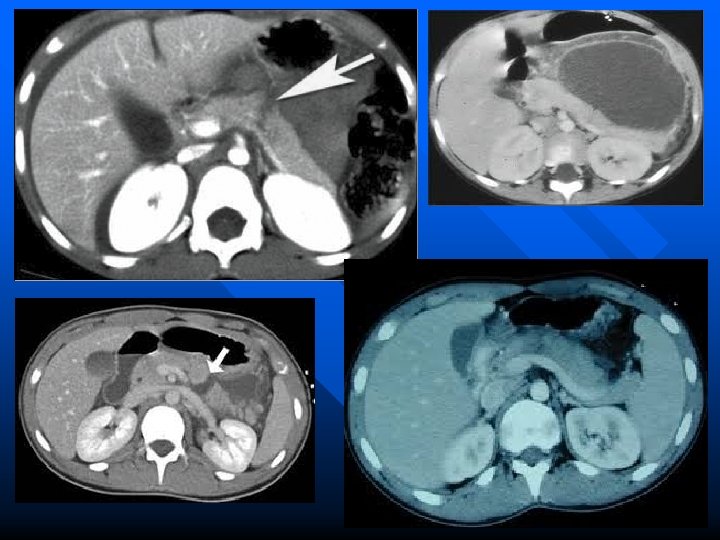
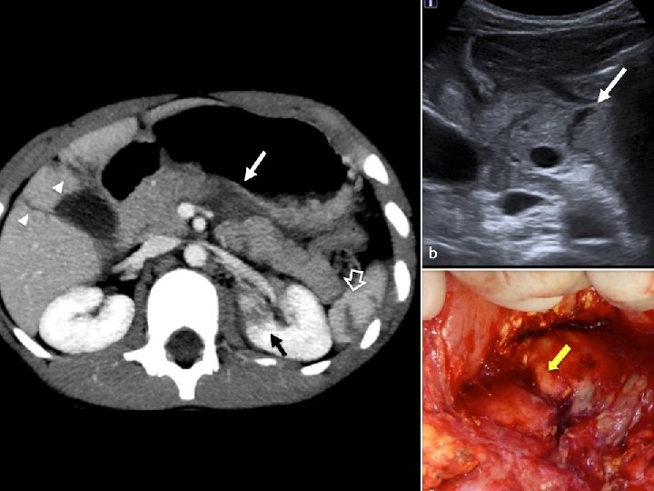
Radiographic Evaluation n Abdominal ultrasound – May be of limited value due to the presence of air and fluid within the bowel overlying the pancreas – May detect pancreatic edema n CT scan – Currently the most widely accepted and sensitive method used to confirm the diagnosis n MRI – Holds great promise – Results are equal to that of CT scan

Clinical Course It is possible to predict the severity of an attack using routinely available clinical and laboratory determinations n These tests are valuable in guiding therapy of patients n

Early Prognostic Signs of Acute Pancreatitis Criteria for Pancreatitis Not Due to Gallstones At admission Age over 55 years White blood count > 16, 000 Blood glucose > 200 mg Serum lactate dehydrogenase (LDH) > 400 AST > 250 During Initial 48 Hours Hematocrit fall > 10 percentage points BUN elevation > 5 Serum calcium fall to < 8 Arterial oxygen concentration < 60 Base deficit > 4 Estimated fluid sequestration > 6 l

Early Prognostic Signs of Acute Pancreatitis Criteria for Gallstone Pancreatitis At Admission Age over 70 years White blood count > 18, 000 Glucose > 220 Serum lactate dehydrogenase (LDH) > 400 AST > 250 During Initial 48 Hours Hematocrit fall > 10 percentage points BUN elevation > 2 Serum calcium fall to < 8 Base deficit > 5 Estimated fluid sequestration > 4 l

Nonoperative Management n n Standard therapy includes IV fluids, electrolyte replacement and pain medication (meperidinedrug of choice) (MSO 4 -causes sphincter of Oddi spasm) NG tube is reserved for patients with an ileus Patients with severe pancreatitis often require nutritional support via parenteral alimentation (NPO) Antibiotic administration for prevention of septic complications

Operative Management n Is indicated in four specific circumstances – Uncertainty of the diagnosis – Treatment of secondary pancreatic infections – Correction of associated biliary tract disease – Progressive clinical deterioration despite optimal supportive care

Operative Management n Uncertainty of clinical diagnosis – No single test is 100 % accurate in diagnosing acute pancreatitis – Occasionally it may be difficult to exclude another diagnosis that mimics acute pancreatitis » In these situations exploratory laparotomy may be indicated to exclude surgically correctable disease » If cholelithiasis is the cause this can be surgically corrected

Operative Management n Treatment of secondary pancreatic infections – Pancreatic abscesses, infected pancreatic pseudocyst and pancreatic necrosis with infection (three serious and life-threatening complications) – Their frequency of occurrence is in direct proportion to the severity of pancreatitis – In patients with six or more prognostic signs over half will develop a pancreatic septic complication – The development of pancreatic septic complications should be suspected in patients in whom pancreatitis fails to resolve within the first week to 10 days
 » Reduces")
Operative Management n Correction of associated biliary tract disease – Cholecystectomy (early) » Reduces overall length of stay » Eliminates the need for a second hospitalization

Operative Management n Deterioration of clinical status – In patients who fail to respond to nonoperative supportive care, operative intervention has been advocated » This is the most controversial indication for surgical therapy Some physicians recommend removing necrotic tissue n Others recommend total pancreatectomy n

Chronic Pancreatitis
, cystic fibrosis,")
Chronic Pancreatitis n Etiology – Is associated with alcohol abuse (most common), cystic fibrosis, congenital anomalies of the pancreatic duct and trauma to the pancreas – The exact mechanism of alcohol induced disease is unknown

Chronic Pancreatitis n Clinical presentation – Incidence is approximately 4 per 100, 000 – The typical patient presents with a history of alcohol abuse in the fourth or fifth decade of life – Abdominal pain is the feature that prompts consultation – The pain is commonly epigastric in location but may be localized to the right or left of midline – Radiation to the back is common – Anorexia and weight loss may be present – Insulin-dependent diabetes occurs in up to one-third of these patients – Up to 25% have steatorrhea (implies reduced pancreatic exocrine function)

Chronic Pancreatitis n Diagnosis – Is usually suspected on clinical findings – Routine laboratory tests are rarely helpful – Radiographic evaluation may reveal pancreatic calcifications on plain films – CT scan is useful in evaluating the size and texture of the pancreas – Endoscopic retrograde pancreatography – Pancreatic endocrine function

Chronic Pancreatitis n Nonoperative management – Control of abdominal pain » Can be a problem (drug dependency) » In some patients total abstinence from alcohol relieves the pain » Dietary changes are also recommended – Treatment of endocrine insufficiency – Treatment of exocrine insufficiency

Chronic Pancreatitis n Operative management – Ampullary procedures » Designed to eliminate pancreatitis by preventing bile reflux into the pancreatic duct (results have not been favorable) – Ductal drainage procedures » Designed to decompress the pancreatic duct in a retrograde manner n Pancreaticojejunostomy (success rates of 60 -90%) – Ablative procedures (last step) » Pancreatectomy (total or subtotal) n Most obtain adequate pain relief however these procedures can cause IDDM

Disruptions of the Pancreatic Duct In adults, the most common cause is alcoholic pancreatitis n In children the most common cause is neoplasms n Disruptions of the main pancreatic duct cause external or internal pancreatic fistulas n

Disruptions of the Pancreatic Duct n External pancreatic fistula – May occur as a result of pancreatic operations (25% of cases) – Fistulas that drain < 200 ml per day are classified as low output fistulas – Complications include sepsis, fluid and electrolyte abnormalities and excoriation – Parenteral nutrition is utilized to avoid pancreatic stimulation by oral intake – Most pancreatic fistulas close with nonoperative management
 – Represents 75%")
Disruptions of the Pancreatic Duct n Internal pancreatic fistula (pancreatic pseudocyst) – Represents 75% of cystic lesions of the pancreas – Electrolyte concentrations in the pseudocyst fluid are equivalent to those in plasma – Patients present most often with upper abdominal pain, early satiety, nausea and vomiting – An abdominal mass is present in less than half – Laboratory findings are nonspecific – CT scan of the abdomen is the favored study and initial assessment

Neoplasms of the Pancreas n n n n The fifth most common cause of cancer death 90% of patients die within the first year after diagnosis The five-year survival rate is 1% More common in blacks, smokers and in males Appears to be linked to the presence of diabetes mellitus and possibly past history of chronic pancreatitis and long-term high-fat diets Over 90% of malignant pancreatic exocrine tumors are duct cell adenocarcinomas The most common site of origin is in the head of the pancreas

Periampullary Adenocarcinoma n n Difficult to differentiate from three other malignant periampullary neoplasms: ampullary carcinoma, duodenal carcinoma and carcinoma of the distal common bile duct Most common clinical features are jaundice, weight loss and abdominal pain Laboratory abnormalities include elevated serum bilirubin, alkaline phosphatase, CEA and CA 19 -9 Radiographic studies-upper barium series may be positive with large tumors, ultrasound may be of benefit and CT scan provides better accuracy of diagnosis

Periampullary Adenocarcinoma n Management – Nonoperative therapy » Is recommended in patients with documented distant metastases » Unresectable local disease » Chronic debilitating illnesses

Periampullary Adenocarcinoma n Management – Operative therapy » The Whipple procedure n n The gallbladder, common bile duct, entire duodenum, head of the pancreas, pylorus and distal stomach are removed (five-year survival rate approximates 15 -25%) Care is taken to assess distant intra-abdominal metastasis including lymph nodes » Palliative surgery n n Designed for patients with unresectable disease to alleviate obstruction and tumor associated pain Biliary-enteric bypass » Adjuvtant therapy n n The data indicates that chemotherapy alone has no role in periampullary carcinoma Combination therapy has slightly better results (20 months)

Adenocarcinoma of the Body and Tail of the Pancreas n n n Represents up to 30% of all cases of pancreatic carcinoma The tumors in this location usually grow quite large prior to becoming symptomatic These tumors do not cause early obstructive jaundice or GI obstructive symptoms Clinical presentation is usually weight loss and pain (90% of patients) Physical exam findings are often nonspecific CT scan is the best study for detection of primary and metastatic disease

Other Pancreatic Tumors n Cystadenocarcinoma of the pancreas – Generally seen in females between the ages of 40 -60 – Not commonly seen n Acinar-cell carcinoma – A rare malignancy with no sexual predominance n Benign neoplasms of the exocrine pancreas – Cystadenoma – Solid and papillary neoplasms of the pancreas

Endocrine Tumors Rare with an incidence of five per one million n Endocrine tumors are named according to the major hormone produced by the tumor n Malignancy is determined by the presence of local invasion, spread to regional lymph nodes or hepatic/distant metastasis n Tumor localization is best identified using CT scan’s/MRI n

Insulinoma n The most common endocrine tumor of the pancreas – Symptoms include: hypoglycemia at fasting, documentation of blood glucose levels of < 50 and relief of symptoms following administration of glucose – Detection methods: blood glucose and insulin levels are sampled every 4 -6 hours after a 72 hour fast – Treatment: surgery-most are benign – Pharmacologic therapy may be useful in patients with residual tumor following resection (Diazoxide)
 n Symptoms include: – Primary peptic ulceration in unusual locations –")
Gastrinoma (Zollinger-Ellison Syndrome) n Symptoms include: – Primary peptic ulceration in unusual locations – Gastric acid hypersecretion despite adequate therapy – Identification of a islet cell tumor of the pancreas n n When a gastrinoma is suspected, fasting serum gastrin levels should be obtained Patient management: – Control of gastric acid hypersecretion – Alteration of the natural history of the gastrinoma (tumor localization, assessment of metastatic disease and tumor resection)
 – A pancreatic islet cell tumor associated")
Other Endocrine Tumors n Verner-Morrison syndrome (VIPoma) – A pancreatic islet cell tumor associated with severe watery diarrhea, hypokalemia and either achlorhydria or hypochlorhydria n Glucagonoma – Hallmarks are mild diabetes and severe dermatitis n Somatostatinoma – Presents as gallstones, diabetes and steatorrhea – A rare endocrine tumor n Nonfunctional islet cell tumors

Pancreatic Lymphoma n n n Involvement of the pancreas with non-Hodgkin’s lymphoma is an unusual neoplasm It usually presents with weight loss abdominal pain and may include jaundiced and symptoms of gastric outlet obstruction The most common physical finding is a palpable abdominal mass Abdominal CT scan may suggest the diagnosis (large soft tissue mass in the vicinity of the pancreas) Diagnosis is confirmed by needle biopsy

Pancreatic Trauma n The pancreas is injured in less than 2% of patients with abdominal trauma – Two-thirds are associated with penetrating abdominal trauma – If the pancreas is injured usually adjacent organs and major vascular structures are also injured – The majority of fatal cases is due to damage from nearby vascular structures – The second most common cause of death involves intraabdominal sepsis – In blunt abdominal trauma, the extent and location of pancreatic injury is determined by the mechanism of injury and location of impact

Diagnosis n n n No laboratory test is sufficiently accurate for the specific diagnosis of pancreatic injury Amylase is elevated in most patients with blunt trauma and only slightly elevated with penetrating injuries Peritoneal lavage is inaccurate because the pancreas is retroperitoneal Chest and abdominal films are often not helpful CT scan of the abdomen is gaining acceptance in evaluating pancreatic injury

Management n n n If the patients have stable vital signs and lack a specific indication for exploration they are treated with observation and followed for complications such as abscess, pseudocyst or phlegmon If the patients undergo laparotomy for other reasons the pancreas must be assessed utilizing a Kocher’s maneuver The goal of operative therapy should include control of hemorrhage, debridement of nonviable tissue and adequate drainage of exocrine secretions

Categorization of Pancreatic Injury Pancreatic contusion without capsular rupture n Pancreatic capsular and parenchymal rupture without injury to the main pancreatic duct n Severe pancreatic parenchymal injury with rupture of the main pancreatic duct n Combined severe pancreatic and duodenal injuries n

Categorization of Pancreatic Injury n Class I injury – Is treated by external drainage alone – This prevents occult capsular disruptions that could potentially cause accumulation of pancreatic secretions that will eventually cause a pseudocyst or abscess – Drains are usually left in place until oral intake is reestablished n Class II injury – Are treated by cautious debridement of devitalized tissue, adequate hemostasis and closure of major capsular disruptions – External drainage is also recommended

Categorization of Pancreatic Injury n Class III injury – These injuries require individualized treatment based on their location and injuries to adjacent structures – Injuries to the body and tail are best treated by distal pancreatectomy – Drains are also placed n Class IV injury – Mortality approaches 45% due to frequently associated adjacent visceral or vascular injuries – Treatment is individualized based on the extent of damage

The End
- Slides: 54