PALATINE TONSILS Presented by Dr Sushma Tomar Associate

PALATINE TONSILS Presented by : - Dr. Sushma Tomar Associate Professor Department of

• There are two palatine tonsils. Introduction • Each palatine tonsil is a mass of lymphoid tissue. Location • In tonsillar fossa, which is situated in the lateral wall of oropharynx between anterior and posterior
. Posterior- Posterior faucial pillar")
Boundaries of Tonsillar Fossa Anterior- Anterior faucial pillar (palatoglossal arch). Posterior- Posterior faucial pillar (palatopharyngeal arch). Apex- Soft palate. Base- Dorsal surface of posterior 1/3 rd of tongue. Lateral wall [Tonsillar Bed]Superior Constrictor Muscle (mainly).

Tonsillar Bed v • • • Following structures form the tonsillar bed ( from inside outwards): Pharyngobasilar fascia. Superior Constrictor muscle. Buccopharyngeal fascia.

Presenting Parts • 2 surfaces- Medial & Lateral • 2 borders- Anterior & Posterior • 2 Poles- Upper & Lower Medial Surface • It bulges into oropharynx. • It is covered by epithelium. • It has crypts. • There are ~ 12 -15 crypts. Crypta Magna • A very large and deep crypt located near upper pole. • It represents the remnant of 2 nd pharyngeal pouch.

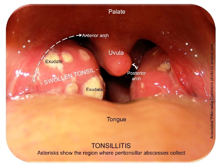
Lateral Surface • It is covered by fibrous capsule. Peritonsillar Space • A space between fibrous capsule and tonsillar bed. • It is filled with loose areolar tissue. • It is the site of collection of pus in peritonsillar abscess. • During tonsillectomy, tonsil is dissected in this plane. Internal Carotid Artery is ~2. 5 cm posterolateral to the

Lateral Surface contd… v Superior constrictor separates the lateral surface from following structures: • Facial artery and its ascending palatine and tonsillar branches. • Styloglossus muscle. • Glossopharyngeal nerve. • Angle of mandible. • Medial Pterygoid muscle. • Submandibular salivary gland. Facial Artery

Presenting Parts contd… Anterior Border • Passes underneath the palatoglossal arch. Posterior Border • Passes underneath the palatopharyngeal arch. Upper Pole- extends up into the soft palate. Lower Pole • It is attached to the tongue by a band of fibrous tissue called suspensory ligament of tonsil.

Arterial Supply v • • v v • Facial Artery. Tonsillar branch. Ascending Palatine branch. Lingual Artery. Dorsalis Linguae branches. Ascending Pharyngeal Artery. Maxillary Artery. Greater Palatine Branch. Greater (Descending) Pa branch Dorsal Lingual branch

Venous Drainage • Paratonsillar Vein drains into pharyngeal venous plexus.
![Lymphatic Drainage • Upper deep cervical lymph nodes [mainly Jugulo-digastric nodes]. • Jugulo-digastric nodes](http://slidetodoc.com/presentation_image_h2/163a6509e1f9e885737bf0e39d9cc91d/image-12.jpg "Lymphatic Drainage • Upper deep cervical lymph nodes [mainly Jugulo-digastric nodes]. • Jugulo-digastric nodes")
Lymphatic Drainage • Upper deep cervical lymph nodes [mainly Jugulo-digastric nodes]. • Jugulo-digastric nodes are called ‘Tonsillar Lymph Nodes’.

Nerve Supply v Glossopharyngeal nerve. v Pterygopalatine ganglion • Lesser palatine branches.

Applied Aspects Acute Tonsillitis • Palatine tonsils are frequent sites of acute infection. Age group • School-going children. Etiology • Mostly viral. Acute Follicular Tonsillitis • Infection spreads into crypts. • Crypts become filled with purulent material, which presents at the opening of the crypt as yellowish spots.

Applied Aspects contd… Tonsillectomy • Surgical removal of tonsil. • If paratonsillar vein gets damaged during tonsillectomy, severe bleeding occurs from tonsillar fossa. • To check bleeding, blood clots should be removed because they interfere with retraction of walls of vein. • Blood clots prevent the contraction of surrounding muscles. • After tonsillectomy,

- Slides: 16