PALATE Presented by Dr Sushma Tomar Associate Professor

PALATE Presented by : - Dr. Sushma Tomar Associate Professor Department of

Introduction L. palate=roof of mouth • It is a partition between the nasal and oral cavities. Parts- 2 • Hard palate. • Soft palate.

Hard Palate

Introduction • Forms anterior 4/5 th of the palate. Gums Anterior 4/5 th Formation. Anterior 2/3 rd – by palatine processes of maxillae. Posterior 1/3 rd – by horizontal plates of palatine bones. Alveolar Arch • It becomes continuous with maxillary alveolar arches and gums anterolaterally. • Its posterior border provides Posterior border

Introduction contd… • Its superior surface forms the floor of nasal cavities. • Its inferior surface forms the roof of oral cavity.

Inferior Surface v Presents the following features: • Incisive fossa. • Greater palatine foramen. • Lesser palatine foramina. Incisive Fossa • Posterior nasal spine. • Palatine crest. • Masticatory mucosa. Palatine Crest Posterior Nasal Spine

Incisive Fossa • A small pit anteriorly in the midline. • It has openings of incisive canals in the form of incisive foramina. • Each incisive canal transmits Nasopalatine nerve and Greater palatine vessels. Incisive Fossa

Greater Palatine Foramen • It lies in the posterolateral corner of hard palate, medial to last molar tooth. • It transmits Greater palatine nerve and vessels.

Lesser Palatine Foramina • 1 -3 in number. Location- just behind the Greater Palatine foramen. • They are in the pyramidal process of palatine bone. • They transmit lesser palatine nerve and vessels.

Posterior Nasal Spine • A conical projection in the median plane on posterior border of hard palate. Palatine Crest • A curved ridge near the posterior border of hard palate. Posterior Nasal Palatine Crest. Spine

Masticatory Mucosa • It is the mucous membrane lining the hard palate. • It is firmly adherent with the periosteum by multiple Sharpey’s fibers. • It presents transverse masticatory ridges on either side of midline. • A narrow ridge extending anteroposteriorly in the midline from a papilla overlying the incisive fossa is known as palatine raphe.

Arterial Supply • Greater palatine arteries. Venous drainage • Pterygoid venous plexus. • Pharyngeal venous plexus.

Nerve Supply • Greater palatine nerves. • Nasopalatine nerves.

Lymphatic Drainage • Upper deep cervical lymph nodes. • Retropharyngeal lymph nodes.

Soft Palate

Introduction • A mobile muscular flap hanging down from the posterior border of hard palate into the pharyngeal cavity. • It separates the nasopharynx from oropharynx, when abuts on the Passavant’s ridge. SOFT PALATE
 • Posterior")
External Features • 2 surfaces • 2 borders Surfaces • Anterior (Oral) • Posterior Borders • Superior • Inferior Superior Border Anterior Surface Median Raphe Inferior Border Anterior (Oral) Surface • Concave. • Has a median raphe. Posterior Surface Posterior (Oral) Surface • Convex. Anterior Surface

Borders Superior Border • Attached to posterior border of hard palate. Inferior Border • Free. • Forms the anterior boundary of pharyngeal isthmus. • A small, conical, tongue- like projection hanging down from its middle is called uvula.

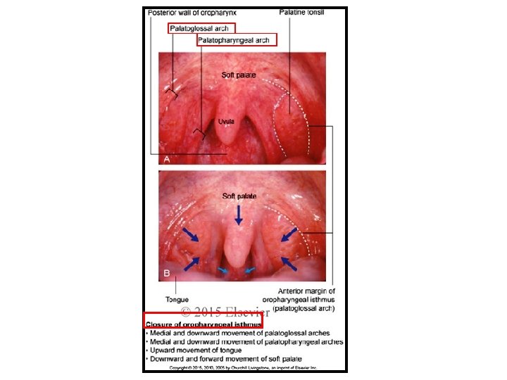
Uvula v On each side from the base of uvula, 2 curved folds of mucous membrane extend laterally and downwards: • Anterior fold (Palatoglossal fold). • Posterior fold ( Palatopharyngeal fold). Anterior fold (Palatoglossal fold) • Merges inferiorly with the side of the tongue ( at the junction of anterior 2/3 rd and posterior 1/3 rd ). • It contains palatoglossus muscle. • It forms the lateral boundary of oropharyngeal isthmus and anterior boundary of tonsillar fossa. Posterior fold ( Palatopharyngeal fold) • Merges inferiorly with the lateral wall of pharynx. • It contains palatopharyngeus Uvula
. • Levator")
v 5 pairs of muscles: Muscles • Tensor palati (Tensor Veli Palatini). • Levator palati (Levator Veli Palatini). • Palatoglossus. • Palatopharyngeus • Musculus uvulae. • All muscles are extrinsic except Musculus uvulae, which are intrinsic.

Tensor Palati • It is a thin triangular muscle. Origin • Lateral aspect of cartilaginous part of auditory tube. • Adjoining part of greater wing of sphenoid including its spine. Insertion • Muscle descends, converges to form a tendon. • The tendon hooks round the pterygoid hamulus and then expands to form the palatine aponeurosis. v Palatine aponeurosis attaches to: • Posterior border of hard palate. • Inferior surface of hard palate behind the palatine crest. Actions • Tightens the soft palate. • Helps in opening the auditory tube.

Levator Palati • A cylindrical muscle lying deep to Tensor palati. Origin • Medial aspect of cartilaginous part of auditory tube. • Adjoining part of petrous part of temporal bone ( inferior surface of its apex anterior to carotid canal). Insertion • Muscle runs downwards and medially and spreads out to be inserted on the upper surface of palatine aponeurosis. Actions • Elevates the soft palate to close the pharyngeal isthmus. Cartilaginous part of Auditory tu
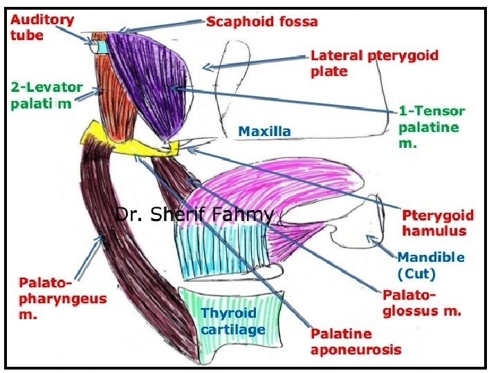

Musculus Uvulae • A longitudinal muscle strip. • One on either side of median plane within the palatine aponeurosis. Origin • Posterior nasal spine. • Palatine aponeurosis. Insertion • Mucous membrane of uvula. Actions • Pulls the uvula forwards to its own side.

Functions of Soft Palate • During swallowing, closes the pharyngeal isthmus to separate the oropharynx from nasopharynx, thereby prevents the entry of food into the nasopharynx and nasal cavities, and closes the oropharyngeal isthmus to prevent the regurgitation of food contents into the oral cavity. Closure of Pharyngeal isthmus [Soft palate is raised up (Levator Palati) and makes contact with the posterior pharyngeal wall (contraction of Palatopharyngeus)] Closure of Oropharyngeal isthmus [Soft palate is pulled down,

Functions of Soft Palate contd… • During chewing, closes the oropharyngeal isthmus to isolate the oral cavity from oropharynx so that breathing is not affected. • Helps to modify the quality of voice by varying the degree of closure of pharyngeal isthmus. • During sneezing, prevents the damage of nasal mucosa, by appropriately dividing and directing the blast of air through both nasal and oral cavities. • During coughing, prevents the entry of sputum into the nasal cavities by directing it into oral cavity.

Arterial Supply v Maxillary artery • Lesser palatine branches. v Facial artery • Ascending palatine branch. v Ascending pharyngeal artery • Palatine branches. MA- Maxillary Artery, LPA- Lesser Palatine Arteries, FA- Facial Artery, APal. AAscending Palatine Artery, APA- Ascending Pharyngeal Aretry CCA- Common Carotid Artery, ECA- External Carotid Artery, DPA-Descending Palatine Artery, GPA- Greater Palatine Artery

Venous Drainage Into: • Pharyngeal venous plexus. • Pterygoid venous plexus.

Lymphatic Drainage • Retropharyngeal lymph nodes. • Upper deep cervical lymph nodes.

Nerve Supply Motor supply • All muscles are supplied by cranial root of Accesssory nerve via pharyngeal plexus except Tensor palati. • Tensor palati is supplied by nerve to Medial Pterygoid. Sensory supply. General Sensory • By Lesser palatine nerves. • Glossopharyngeal nerve.

Applied Aspects Gag Reflex • It is a protective reflex, in response to stimulation of mucous membrane of oropharynx. • Characterized by reflex contraction of pharyngeal and palatal muscles. Afferent limb. Glossopharyngeal nerve. Efferent limb • Vagus nerve.

- Slides: 33