
Pakistan Society Of Chemical Pathologists Distance Learning Programme








- Slides: 55
Pakistan Society Of Chemical Pathologists Distance Learning Programme In Chemical Pathology Lesson No 9 Interpretation of Electrolytes and Arterial Blood Gasses Reports By Surg Commodore Aamir Ijaz, FCPS, FRCP (Edin) Prof of Pathology / Consultant Chemical Pathologist BUM&DC and PNS SHIFA Karachi Dr Muhammad Anwar Magsi, MPhil Assoc Prof of Pathology, Bolan Med College, Quetta (Trainee FCPS-Part II PNS SHIFA Karachi Dr Humaira Asif, FCPS Assist Prof of Pathology, BUM&DC Karachi 11/6/2020 6: 17 AM 2
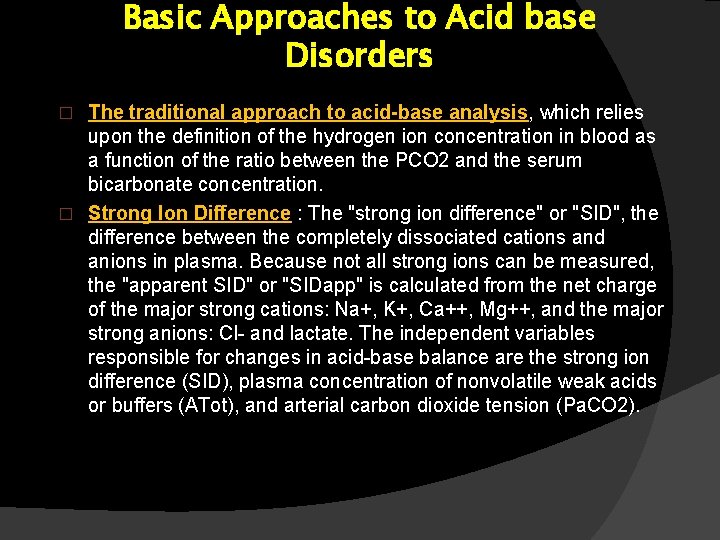
Basic Approaches to Acid base Disorders The traditional approach to acid-base analysis, which relies upon the definition of the hydrogen ion concentration in blood as a function of the ratio between the PCO 2 and the serum bicarbonate concentration. � Strong Ion Difference : The "strong ion difference" or "SID", the difference between the completely dissociated cations and anions in plasma. Because not all strong ions can be measured, the "apparent SID" or "SIDapp" is calculated from the net charge of the major strong cations: Na+, K+, Ca++, Mg++, and the major strong anions: Cl- and lactate. The independent variables responsible for changes in acid-base balance are the strong ion difference (SID), plasma concentration of nonvolatile weak acids or buffers (ATot), and arterial carbon dioxide tension (Pa. CO 2). �
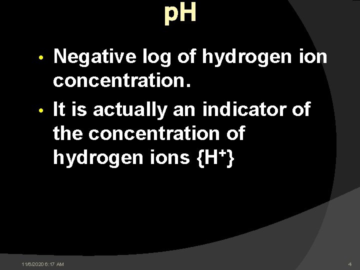
p. H Negative log of hydrogen ion concentration. • It is actually an indicator of the concentration of hydrogen ions {H+} • 11/6/2020 6: 17 AM 4
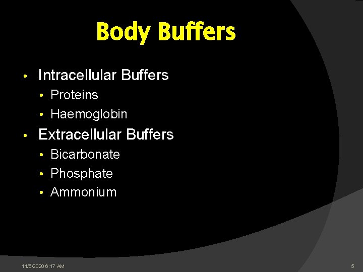
Body Buffers • Intracellular Buffers • Proteins • Haemoglobin • Extracellular Buffers • Bicarbonate • Phosphate • Ammonium 11/6/2020 6: 17 AM 5
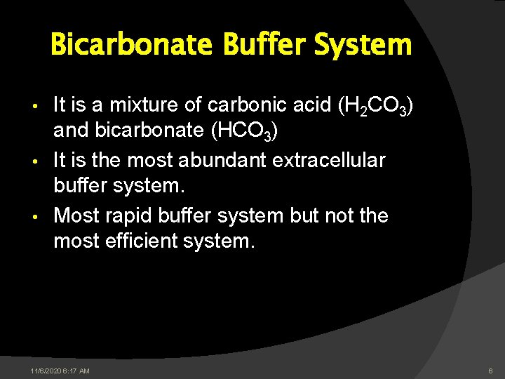
Bicarbonate Buffer System It is a mixture of carbonic acid (H 2 CO 3) and bicarbonate (HCO 3) • It is the most abundant extracellular buffer system. • Most rapid buffer system but not the most efficient system. • 11/6/2020 6: 17 AM 6
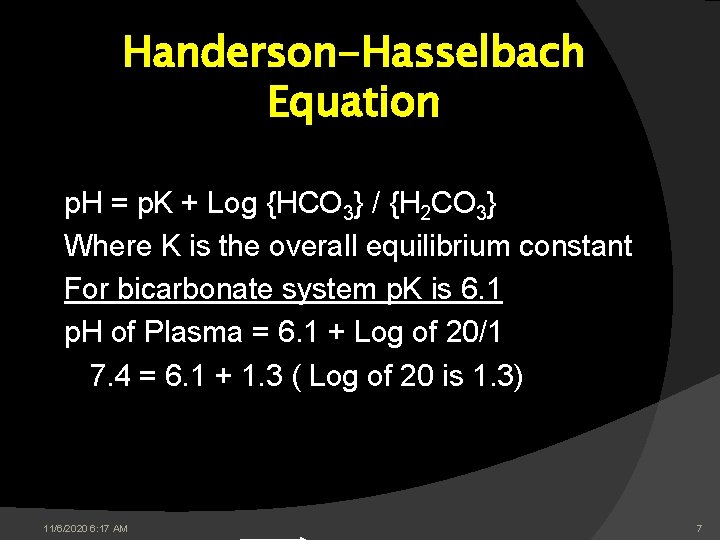
Handerson-Hasselbach Equation p. H = p. K + Log {HCO 3} / {H 2 CO 3} Where K is the overall equilibrium constant For bicarbonate system p. K is 6. 1 p. H of Plasma = 6. 1 + Log of 20/1 7. 4 = 6. 1 + 1. 3 ( Log of 20 is 1. 3) 11/6/2020 6: 17 AM 7
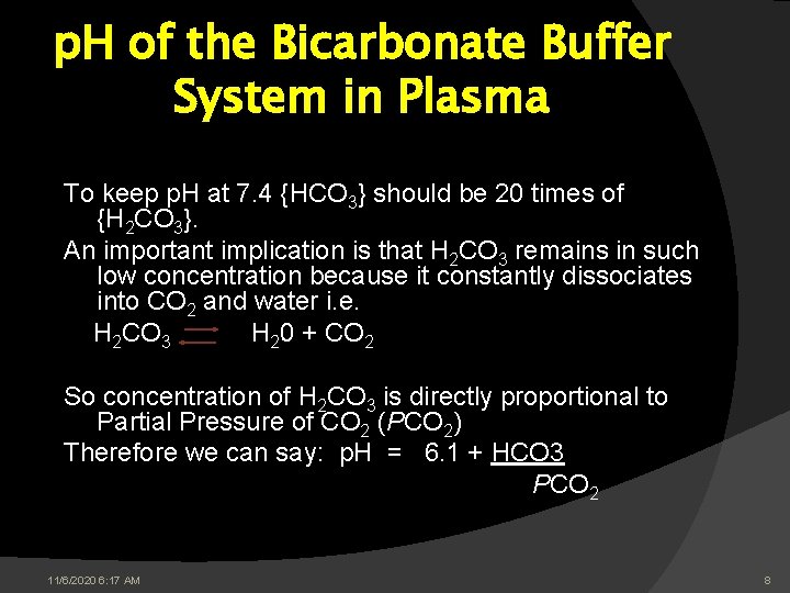
p. H of the Bicarbonate Buffer System in Plasma To keep p. H at 7. 4 {HCO 3} should be 20 times of {H 2 CO 3}. An important implication is that H 2 CO 3 remains in such low concentration because it constantly dissociates into CO 2 and water i. e. H 2 CO 3 H 20 + CO 2 So concentration of H 2 CO 3 is directly proportional to Partial Pressure of CO 2 (PCO 2) Therefore we can say: p. H = 6. 1 + HCO 3 PCO 2 11/6/2020 6: 17 AM 8
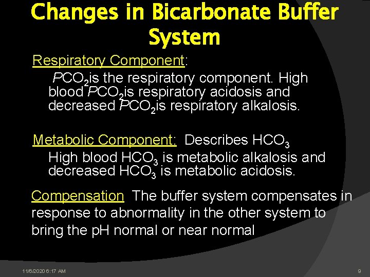
Changes in Bicarbonate Buffer System Respiratory Component: PCO 2 is the respiratory component. High blood PCO 2 is respiratory acidosis and decreased PCO 2 is respiratory alkalosis. Metabolic Component: Describes HCO 3 High blood HCO 3 is metabolic alkalosis and decreased HCO 3 is metabolic acidosis. Compensation: The buffer system compensates in response to abnormality in the other system to bring the p. H normal or near normal 11/6/2020 6: 17 AM 9
Laboratory Investigations for Acid Base Disorders ○ For Biochemical Diagnosis: �Plasma Bicarbonate. �Blood Gases: - p. H - PCO 2 - PO 2 11/6/2020 6: 17 AM 10
Laboratory Investigations For Acid Base Disorders (Cont) For Aetiological diagnosis: • Electrolytes (Na+ and K+) • Renal Function Tests • Plasma Glucose • Plasma Lactate • Urine and Plasma Ketones 11/6/2020 6: 17 AM 11
ABG Analysis .
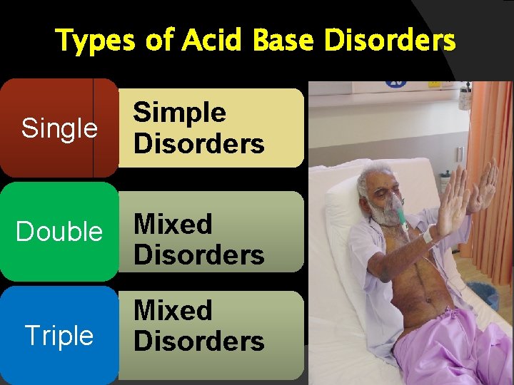
Types of Acid Base Disorders Single Simple Disorders Double Mixed Disorders Triple Mixed Disorders
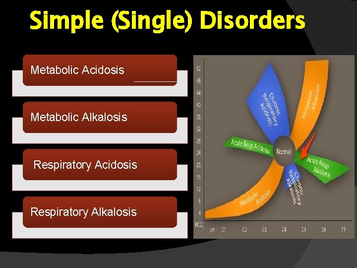
Simple (Single) Disorders Metabolic Acidosis Metabolic Alkalosis Respiratory Acidosis Respiratory Alkalosis
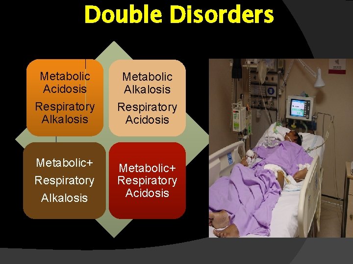
Double Disorders Metabolic Acidosis Respiratory Alkalosis Metabolic Alkalosis Respiratory Acidosis Metabolic+ Respiratory Alkalosis Metabolic+ Respiratory Acidosis
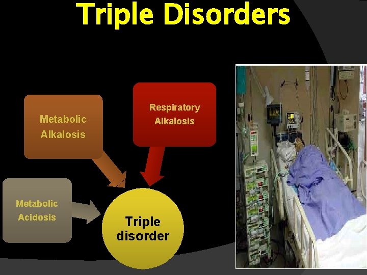
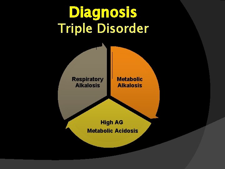
Triple Disorders Respiratory Metabolic Alkalosis Metabolic Acidosis Triple disorder
Interpretation of ABG Report Vital for Emergency Management • Instant Report and Instant Interpretation • Immediate Correction •
How to Interrupt Quickly Good Understanding of Pathophysiology • Softwares • Mobile Apps •
One Minute Decoder Can works in tandem with Mobile Apps (or Equations) For : • Identification of Primary Disorder • Evaluation of Compensatory Process
Salient Features of ‘One Minute Decoder’ Question 1: Acidosis or Alkalosis ? • Question 2: Primary disorder is Metabolic or Respiratory ? ? • Question 3: Single or Double disorder ? ? ? •
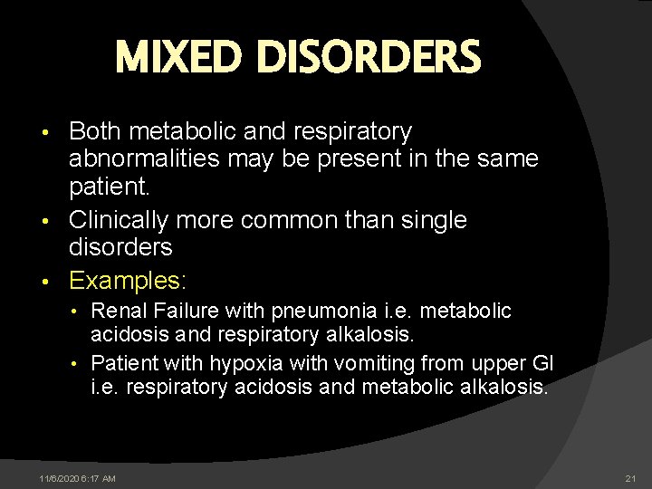
MIXED DISORDERS Both metabolic and respiratory abnormalities may be present in the same patient. • Clinically more common than single disorders • Examples: • • Renal Failure with pneumonia i. e. metabolic acidosis and respiratory alkalosis. • Patient with hypoxia with vomiting from upper GI i. e. respiratory acidosis and metabolic alkalosis. 11/6/2020 6: 17 AM 21
Interpreting ABG Report • • • Pathophysiological principles By “One Minute Decoder” By Equations Flenley’s Graph Electronic Calculators (Skyscape for desktop, Smart phones etc). 11/6/2020 6: 17 AM 22
Decoding an ABG Report 11/6/2020 6: 17 AM 23
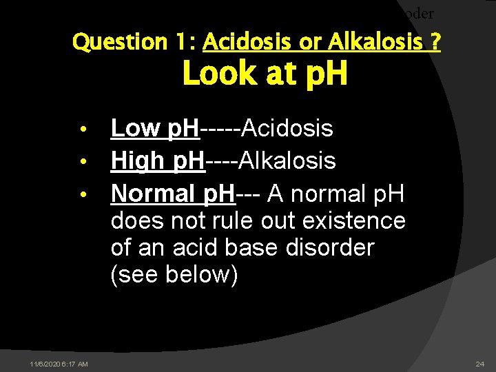
The Decoder Question 1: Acidosis or Alkalosis ? Look at p. H Low p. H-----Acidosis • High p. H----Alkalosis • Normal p. H--- A normal p. H does not rule out existence of an acid base disorder (see below) • 11/6/2020 6: 17 AM 24
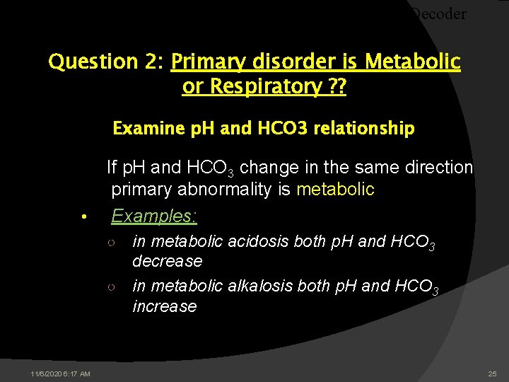
The Decoder Question 2: Primary disorder is Metabolic or Respiratory ? ? Examine p. H and HCO 3 relationship If p. H and HCO 3 change in the same direction primary abnormality is metabolic • Examples: in metabolic acidosis both p. H and HCO 3 decrease ○ in metabolic alkalosis both p. H and HCO 3 increase ○ 11/6/2020 6: 17 AM 25
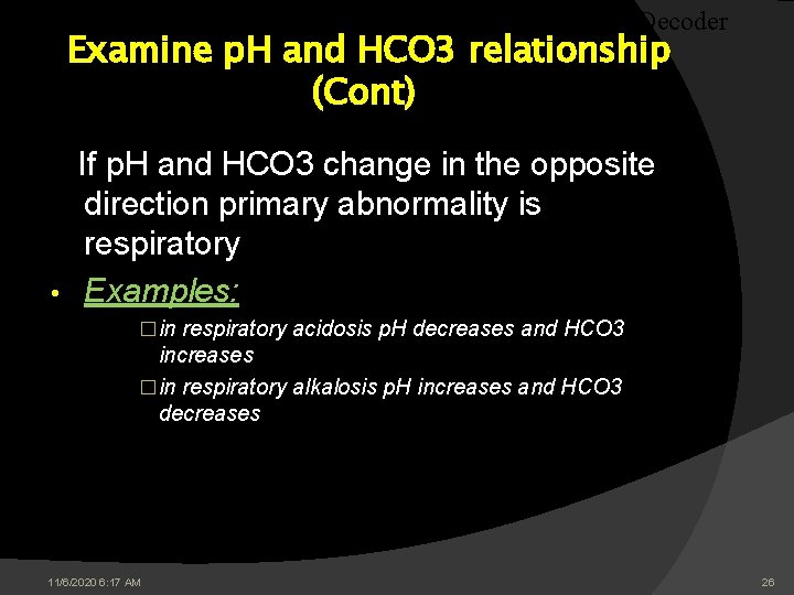
The Decoder Examine p. H and HCO 3 relationship (Cont) If p. H and HCO 3 change in the opposite direction primary abnormality is respiratory • Examples: �in respiratory acidosis p. H decreases and HCO 3 increases �in respiratory alkalosis p. H increases and HCO 3 decreases 11/6/2020 6: 17 AM 26
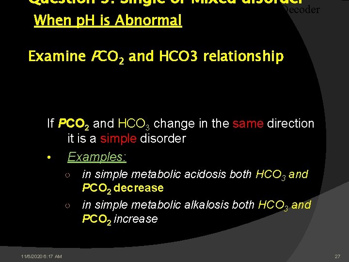
Question 3: Single or Mixed disorder When p. H is Abnormal The Decoder Examine PCO 2 and HCO 3 relationship If PCO 2 and HCO 3 change in the same direction it is a simple disorder • Examples: in simple metabolic acidosis both HCO 3 and PCO 2 decrease ○ in simple metabolic alkalosis both HCO 3 and PCO 2 increase ○ 11/6/2020 6: 17 AM 27
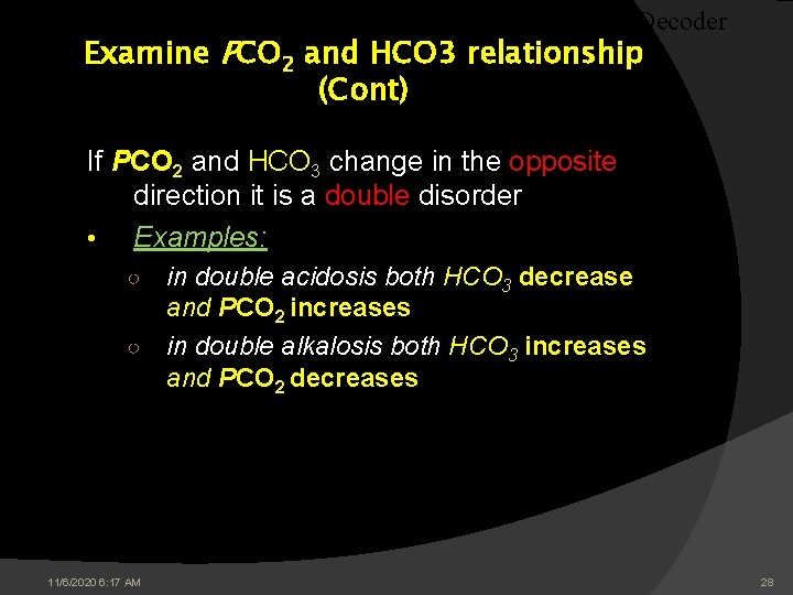
The Decoder Examine PCO 2 and HCO 3 relationship (Cont) If PCO 2 and HCO 3 change in the opposite direction it is a double disorder • Examples: in double acidosis both HCO 3 decrease and PCO 2 increases ○ in double alkalosis both HCO 3 increases and PCO 2 decreases ○ 11/6/2020 6: 17 AM 28
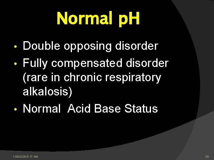
Normal p. H Double opposing disorder • Fully compensated disorder (rare in chronic respiratory alkalosis) • Normal Acid Base Status • 11/6/2020 6: 17 AM 29
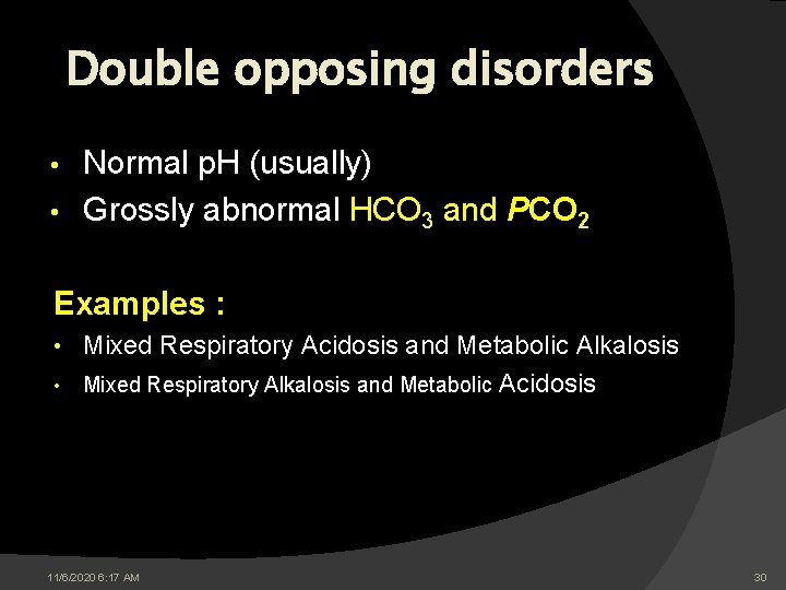
Double opposing disorders Normal p. H (usually) • Grossly abnormal HCO 3 and PCO 2 • Examples : • • Mixed Respiratory Acidosis and Metabolic Alkalosis Mixed Respiratory Alkalosis and Metabolic Acidosis 11/6/2020 6: 17 AM 30
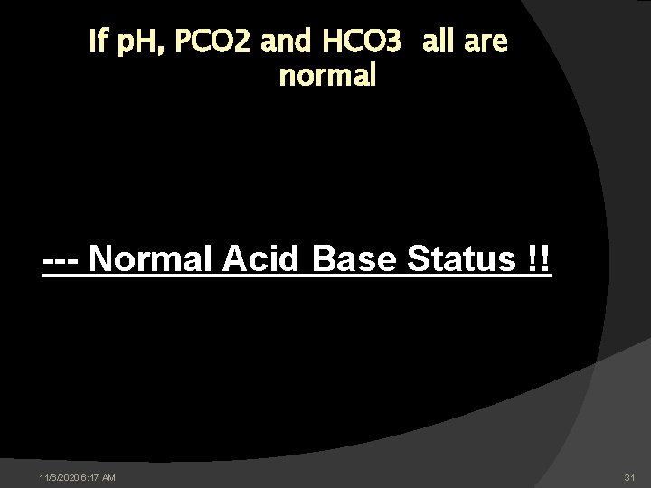
If p. H, PCO 2 and HCO 3 all are normal --- Normal Acid Base Status !! 11/6/2020 6: 17 AM 31
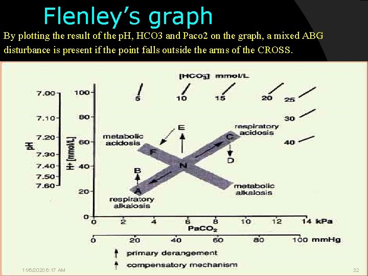
Flenley’s graph By plotting the result of the p. H, HCO 3 and Paco 2 on the graph, a mixed ABG disturbance is present if the point falls outside the arms of the CROSS. 11/6/2020 6: 17 AM 32
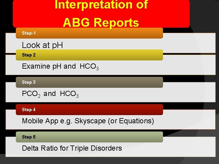
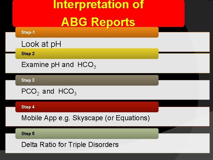
Interpretation of ABG Reports Step-1 Look at p. H Step 2 Examine p. H and HCO 3 Step 3 PCO 2 and HCO 3 Step 4 Mobile App e. g. Skyscape (or Equations) Step 5 Delta Ratio for Triple Disorders
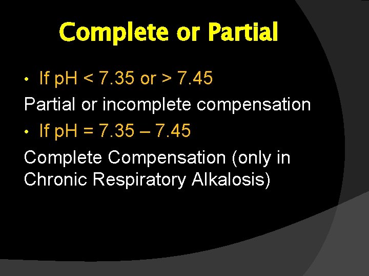
Complete or Partial If p. H < 7. 35 or > 7. 45 Partial or incomplete compensation • If p. H = 7. 35 – 7. 45 Complete Compensation (only in Chronic Respiratory Alkalosis) •
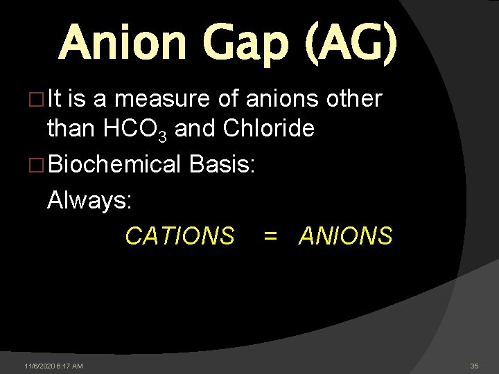
Anion Gap (AG) � It is a measure of anions other than HCO 3 and Chloride � Biochemical Basis: Always: CATIONS = ANIONS 11/6/2020 6: 17 AM 35
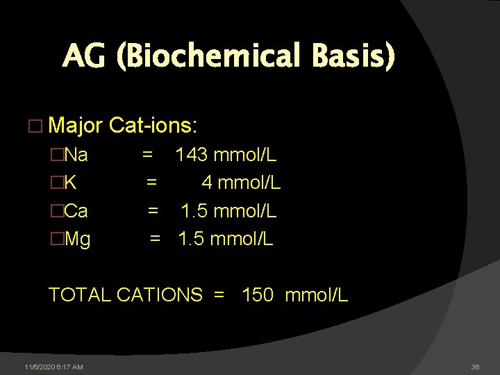
AG (Biochemical Basis) � Major Cat-ions: �Na = 143 mmol/L �K = 4 mmol/L �Ca = 1. 5 mmol/L �Mg = 1. 5 mmol/L TOTAL CATIONS = 150 mmol/L 11/6/2020 6: 17 AM 36
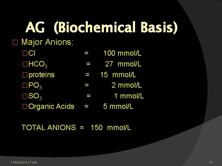
AG (Biochemical Basis) � Major Anions: �Cl = 100 mmol/L �HCO 3 = 27 mmol/L �proteins = 15 mmol/L �PO 3 = 2 mmol/L �SO 3 = 1 mmol/L �Organic Acids = 5 mmol/L TOTAL ANIONS = 150 mmol/L 11/6/2020 6: 17 AM 37
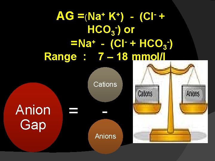
AG =(Na+ K+) - (Cl- + HCO 3 -) or =Na+ - (Cl- + HCO 3 -) Range : 7 – 18 mmol/l Cations Anion Gap = Anions
Anion Gap: �It is only a lab derived index �There can Never be an anion gap in any condition because electro-neutrality is always maintained in the plasma 11/6/2020 6: 17 AM 39
AG corrected for low albumin (AG corr) Albumin accounts for about 90% of the mean value AG and albumin is reversely correlated with HCO 3 • When the level of serum albumin decreases, AG will decrease equally • AG is concealed by hypoalbuminemia • HCO 3 will increase accordingly (low Alb alkalosis) •
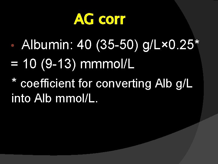
AG corr Albumin: 40 (35 -50) g/L× 0. 25* = 10 (9 -13) mmmol/L * coefficient for converting Alb g/L • into Alb mmol/L.
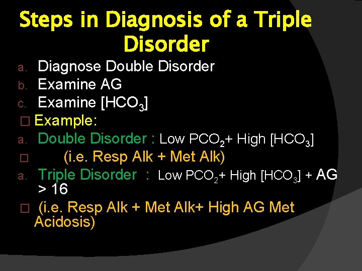
Steps in Diagnosis of a Triple Disorder a. Diagnose Double Disorder b. Examine AG c. Examine [HCO 3] � Example: a. Double Disorder : Low PCO 2+ High [HCO 3] (i. e. Resp Alk + Met Alk) a. Triple Disorder : Low PCO 2+ High [HCO 3] + AG � > 16 � (i. e. Resp Alk + Met Alk+ High AG Met Acidosis)
Diagnosis Triple Disorder Respiratory Alkalosis Metabolic Alkalosis High AG Metabolic Acidosis
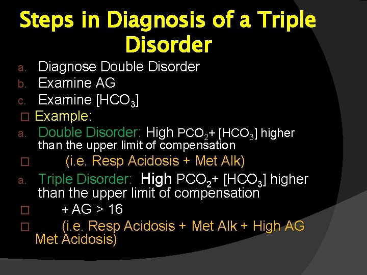
Steps in Diagnosis of a Triple Disorder a. b. c. � a. Diagnose Double Disorder Examine AG Examine [HCO 3] Example: Double Disorder: High PCO 2+ [HCO 3] higher than the upper limit of compensation (i. e. Resp Acidosis + Met Alk) a. Triple Disorder: High PCO 2+ [HCO 3] higher than the upper limit of compensation � + AG > 16 � (i. e. Resp Acidosis + Met Alk + High AG Met Acidosis) �
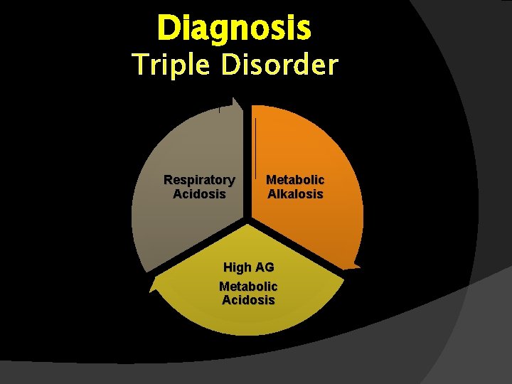
Diagnosis Triple Disorder Respiratory Acidosis Metabolic Alkalosis High AG Metabolic Acidosis
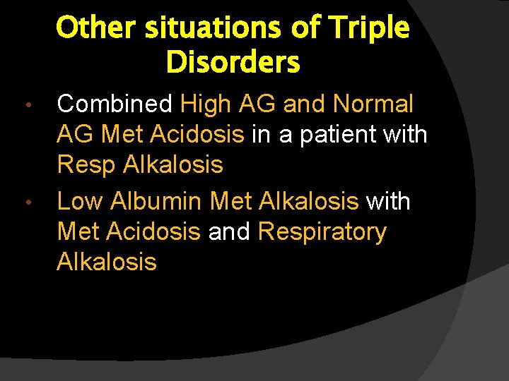
Other situations of Triple Disorders Combined High AG and Normal AG Met Acidosis in a patient with Resp Alkalosis • Low Albumin Met Alkalosis with Met Acidosis and Respiratory Alkalosis •
Important Fact �Respiratory Acidosis and Respiratory Alkalosis never occur together in the same patient !!!
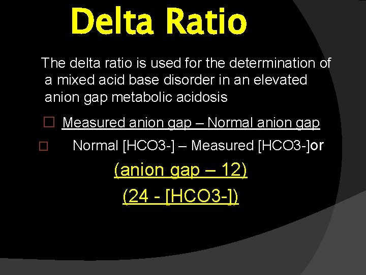
Delta Ratio The delta ratio is used for the determination of a mixed acid base disorder in an elevated anion gap metabolic acidosis � Measured anion gap – Normal anion gap � Normal [HCO 3 -] – Measured [HCO 3 -]or (anion gap – 12) (24 - [HCO 3 -])
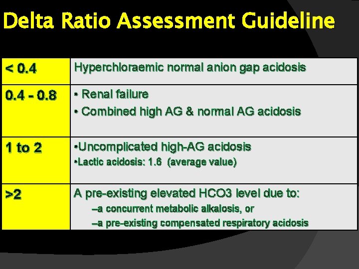
Delta Ratio Assessment Guideline < 0. 4 Hyperchloraemic normal anion gap acidosis 0. 4 - 0. 8 • Renal failure • Combined high AG & normal AG acidosis 1 to 2 • Uncomplicated high-AG acidosis • Lactic acidosis: 1. 6 (average value) >2 A pre-existing elevated HCO 3 level due to: –a concurrent metabolic alkalosis, or –a pre-existing compensated respiratory acidosis
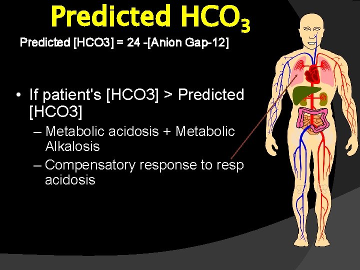
Predicted HCO 3 Predicted [HCO 3] = 24 -[Anion Gap-12] • If patient's [HCO 3] > Predicted [HCO 3] – Metabolic acidosis + Metabolic Alkalosis – Compensatory response to resp acidosis
If patient's [HCO 3] =Predicted [HCO 3] – Simple Metabolic acidosis • If patient's [HCO 3] < Predicted [HCO 3] – Compensatory response to resp alkalosis
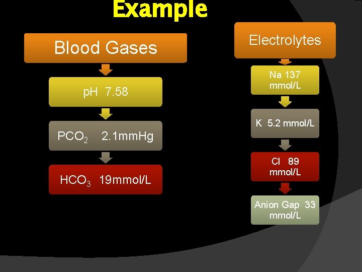
Example Blood Gases p. H 7. 58 Electrolytes Na 137 mmol/L K 5. 2 mmol/L PCO 2 2. 1 mm. Hg HCO 3 19 mmol/L Cl 89 mmol/L Anion Gap 33 mmol/L
Interpretation of ABG Reports Step-1 Look at p. H Step 2 Examine p. H and HCO 3 Step 3 PCO 2 and HCO 3 Step 4 Mobile App e. g. Skyscape (or Equations) Step 5 Delta Ratio for Triple Disorders
THANK YOU