
PAIN PATHOPHYSIOLOGY MANAGEMENT DR MANSOOR AQIL B Sc
PAIN PATHOPHYSIOLOGY & MANAGEMENT DR. MANSOOR AQIL B. Sc. , M. B. B. S. , F. C. P. S ASSOCIATE PROFESSOR, KING SAUD UNIVERSITY & CONSULTANT KING KHALID UNIVERSITY HOSPITAL, RIYADH.




PAIN l Word pain is derived from Latin word “Poena”, meaning penalty, suffering or punishment
PAIN l An unpleasant sensory and emotional experience associated with actual or potential tissue damage or described in terms of such damage. (International association of study of pain)
CLASSIFICATION OF PAIN
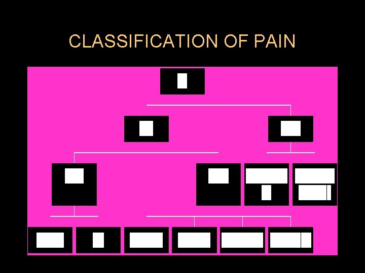
TYPES OF PAIN According to duration l Acute l Chronic
TYPES OF PAIN According to Etiology l Postoperative OR l Cancer pain
TYPES OF PAIN According to Type of the organ affected l Toothache l Earache l Headache l Low backache
TYPES OF PAIN (According to Pathophysiology) l Nociceptive; Due to activation or sensitization of peripheral nociceptors. l Neuropathic: Due to injury or acquired abnormalities of peripheral or CNS.
ACUTE PAIN l Caused by noxious stimulation due to injury, a disease process or abnormal function of muscle or viscera It is nearly always nociceptive l Nociceptive pain serves to detect, localize and limit the tissue damage. l
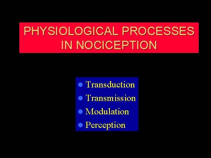
PHYSIOLOGICAL PROCESSES IN NOCICEPTION l Transduction l Transmission l Modulation l Perception
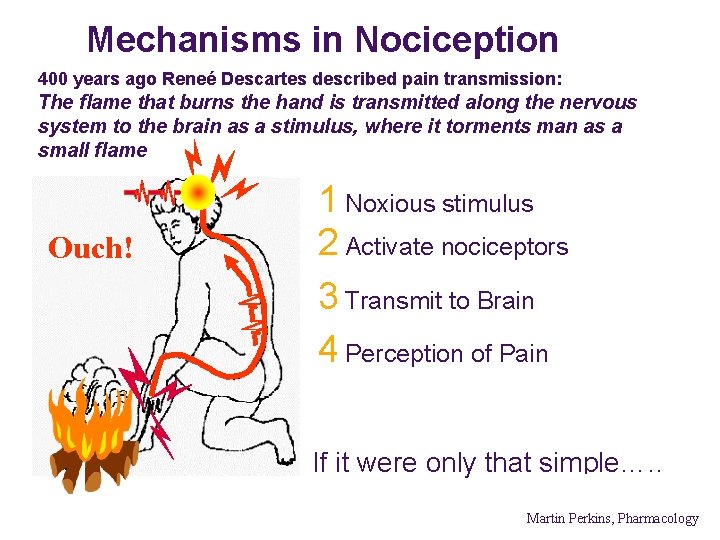
Mechanisms in Nociception 400 years ago Reneé Descartes described pain transmission: The flame that burns the hand is transmitted along the nervous system to the brain as a stimulus, where it torments man as a small flame Ouch! 1 Noxious stimulus 2 Activate nociceptors 3 Transmit to Brain 4 Perception of Pain If it were only that simple…. . Martin Perkins, Pharmacology
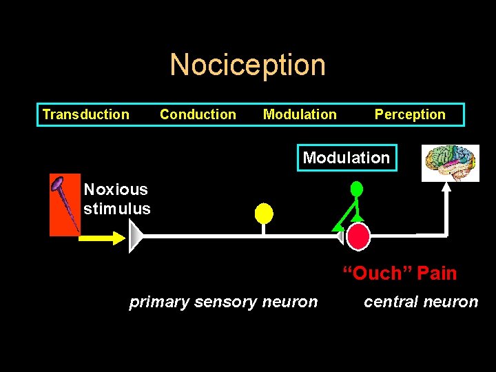
Nociception Transduction Conduction Modulation Perception Modulation Noxious stimulus “Ouch” Pain primary sensory neuron central neuron
TYPES OF ACUTE PAIN l Somatic OR l Visceral
SOMATIC PAIN l Superficial OR l Deep
SUBTYPES OF VISCERAL PAIN l Localized visceral pain l Referred Visceral pain l True Localized parietal pain l Referred parietal pain
TRUE VISCERAL PAIN l Dull, diffuse and in Midline l Frequently associated with sympathetic or parasympathetic activity – – Nausea Vomiting Sweating Changes in HR and BP.
PARIETAL PAIN l Sharp (stabbing sensation) either localized or referred to a distant site. l Phenomenon of visceral pain or parietal pain referred to cutaneous area results from embryologic development and migration.
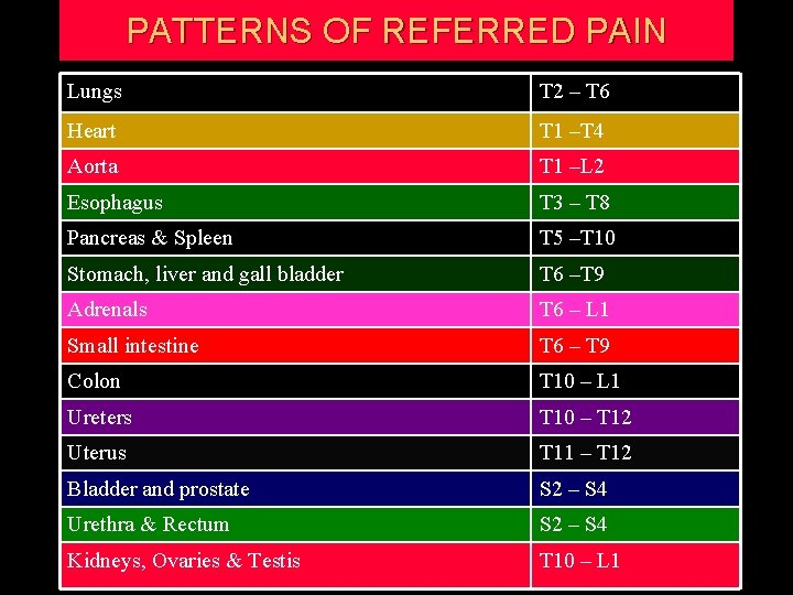
PATTERNS OF REFERRED PAIN Lungs T 2 – T 6 Heart T 1 –T 4 Aorta T 1 –L 2 Esophagus T 3 – T 8 Pancreas & Spleen T 5 –T 10 Stomach, liver and gall bladder T 6 –T 9 Adrenals T 6 – L 1 Small intestine T 6 – T 9 Colon T 10 – L 1 Ureters T 10 – T 12 Uterus T 11 – T 12 Bladder and prostate S 2 – S 4 Urethra & Rectum S 2 – S 4 Kidneys, Ovaries & Testis T 10 – L 1
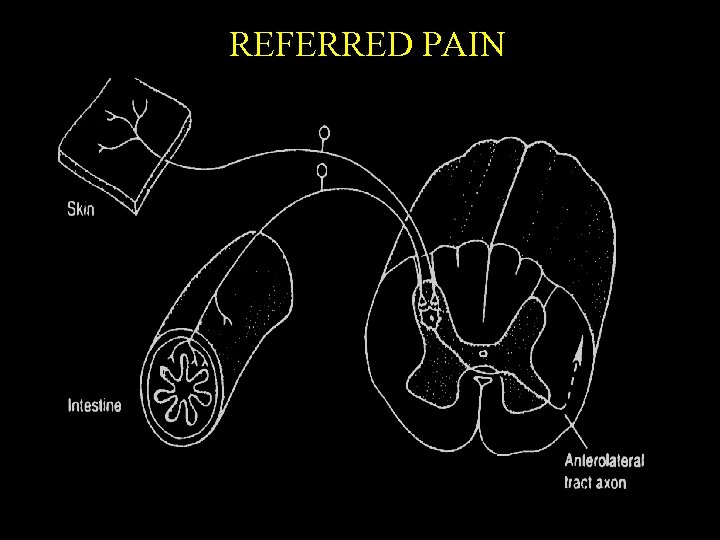
REFERRED PAIN

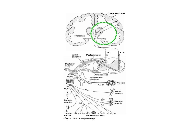
PAIN PATHWAY
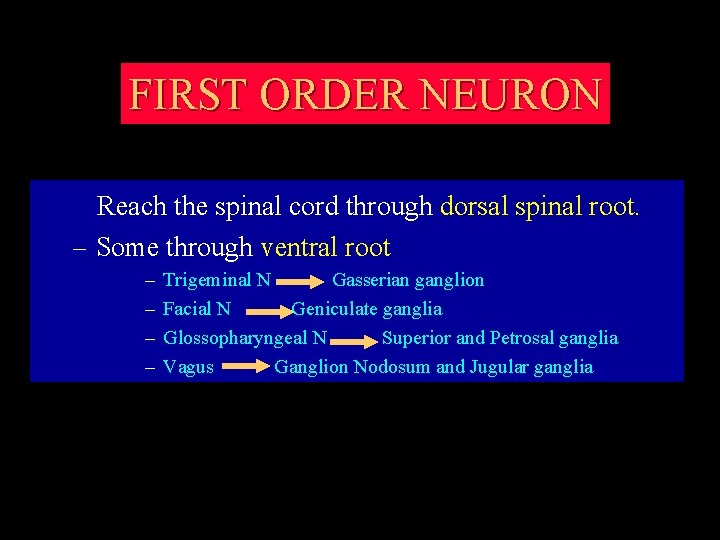
FIRST ORDER NEURON Reach the spinal cord through dorsal spinal root. – Some through ventral root – – Trigeminal N Gasserian ganglion Facial N Geniculate ganglia Glossopharyngeal N Superior and Petrosal ganglia Vagus Ganglion Nodosum and Jugular ganglia
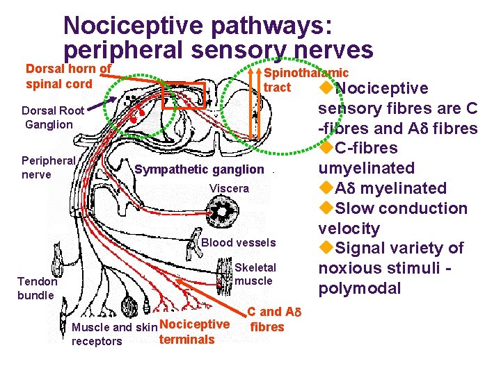
Nociceptive pathways: peripheral sensory nerves Dorsal horn of spinal cord Spinothalamic tract u. Nociceptive Dorsal Root Ganglion Peripheral nerve Sympathetic ganglion Viscera Blood vessels Skeletal muscle Tendon bundle Muscle and skin Nociceptive terminals receptors C and Ad fibres sensory fibres are C -fibres and Ad fibres u. C-fibres umyelinated u. Ad myelinated u. Slow conduction velocity u. Signal variety of noxious stimuli polymodal
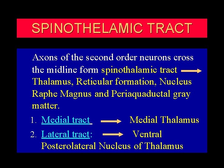
SPINOTHELAMIC TRACT Axons of the second order neurons cross the midline form spinothalamic tract Thalamus, Reticular formation, Nucleus Raphe Magnus and Periaquaductal gray matter. 1. Medial tract Medial Thalamus 2. Lateral tract: Ventral Posterolateral Nucleus of Thalamus
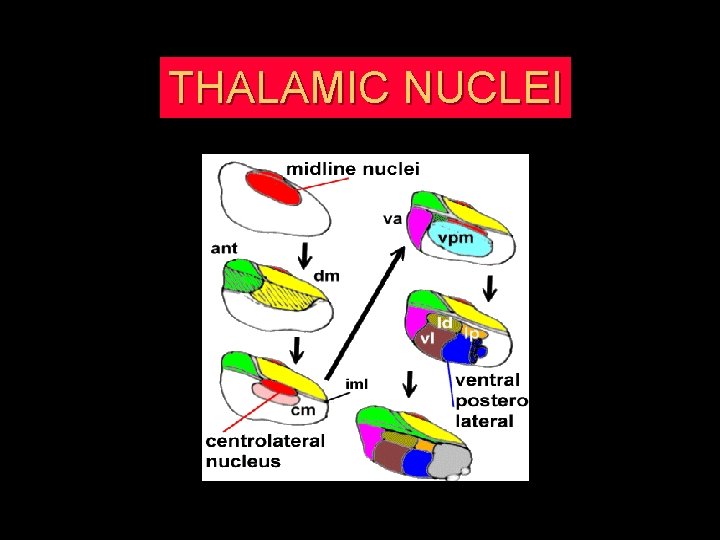
THALAMIC NUCLEI
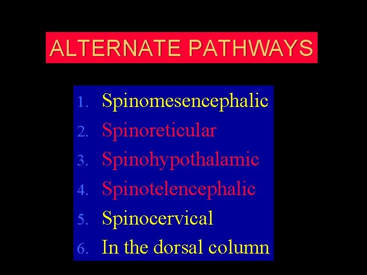
ALTERNATE PATHWAYS 1. 2. 3. 4. 5. 6. Spinomesencephalic Spinoreticular Spinohypothalamic Spinotelencephalic Spinocervical In the dorsal column
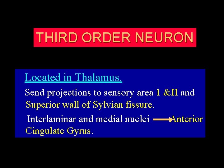
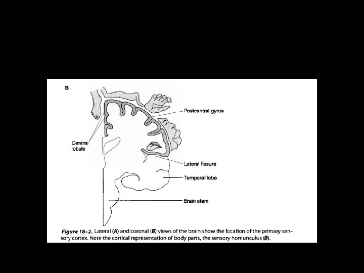
THIRD ORDER NEURON Located in Thalamus. Send projections to sensory area 1 &II and Superior wall of Sylvian fissure. Interlaminar and medial nuclei Anterior Cingulate Gyrus.
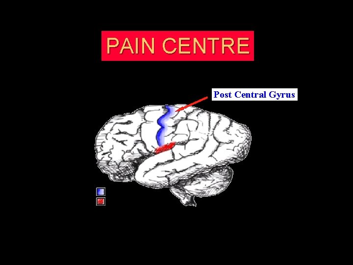
PAIN CENTRE Post Central Gyrus
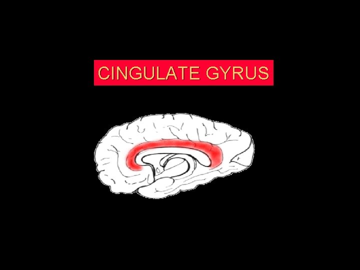
CINGULATE GYRUS


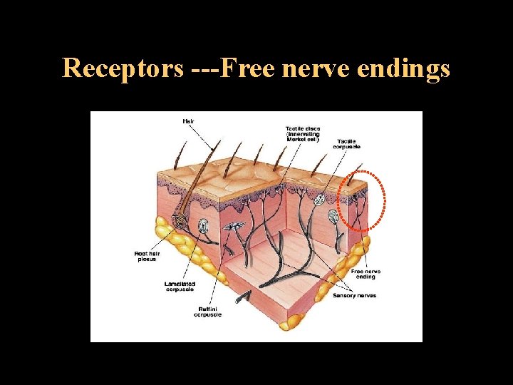
Receptors ---Free nerve endings
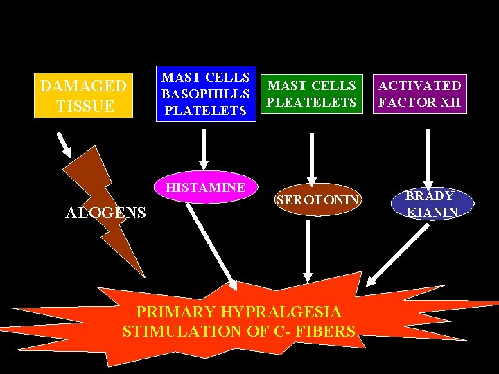
Receptors ---Free nerve endings l Mechanoreceptors: Pinch and Pinprick l Silent Nociceptors: Inflammation l Polymodal mechanoheat nociceptors: Excessive pressure, Extremes of Temperature and Alogens like Bradykinin, Serotonin, Histamine, H, K, Prostaglandins and ATP.
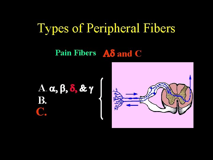
Types of Peripheral Fibers Pain Fibers Ad and C A. a, , d, & g B. C.
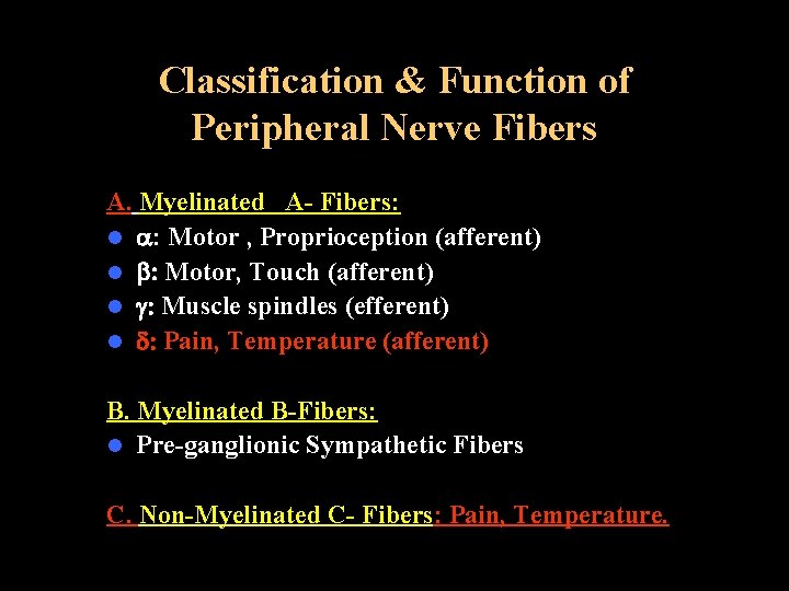
Classification & Function of Peripheral Nerve Fibers A. Myelinated A- Fibers: l a: Motor , Proprioception (afferent) l : Motor, Touch (afferent) l g: Muscle spindles (efferent) l d: Pain, Temperature (afferent) B. Myelinated B-Fibers: l Pre-ganglionic Sympathetic Fibers C. Non-Myelinated C- Fibers: Pain, Temperature.
PHYSIOLOGY OF NOCICEPTION l Fast pain (First pain) l Slow pain (Second pain)
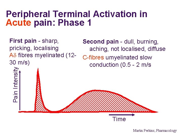
Peripheral Terminal Activation in Acute pain: Phase 1 Pain Intensity First pain - sharp, pricking, localising Ad fibres myelinated (1230 m/s) Second pain - dull, burning, aching, not localised, diffuse C-fibres umyelinated slow conduction (0. 5 - 2 m/s Time Martin Perkins, Pharmacology
MODULATION Martin Perkins, Pharmacology
MODULATION OF PAIN l Peripheral Modulation l Central modulation
PERIPHRAL MODULATION l Primary Hyperalgesia l Secondary Hyperalgesia
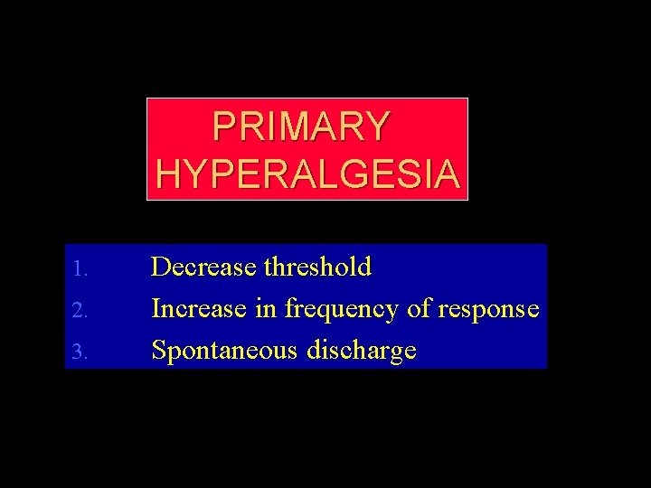
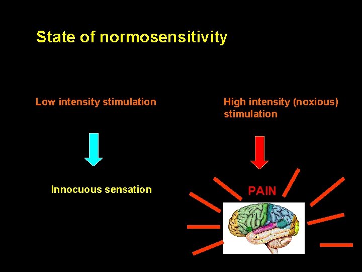
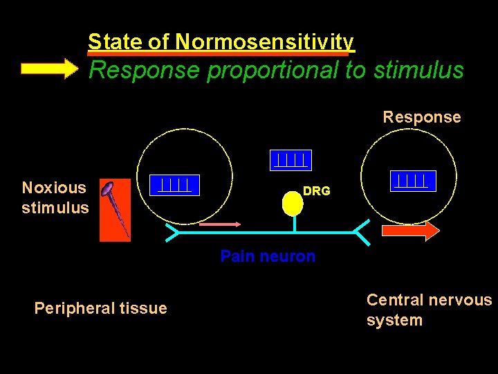
PRIMARY HYPERALGESIA Decrease threshold 2. Increase in frequency of response 3. Spontaneous discharge 1.
State of normosensitivity Low intensity stimulation Innocuous sensation High intensity (noxious) stimulation PAIN
State of Normosensitivity Response proportional to stimulus Response Noxious stimulus DRG Pain neuron Peripheral tissue Central nervous system
DAMAGED TISSUE MAST CELLS BASOPHILLS PLATELETS HISTAMINE ALOGENS MAST CELLS PLEATELETS ACTIVATED FACTOR XII SEROTONIN BRADYKIANIN PRIMARY HYPRALGESIA STIMULATION OF C- FIBERS
SENSITIZARION (STIMULATION) RECEPTOR PC G-RPOTEIN PLC ARACHADONIC ACID COX THROMBOXANE PIP 2 DAG IP 3 PKC LIPOXYGENASE PROSTAGLANDINS LEUKOTRINES RELEASE INTRACELLULAR CALCIUM
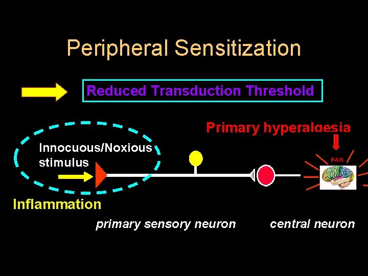
Peripheral Sensitization Reduced Transduction Threshold Primary hyperalgesia Innocuous/Noxious stimulus Inflammation primary sensory neuron central neuron
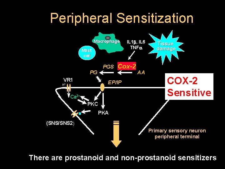
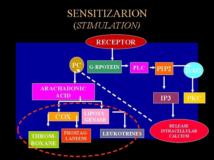
Peripheral Sensitization Macrophage Mast cell PG VR 1 PGS Cox-2 EP/IP H+ Ca 2+ IL 1 , IL 6 TNFa AA Tissue damage COX-2 Sensitive PKC PKA (SNS/SNS 2) Primary sensory neuron peripheral terminal There are prostanoid and non-prostanoid sensitizers
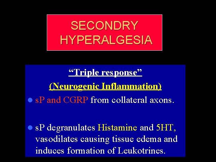
SECONDRY HYPERALGESIA “Triple response” (Neurogenic Inflammation) l s. P and CGRP from collateral axons. l s. P degranulates Histamine and 5 HT, vasodilates causing tissue edema and induces formation of Leukotrines.
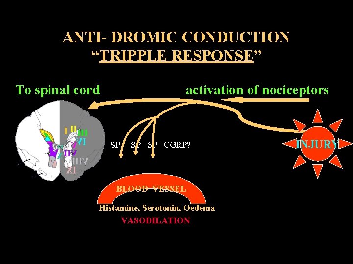
ANTI- DROMIC CONDUCTION “TRIPPLE RESPONSE” To spinal cord activation of nociceptors SP SP CGRP? BLOOD VESSEL Histamine, Serotonin, Oedema VASODILATION INJURY
CENTRAL MODULATION l STIMULATION l INHIBITION
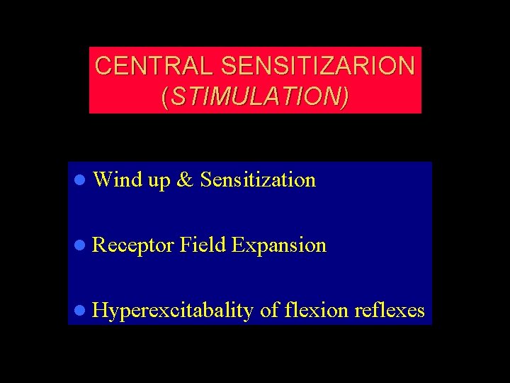
CENTRAL SENSITIZARION (STIMULATION) l Wind up & Sensitization l Receptor Field Expansion l Hyperexcitabality of flexion reflexes
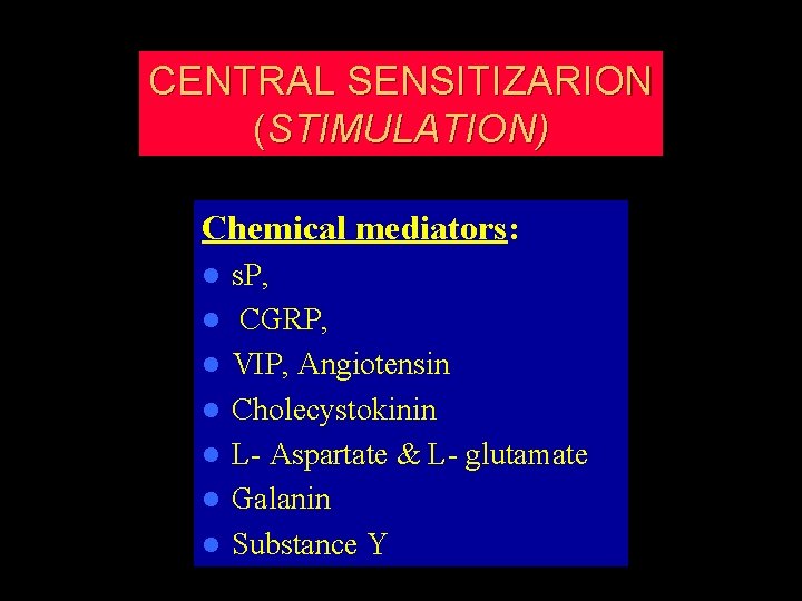
CENTRAL SENSITIZARION (STIMULATION) Chemical mediators: l l l l s. P, CGRP, VIP, Angiotensin Cholecystokinin L- Aspartate & L- glutamate Galanin Substance Y
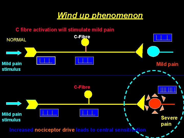
Wind up phenomenon C fibre activation will stimulate mild pain NORMAL C-Fibre Mild pain stimulus Mild pain C-Fibre Mild pain stimulus Increased nociceptor drive leads to central sensitisation Severe pain
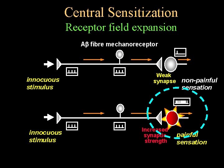
Central Sensitization Receptor field expansion A fibre mechanoreceptor innocuous stimulus Weak synapse Increased synaptic strength non-painful sensation
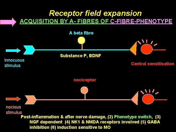
Receptor field expansion ACQUISITION BY A- FIBRES OF C-FIBRE-PHENOTYPE A beta fibre innocuous stimulus Substance P, BDNF Central sensitisation nociceptor noxious stimulus Post-inflammation & after nerve damage, (2) Phenotype switch, (3) NGF dependent (4) NK 1 & NMDA receptors involved (5) GABA inhibition (6) induction sensitive to MO
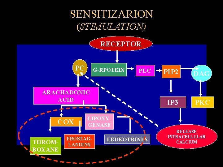
SENSITIZARION (STIMULATION) RECEPTOR PC G-RPOTEIN PLC ARACHADONIC ACID COX THROMBOXANE PIP 2 DAG IP 3 PKC LIPOXYGENASE PROSTAGLANDINS LEUKOTRINES RELEASE INTRACELLULAR CALCIUM
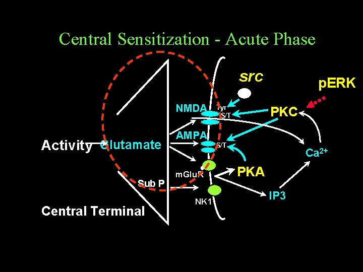
Central Sensitization - Acute Phase src NMDA Activity Glutamate Sub P Central Terminal AMPA m. Glu. R NK 1 Tyr S/T p. ERK PKC S/T Ca 2+ PKA IP 3
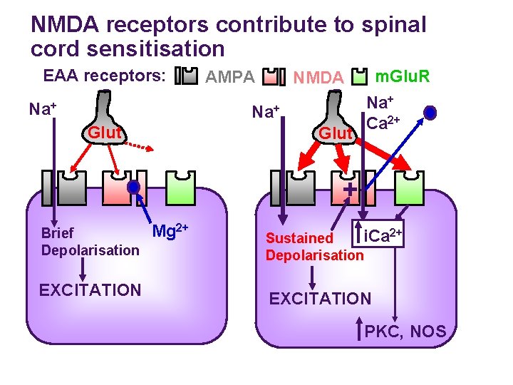
NMDA receptors contribute to spinal cord sensitisation EAA receptors: Na+ AMPA Na+ Glut m. Glu. R NMDA Glut Na+ Ca 2+ + Brief Depolarisation EXCITATION Mg 2+ i. Ca 2+ Sustained Depolarisation EXCITATION PKC, NOS
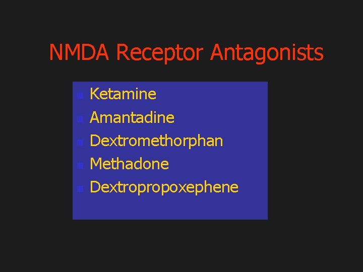
NMDA Receptor Antagonists n n n Ketamine Amantadine Dextromethorphan Methadone Dextropropoxephene
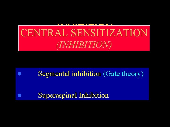
INHIBITION CENTRAL SENSITIZATION (INHIBITION) Segmental inhibition (Gate theory) l Superaspinal Inhibition l
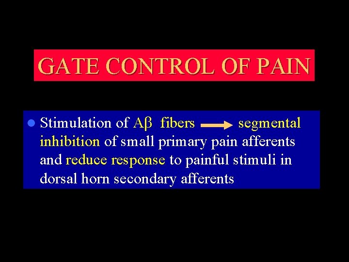
GATE CONTROL OF PAIN l Stimulation of Ab fibers segmental inhibition of small primary pain afferents and reduce response to painful stimuli in dorsal horn secondary afferents
MECHANISM OF ACTION Exact mechanisms of actions are as yet unknown and certainly unproven l 1965 - Melzack and Wall proposed the Gate Theory of Pain – Nociceptive A delta/C fibres project in SC to second-order projection neuron but also send fibres to inhibit an inhibitory interneuron – Large myelinated A alpha neurons in DC send collaterals to activate these same interneurons thereby inhibiting (closing the gate) the pain sensory action potentials l
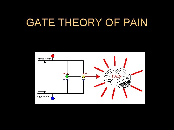
GATE THEORY OF PAIN
l Glycine and GABA are inhibitory transmitters. GABA A and GABAB l Muscimol and Beclofen. l GABAB increases K conductance l GABAA increases Cl conductance l Glycine also increases Cl conductance l Strychnine and Tetanus toxide are Glycine receptor antagonists l Glycine is facilitatory on NMDA receptors l l Adenosine has two types of receptors A 1 and A 2 A 1 inhibits adenyl cyclase and A 2 stimulates adenylcyclase. l A 1 mediate antinociceptive action. l
TRANSCUTANEOUS NERVE STIMULATION (TENS) l Asymmetric biphasic waveform of 12 -20 m. A at 50 -100 Hz via 1000 ohms resistance has proved successful for post operative analgesia
TRANSCUTANEOUS ELECTRICAL NERVE STIMULATION (Tens) l Effective l Over all analgesic effect is modest l Absolutely safe for the fetus l Usually causes electrical interference with fetal heart rate when used concurrently with internal fetal scalp electrode
TENS Advantages l Noninvasive l Patient controlled l No side effects l Non-addictive l Decreased analgesic needs
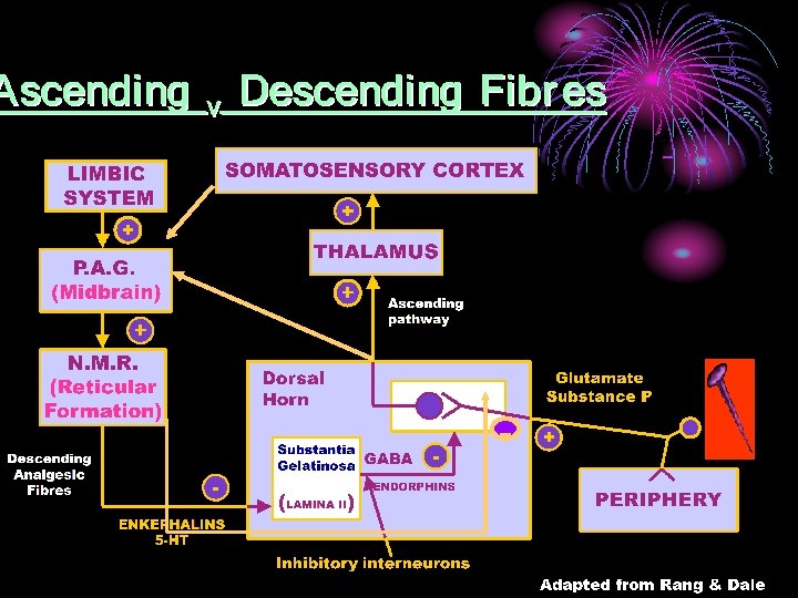
SUPRASPINAL INHIBITION Originate from Cerebral cortex Thalamus Reticular formation of brain stem (Ventro-median Medulla VMM). l Neurotransmitter is Serotonin. l NMR & Locus Ceruleu l l Nor Adrenaline containing fibers from
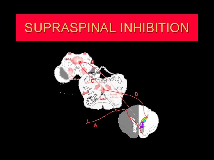
SUPRASPINAL INHIBITION
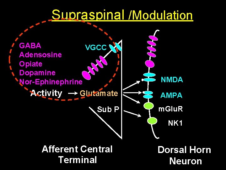
Supraspinal /Modulation GABA VGCC Adensosine Opiate Dopamine Nor-Ephinephrine Activity NMDA Glutamate AMPA Sub P m. Glu. R NK 1 Afferent Central Terminal Dorsal Horn Neuron
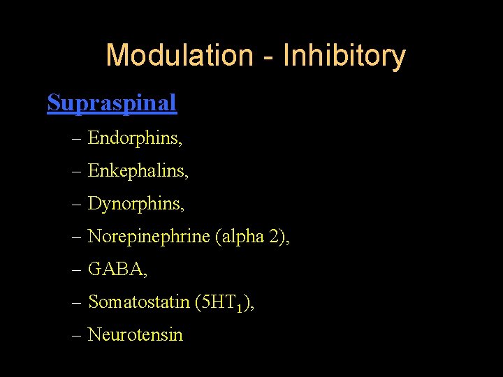
Modulation - Inhibitory Supraspinal – Endorphins, – Enkephalins, – Dynorphins, – Norepinephrine (alpha 2), – GABA, – Somatostatin (5 HT 1), – Neurotensin



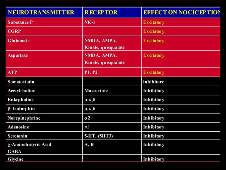
PAIN NEUROTRANSMITTERS
PRE EMPTIVE ANALGESIA
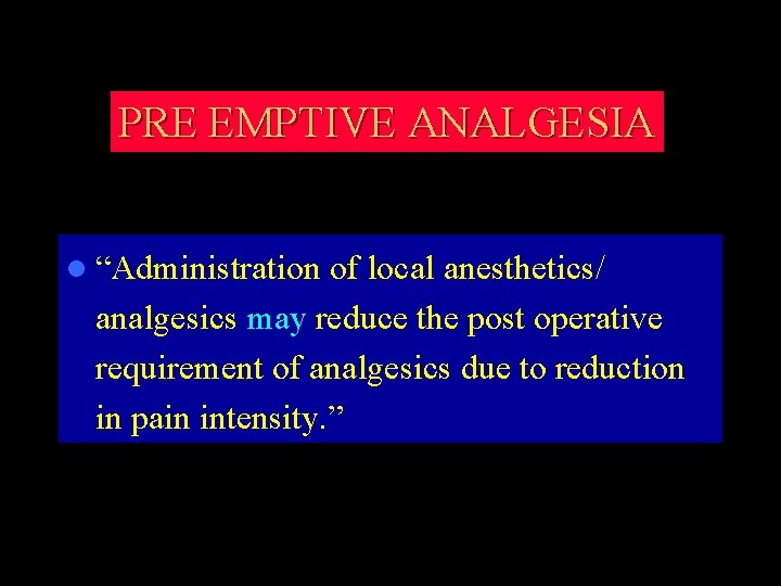
PRE EMPTIVE ANALGESIA l “Administration of local anesthetics/ analgesics may reduce the post operative requirement of analgesics due to reduction in pain intensity. ”
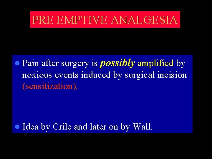
PRE EMPTIVE ANALGESIA l Pain after surgery is possibly amplified by noxious events induced by surgical incision (sensitization). l Idea by Crile and later on by Wall.
PRE EMPTIVE ANALGESIA Promising results from experimental studies • • Prospective studies in humans show conflicting results • No ultimate understanding of the nature of pre-emptive measures needed
SYSTEMIC RESPONCES TO ACUTE PAIN
Cardiovascular effects · Tachycardia · Hypertension · Increased SVR
RESPIRATORY SYSTEM l O 2 demand consumption l M. V l Splinting and Guarding and decreased chest excursion l Atelactasis, increased shunting, hypoxemia l V. C, retention of secretions and chest infection
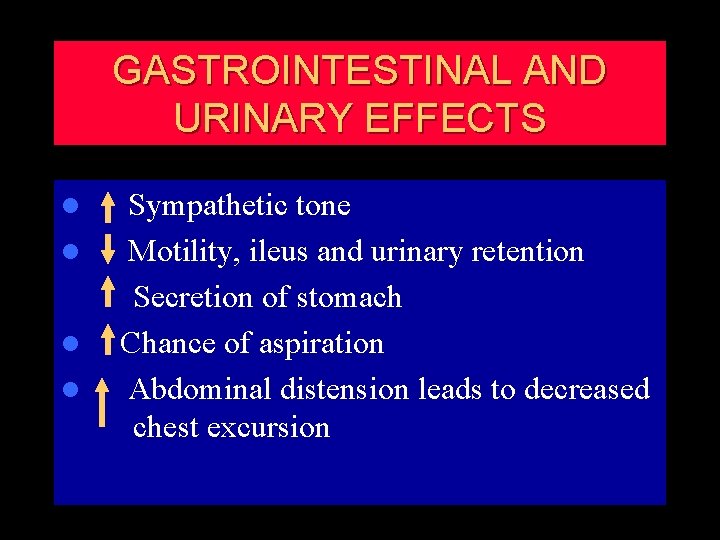
GASTROINTESTINAL AND URINARY EFFECTS l Sympathetic tone l Motility, ileus and urinary retention Secretion of stomach l Chance of aspiration l Abdominal distension leads to decreased chest excursion
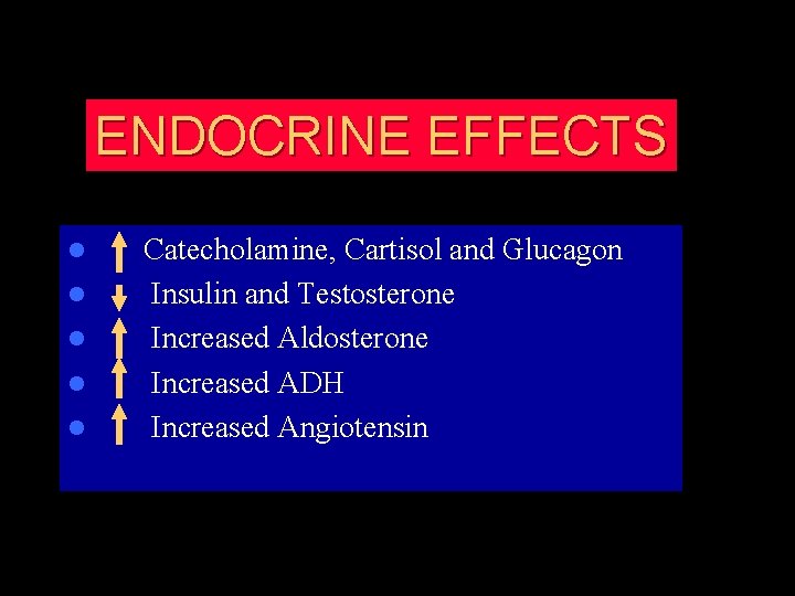
ENDOCRINE EFFECTS l l l Catecholamine, Cartisol and Glucagon Insulin and Testosterone Increased Aldosterone Increased ADH Increased Angiotensin
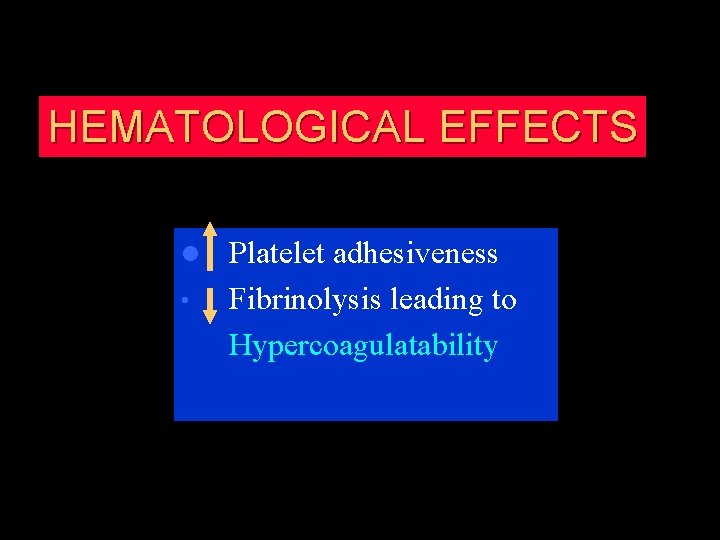
HEMATOLOGICAL EFFECTS Platelet adhesiveness • Fibrinolysis leading to Hypercoagulatability l
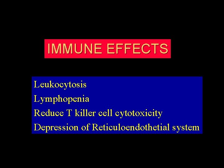
IMMUNE EFFECTS Leukocytosis Lymphopenia Reduce T killer cell cytotoxicity Depression of Reticuloendothetial system
GENERAL SENSE OF WELL-BEING l Anxiety l Sleep disturbances l Depression



PAl. N MEASUREMENT l Descriptive scales such as – Mild. – Moderate. – Severe l. Generally unsatisfactory.
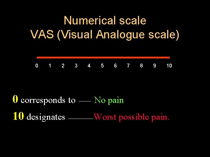
Numerical scale VAS (Visual Analogue scale) 0 1 2 3 4 5 6 7 8 9 10 0 corresponds to No pain 10 designates Worst possible pain.
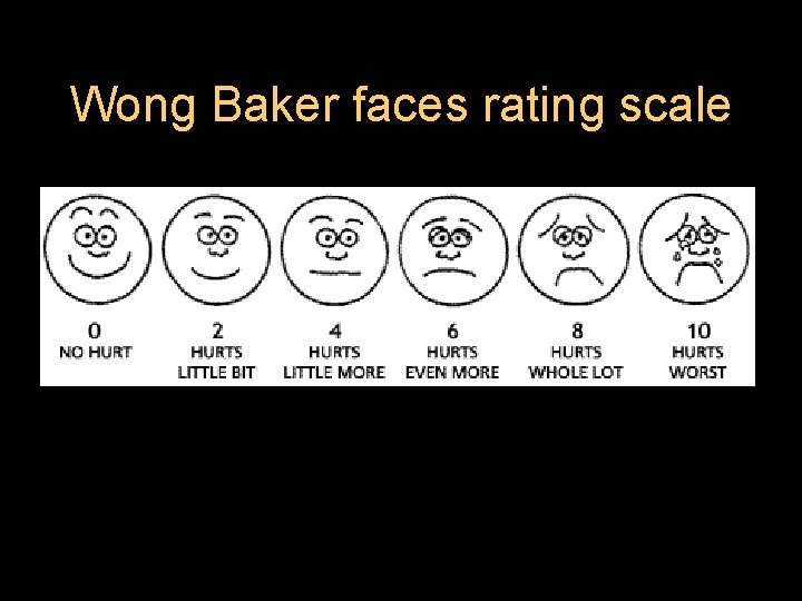
Wong Baker faces rating scale
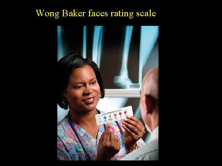
Wong Baker faces rating scale
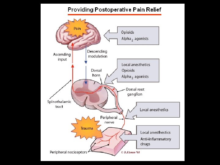
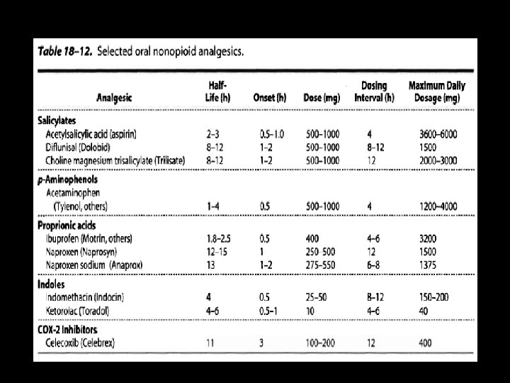
POSTOPERATIVE PAl. N OUTPATIENTS 1 : Oral Analgesics Cyclooxygenase Inhibitors l Opioids l 2. Infiltration of Local Anesthetic
POSTOPERATIVE PAl. N INPATIENTS 1. Opioids l Subcutaneous & Intramuscular Injections l Patient-Controlled Analgesia 2. Peripheral Nerve Blocks

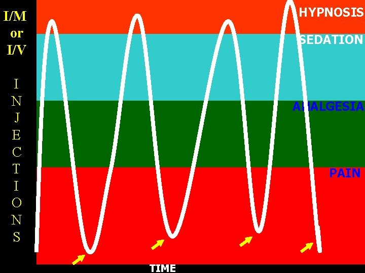
HYPNOSIS I/M or I/V SEDATION I N J E C T I O N S ANALGESIA PAIN TIME
POSTOPERATIVE PAl. N INPATIENTS 3. Central Neuraxial Blockade & Intraspinal drugs l Local Anesthetics l Opioids l Local Anesthetic & Opioid Mixtures
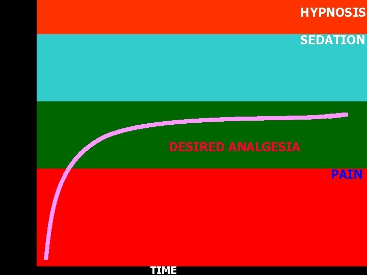
HYPNOSIS SEDATION DESIRED ANALGESIA PAIN TIME
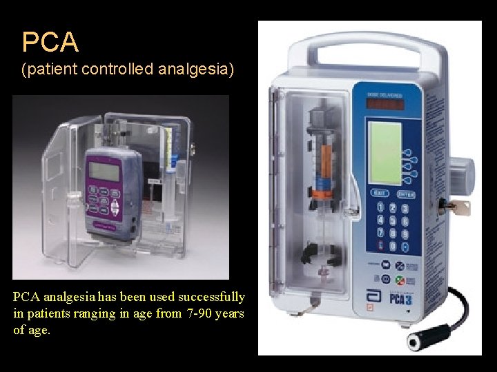
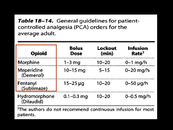
PCA (patient controlled analgesia) PCA analgesia has been used successfully in patients ranging in age from 7 -90 years of age.
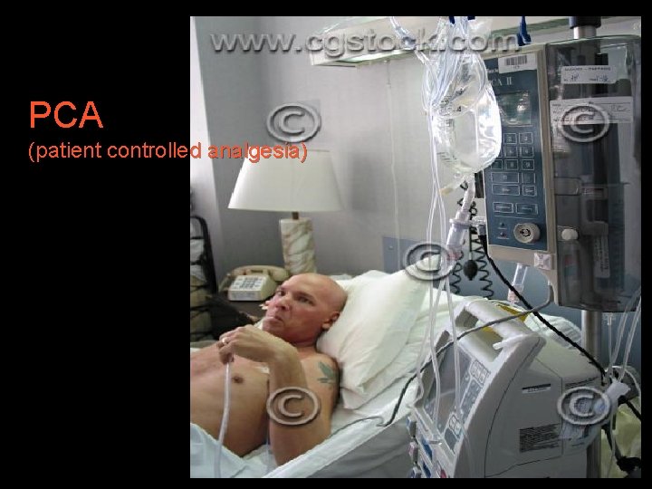
PCA (patient controlled analgesia)
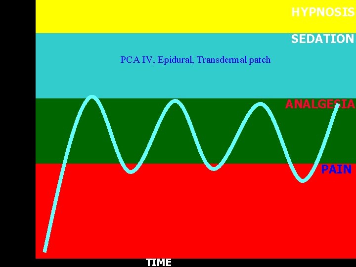
HYPNOSIS SEDATION PCA IV, Epidural, Transdermal patch ANALGESIA PAIN TIME
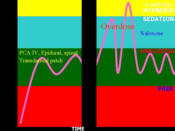
Overdose PCA IV, Epidural, spinal Transdermal patch Anesthesia HYPNOSIS SEDATION Naloxone ANALGESIA PAIN TIME
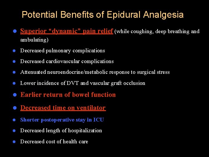
Potential Benefits of Epidural Analgesia l Superior “dynamic” pain relief (while coughing, deep breathing and ambulating) l Decreased pulmonary complications l Decreased cardiovascular complications l Attenuated neuroendocrine/metabolic response to surgical stress l Lower incidence of DVT and vascular graft occlusion l Earlier return of bowel function l Decreased time on ventilator l Shorter postoperative stay in ICU l Decreased length of hospitalization l Decreased cost of health care



Chronic pain
PATHOPHYSIOLOGY OF CHRONIC PAIN l Chronic pain may be caused by a combination of 1. Peripheral, 2. Central, 3. Or psychological mechanisms. l Sensitization of nociceptors plays a major role
EVALUATING THE PATIENT WITH PAl. N Why?
EVALUATING THE PATIENT WITH PAl. N l Acute pain is primarily therapeutic l Chronic pain additionally involves investigative measures.
EVALUATING THE PATIENT WITH PAl. N A written questionnaire – Nature of the pain, – Onset – Duration, – Previous medication and treatments. Diagrams can be useful in defining patterns of radiation.
Investigations l Plain radiographs, l Computed tomography (CT), l Magnetic resonance imaging (MRI), l Bone scans. l MRI is particularly useful for soft tissue analysis and can show nerve compression.
l ELECTROMYOGRAPHY & NERVE CONDUCTION STUDIES l For confirming the diagnosis of entrapment syndromes, radicular syndromes, neural trauma, and polyneuropathies l Can often distinguish between neurogenic and myogenic disorders
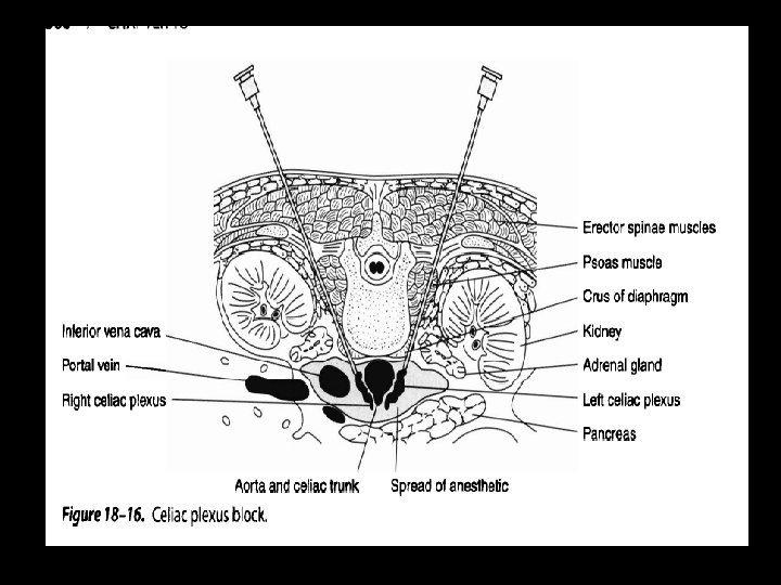
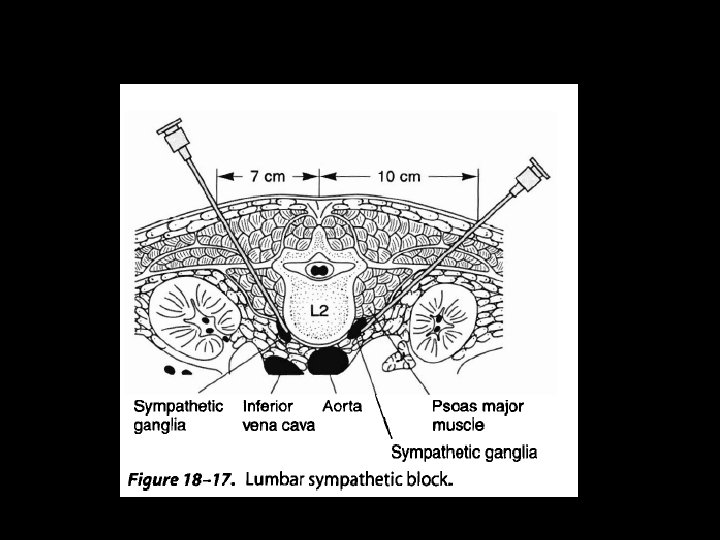
DIAGNOSTIC & THERAPEUTIC NEURAL BLOCKADE l Can be useful in delineating pain mechanisms, but, more importantly, it plays a major role in the management of patients with acute or chronic pain. l SOMATIC BLOCKS – Trigeminal Nerve Blocks – Facial Nerve Block – Glossopharyngeal Block – Cervical Paravertebral Nerve Block
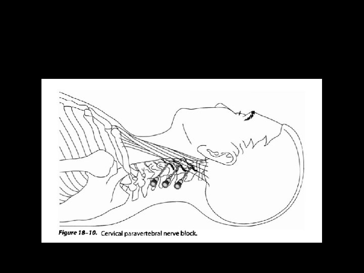
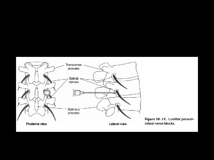
l SYMPATHETIC BLOCKS – Cervicothoracic (Stellate) Block A. INDICATIONS l This block is often used in patients with head, neck, arm, and upper chest pain. l – Intravenous Regional Sympathetic Blockade l A Bier block utilizing guanethidine l (20 -40 mg) can selectively interrupt sympathetic innervation to an extremity. l
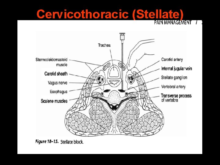
Cervicothoracic (Stellate) Block
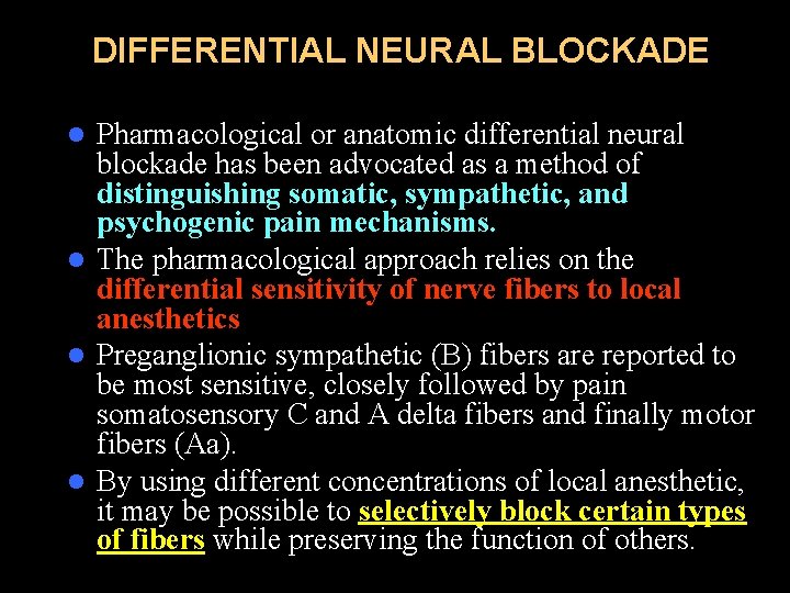
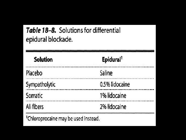
DIFFERENTIAL NEURAL BLOCKADE Pharmacological or anatomic differential neural blockade has been advocated as a method of distinguishing somatic, sympathetic, and psychogenic pain mechanisms. l The pharmacological approach relies on the differential sensitivity of nerve fibers to local anesthetics l Preganglionic sympathetic (B) fibers are reported to be most sensitive, closely followed by pain somatosensory C and A delta fibers and finally motor fibers (Aa). l By using different concentrations of local anesthetic, it may be possible to selectively block certain types of fibers while preserving the function of others. l
PHARMACOLOGICAL INTERVENTIONS Pharmacological interventions in pain management include – – – – – COX inhibitors, Opioids, Antidepressants, Neuroleptic agents, Anticonvulsants, Corticosteroids, Systemic administration of local anesthetics Alpha 2 agonists Botulinum toxin
THERAPEUTIC ADJUNCTS PSYCHOLOGICAL INTERVENTIONS PHYSICAL THERAPY Heat and cold ACUPUNCTURE
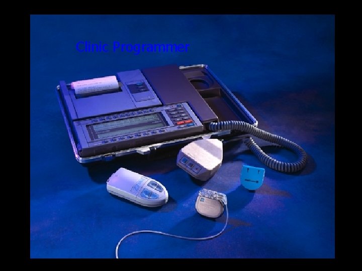
THERAPEUTIC ADJUNCTS ELECTRICAL STIMULATION – Transcutaneous Stimulation – Spinal Cord Stimulation (SCS) Proposed mechanisms include activation of descending modulating systems and inhibition of sympathetic outflow – lntracerebral Stimulation Deep brain stimulation may be used for intractable cancer pain (periaqueductal and periventricular gray areas for nociceptive pain)
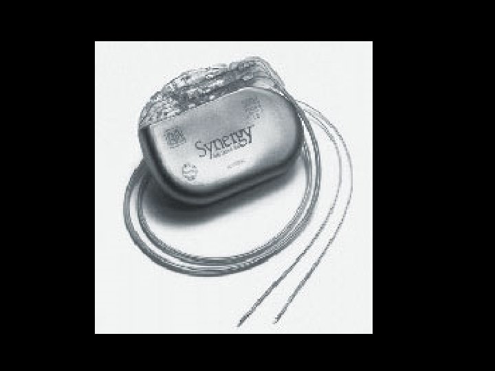
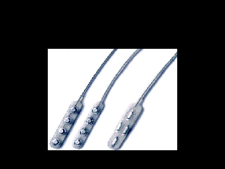
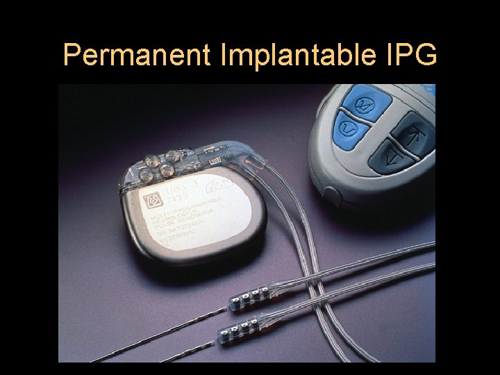
Permanent Implantable IPG
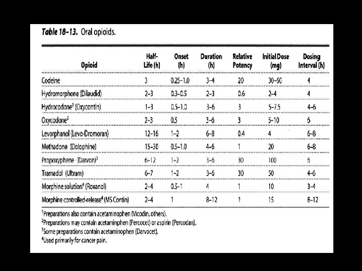
CANCER PAIN l ORAL OPl. Ol. D THERAPY l TRANSDERMAL OPl. Ol. DS l PARENTERAL THERAPY l INTRASPINAL OPl. Ol. DS l NEUROLYTIC TECHNIQUES
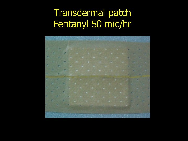
Transdermal patch Fentanyl 50 mic/hr
ALCOHOL & PHENOL NEUROLYTIC BLOCKS l Neurolytic blocks are indicated for patients with severe intractable cancer pain
RADIOFREQUENCY ABLATION & CRYONEUROLYSIS l Percutaneous radio-frequency ablation relies on the heat produced by current flow from an active electrode that is incorporated at the tip of a special needle. The needle is positioned under fluoroscopy. l Electrical stimulation A z (2'Hz for motor responses and 50 Hz for sensory responses) via the electrode and impedance measurement prior to ablation also help confirm correct positioning.
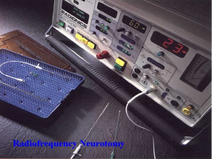
Radiofrequency Neurotomy


Clinic Programmer
- Slides: 152