Packed cell volume PCV Hematocrit Volume of RBCs
 Hematocrit")
Packed cell volume (PCV) Hematocrit

• Volume of RBCs ∕ unit volume of whole blood i. e. percentage of packed cells to the whole blood. • Normal value: for ♂ 40% -54% (47%) ♀ 36%- 47% (42%)

Aim of the experiment Screening test for anemia and polycythemia • PCV↑↑ in polycythemia & dehydration. • PCV↓↓ in anemia. • The color of plasma may give an indication about some illness, red (hemolysis), yellow (jaundice) and milky (hypercholesterolemia).

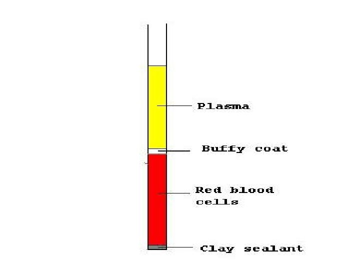
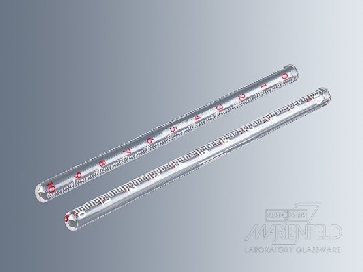
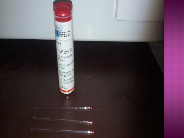
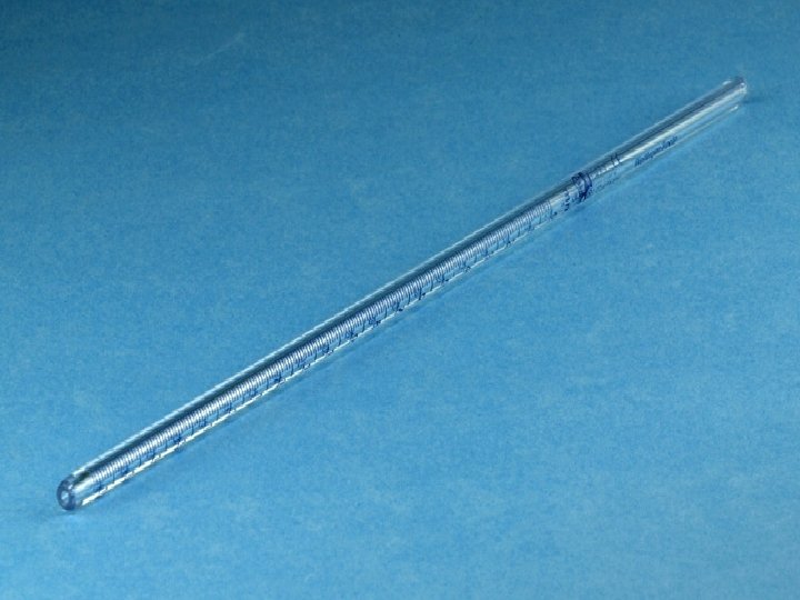
Instruments Special tubes →→ wintrobe hematocrit tube or Capillary tube • Sample of blood + anti coagulant. • Centrifuge the tube for 30 mint at 3000 rev. /min. • The blood separated into 3 layers: • Top layer (plasma) clear. • Middle thin layer Buffy coat less than 1 mm in thickness (WBCs & platelets). • Dark red color bottom layer (RBCs).

Factors affecting PCV • No. of RBC • Shape of RBC • Plasma volume
 vis the rate of red blood cell in whole blood")
Erythrocyte sedimentation rate (ESR) vis the rate of red blood cell in whole blood descend in a standardized tube in a period of one hour. va non-specific test. v. Its used to monitor the inflammation. v. Useful for monitor some chronic disorder like tuberculosis, autoimmune disease and others.
 and resist")
Mechanism �ESR is determined by the interaction between factors that promote sedimentation(fibrinogen) and resist (negative charge of RBCs - that repel each other) sedimentation. �Normal RBCs settle slowly as they do not form rouleaux or aggregate together. Instead, they gently repel each other due to the negative charge on their surfaces.

• Rouleaux are stacks of many RBCs that become heavier and sediment faster. • Plasma proteins, especially fibrinogen, adhere to the red cell membranes and neutralize the surface negative charges, promoting cell adherence and rouleaux formation

Factors affecting ESR v No. of RBCs ↑↑PCV will lead to reoulox phenomena that lead to↓↓ESR. v Shape of RBC The size and number of RBCs that show alterations in their bioconcavity, like spherocyte and sickle cells.
. v")
v Fibrinogen plasma protein. (↑↑fibrinogen lead to ↑↑reoulox phenomena that lead to ↑↑ESR). v Technical factors Temperature (18 -25 C) higher temperature cause false high results due to reduction in plasma viscosity Vibration can reduce the ESR

Some conditions with low ESR • Polycythemia • Sickle cell anemia • Sever Leukocytosis

Some conditions with ESR High • • Tuberculosis. Malignancies. Severe anemia. Multiple myeloma

Normal Value Of ESR • • • Adult females 0 -20 mm/h Adult males 0 -15 mm/hr Children (<10) 0 -10 mm/hr
 of the disease.")
Aim of the experiment Responsible for prognosis (follow up) of the disease.
, heparin or")
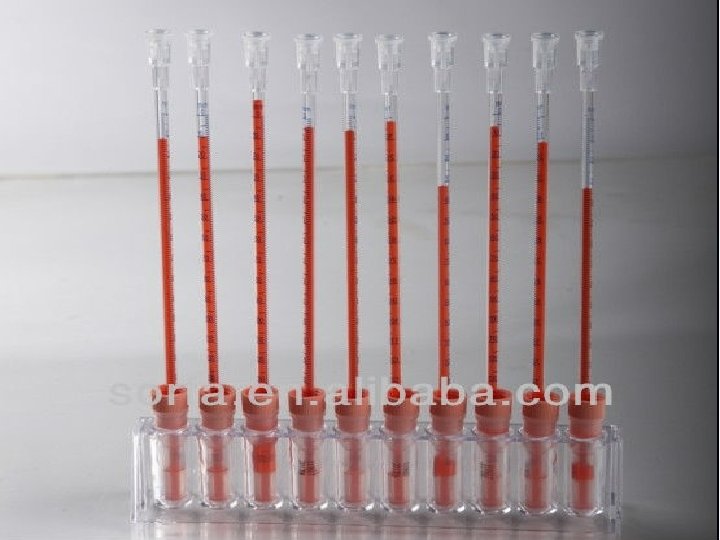
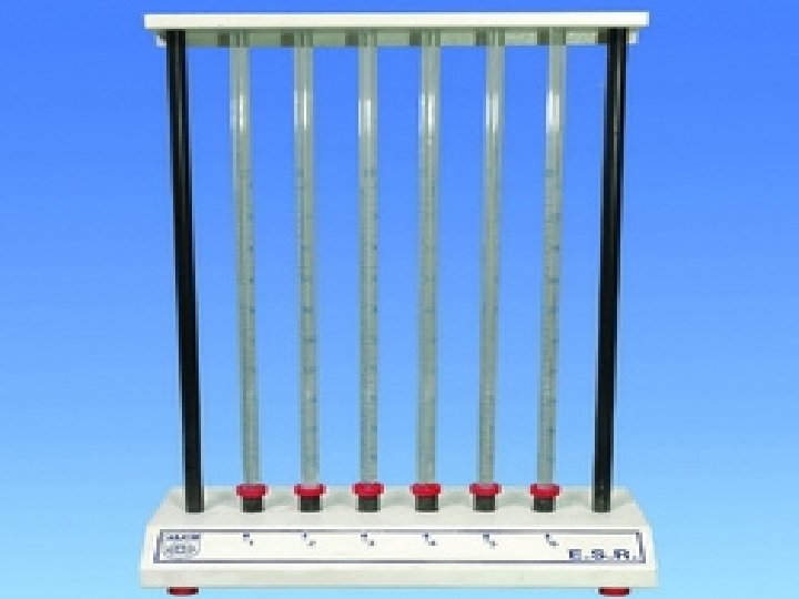
Procedure 1. Blood collect from vein puncture and mixed with anticoagulant (EDTA), heparin or sodium oxalate 2. place blood in narrow vertical tube (westergren tube) 3. Calculate the RBCs have falls in one hour

• When blood mix with an anti coagulant & allow to stand vertically in a special tube (westergren tube) for a period of time (one hour). • The RBCs will settle down (sediment) leaving clear plasma layer above. • The rate at which RBCs sediment is ESR and it is measured by the distance in which the RBCs are settle down in mm. /period of time which is usually one hour.
? settle down Aggregation of RBCs one over the top of other,")
Why RBCs (sediment)? settle down Aggregation of RBCs one over the top of other, the surface area reduce & their weight will increase i. e. reoulox phenomena.
- Slides: 24