Packed cell volume PCV also called hematocrit HCT
 also called hematocrit (HCT) is defined as the percentage of")


- Slides: 5
Packed cell volume (PCV) also called hematocrit (HCT) is defined as the percentage of the volume occupied by RBC to the whole blood volume. The packed cell volume (PCV) can be used as: simple screening test for anaemia, The PCV is about three times the Hb expressed in g/dl. Therefore can be used to calculate Hb concentration It can be used in the calculation of red cell indices. Normal Ranges Adult males = (40% - 52%) Adult females = (37% - 47%)
Hematocrit Method Test Sample Anticoagulated venous blood (k 2 EDTA is recommended, k 3 EDTA cause RBC shrinkage) or capillary blood. Equipment Micro haematocrit centrifuge 75 mm long capillary tubes with an internal diameter of 1 mm. (blue plain capillary with Anticoagulated venous blood and red heparinized capillary tube for the direct collection of capillary blood). Plastic sealer or Bunsen burner. haematocrit reader Procedure Blood samples should be as fresh as possible and well mixed. 1. Using a capillary tube, allow blood to enter the tube by capillary action fill the 3/4 capillary tube leaving about 1_1. 5 cm un filled from one end. Wipe the outside of the tube. 2. Seal the end by pushing into plastic seal two or three times. If heat sealing is used, rotate the dry end of the tube over a fine Bunsen burner flame. 3. Place the tube into one of the centrifuge plate slots, with the sealed end against the rubber gasket of the centrifuge plate. Keep a record of the patient number against the centrifuge plate number. 4. Centrifuge for 5 minutes. This separates the RBCs from plasma and leaves a band of buffy coat consisting of WBCs and platelets.
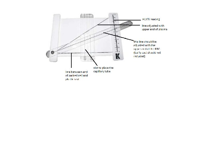
5. Read the PCV in the micro haematocrit reader. The haematocrit result is expressed in a percentage.
Biologic sources of error: If the buffy coat is included in the RBCs when reading the result, the PCV will be falsely elevated. Hemolysis of the specimen cause a falsely decreased result. When the microhematocrit is spun for the correct time period and at the proper speed, a small amount of plasma still remains in the red blood cell portion. This is termed trapped plasma. An increased amount of trapped plasma is found in macrocytic anemia, spherocytosis, thalassemia, hypochromic anemia and sickle cell anemia. The approximate relationship of the hemoglobin level to hematocrit is 1: 3 (± 2), a ratio that may vary with the plasma volume and the cause of the anemia and the effect of that cause on the RBC indices, particularly the mean corpuscular volume (MCV).