
P SH 2 P P P SH 2

- Slides: 34
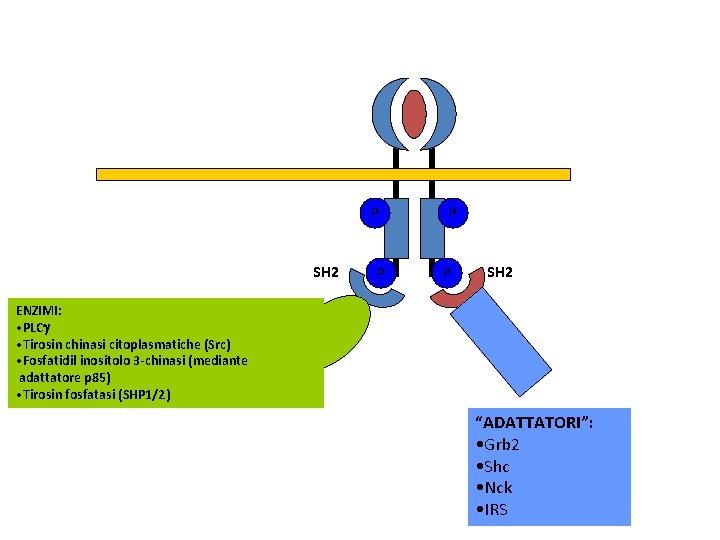
P SH 2 P P P SH 2 ENZIMI: • PLCg • Tirosin chinasi citoplasmatiche (Src) • Fosfatidil inositolo 3 -chinasi (mediante adattatore p 85) • Tirosin fosfatasi (SHP 1/2) “ADATTATORI”: • Grb 2 • Shc • Nck • IRS
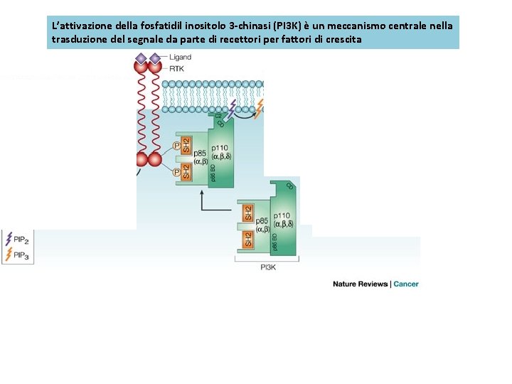
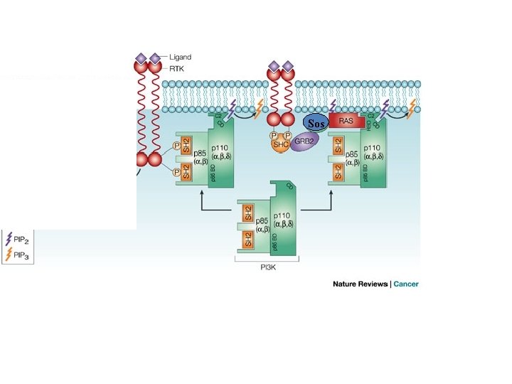
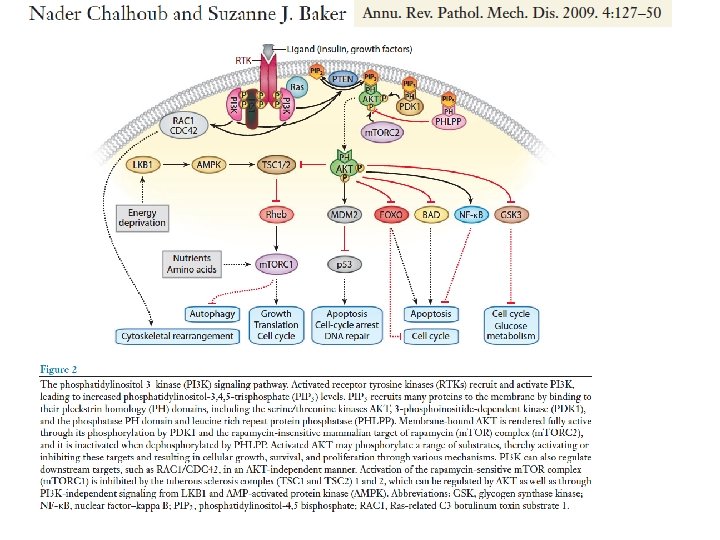
L’attivazione della fosfatidil inositolo 3 -chinasi (PI 3 K) è un meccanismo centrale nella trasduzione del segnale da parte di recettori per fattori di crescita Sos
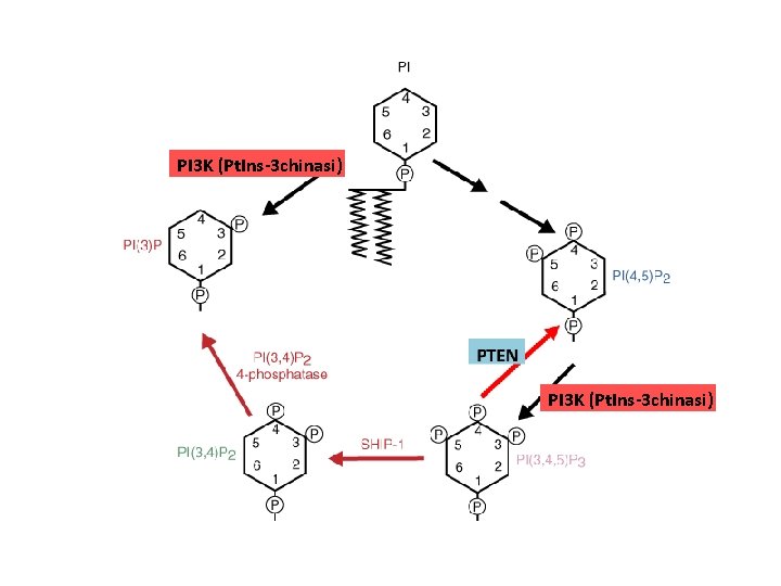
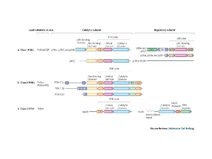
PI 3 K (Pt. Ins-3 chinasi)
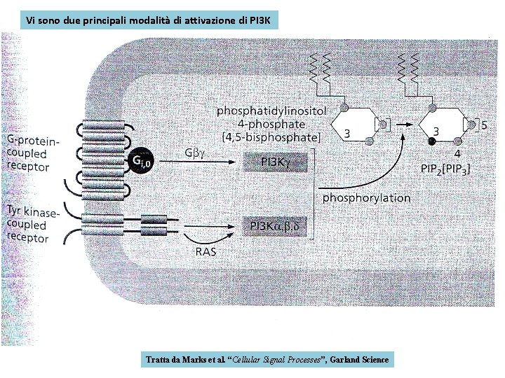
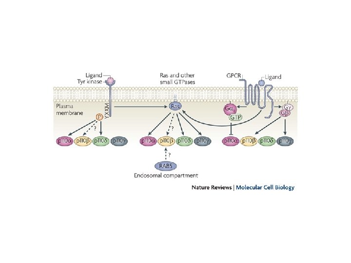
Vi sono due principali modalità di attivazione di PI 3 K Tratta da Marks et al. “Cellular Signal Processes”, Garland Science
Sos
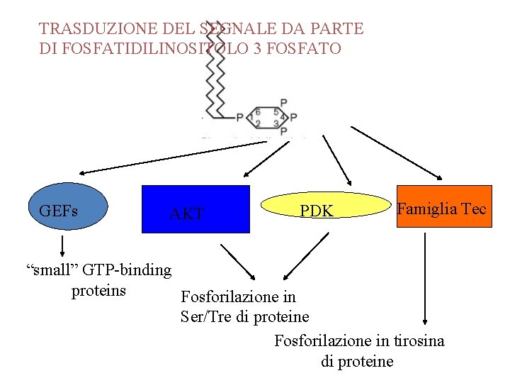
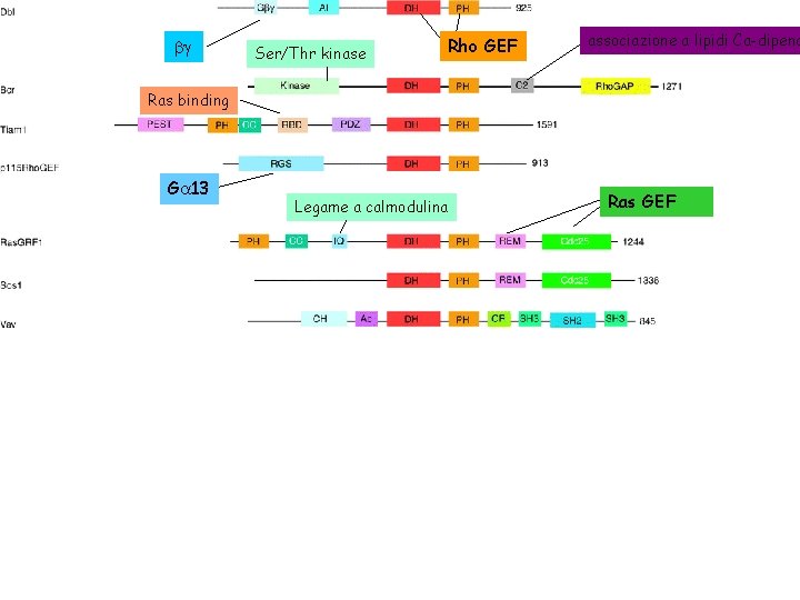
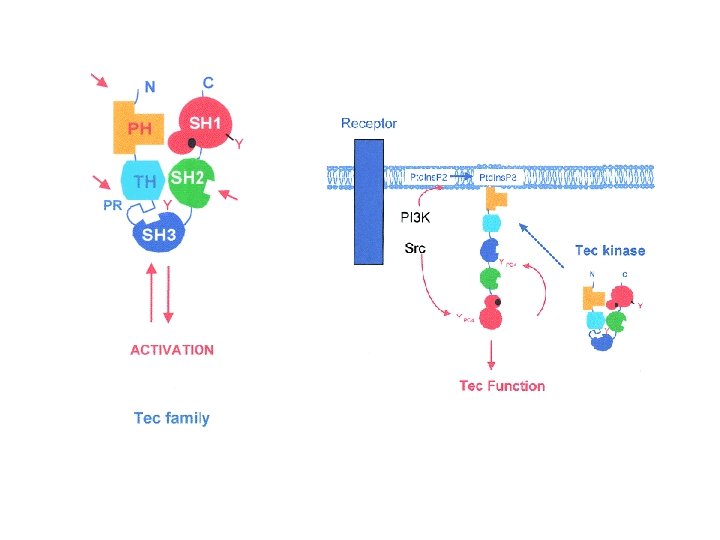
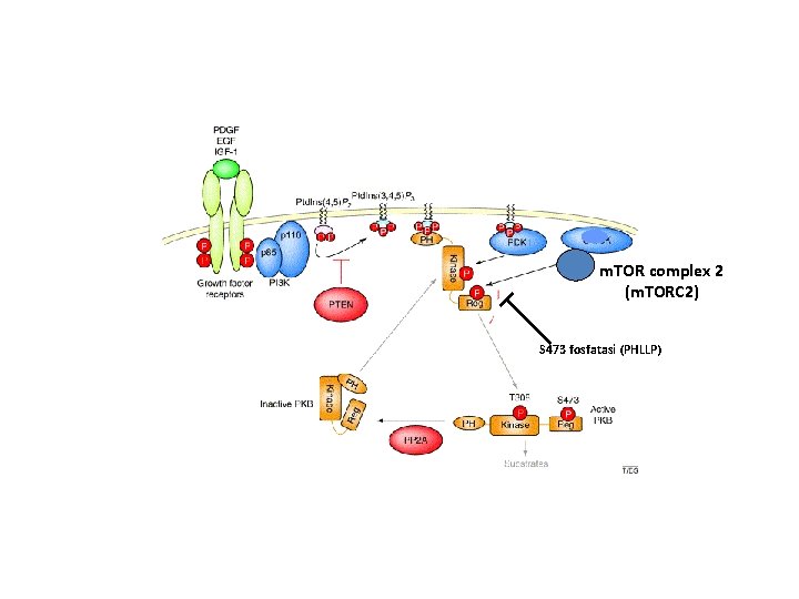
TRASDUZIONE DEL SEGNALE DA PARTE DI FOSFATIDILINOSITOLO 3 FOSFATO GEFs AKT PDK Famiglia Tec “small” GTP-binding proteins Fosforilazione in Ser/Tre di proteine Fosforilazione in tirosina di proteine
bg Ser/Thr kinase Rho GEF associazione a lipidi Ca-dipend Ras binding Ga 13 Legame a calmodulina Ras GEF
m. TOR complex 2 (m. TORC 2) S 473 fosfatasi (PHLLP)
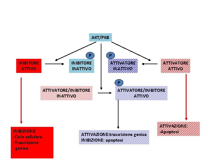
AKT/PKB P INIBITORE ATTIVO INIBITORE INATTIVO P ATTIVATORE INATTIVO ATTIVATORE ATTIVO P ATTIVATORE/INIBITORE INATTIVO INIBIZIONE: -Ciclo cellulare -Trascrizione genica ATTIVATORE/INIBITORE ATTIVO ATTIVAZIONE: trascrizione genica INIBIZIONE: apoptosi ATTIVAZIONE: -Apoptosi
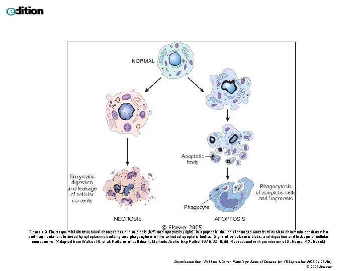
Figure 1 -9 The sequential ultrastructural changes seen in necrosis (left) and apoptosis (right). In apoptosis, the initial changes consist of nuclear chromatin condensation and fragmentation, followed by cytoplasmic budding and phagocytosis of the extruded apoptotic bodies. Signs of cytoplasmic blebs, and digestion and leakage of cellular components. (Adapted from Walker NI, et al: Patterns of cell death. Methods Archiv Exp Pathol 13: 18 -32, 1988. Reproduced with permission of S. Karger AG, Basel. ) Downloaded from: Robbins & Cotran Pathologic Basis of Disease (on 15 September 2005 04: 26 PM) © 2005 Elsevier
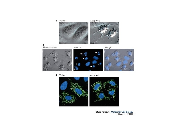
Marzo 2008
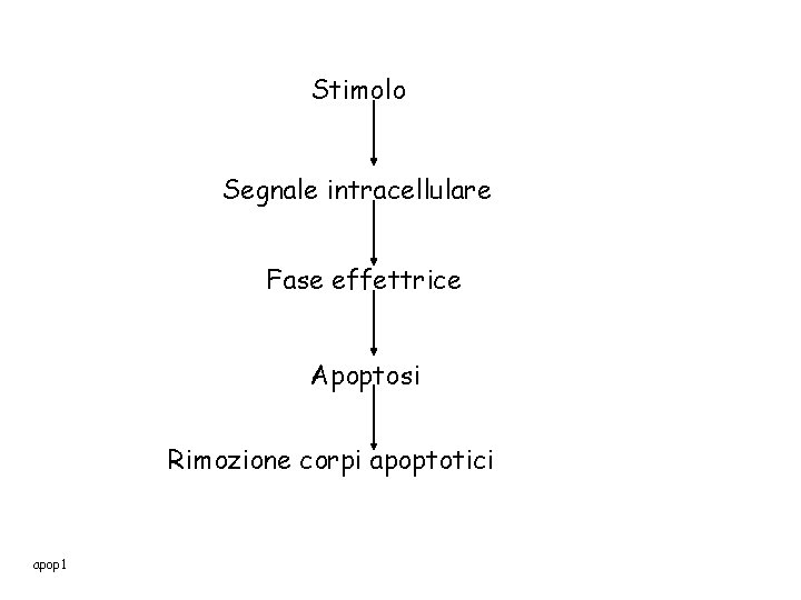
Stimolo Segnale intracellulare Fase effettrice Apoptosi Rimozione corpi apoptotici apop 1
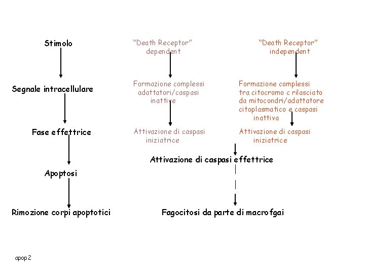
Stimolo Segnale intracellulare Fase effettrice “Death Receptor” dependent “Death Receptor” independent Formazione complessi adattatori/caspasi inattive Formazione complessi tra citocromo c rilasciato da mitocondri/adattatore citoplasmatico e caspasi inattiva Attivazione di caspasi iniziatrice Attivazione di caspasi effettrice Apoptosi Rimozione corpi apoptotici apop 2 Fagocitosi da parte di macrofgai
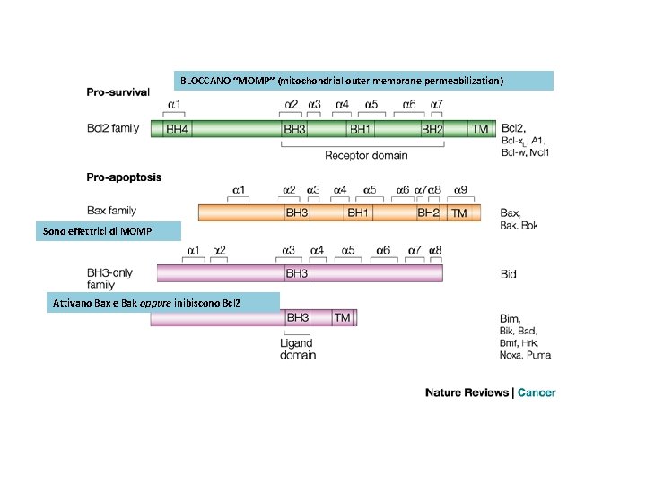
BLOCCANO “MOMP” (mitochondrial outer membrane permeabilization) Sono effettrici di MOMP Attivano Bax e Bak oppure inibiscono Bcl 2
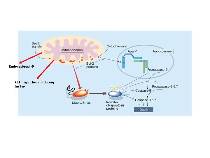
Endonucleasi G AIF: apoptosis inducing factor
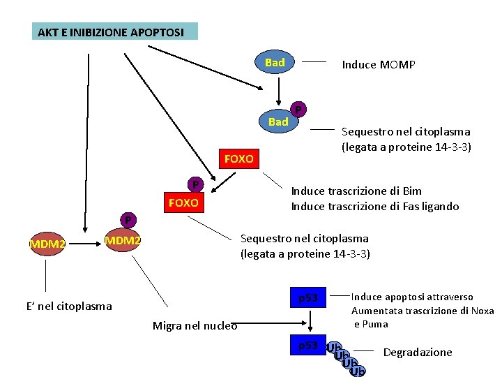
AKT E INIBIZIONE APOPTOSI Bad Induce MOMP P Sequestro nel citoplasma (legata a proteine 14 -3 -3) FOXO P MDM 2 Induce trascrizione di Bim Induce trascrizione di Fas ligando Sequestro nel citoplasma (legata a proteine 14 -3 -3) MDM 2 p 53 E’ nel citoplasma Migra nel nucleo Induce apoptosi attraverso Aumentata trascrizione di Noxa e Puma p 53 Ub Ub Degradazione
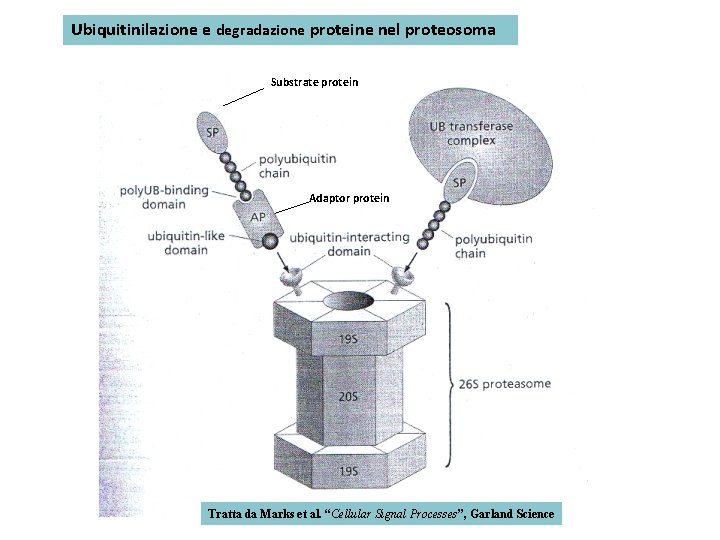
Ubiquitinilazione e degradazione proteine nel proteosoma Substrate protein Adaptor protein Tratta da Marks et al. “Cellular Signal Processes”, Garland Science
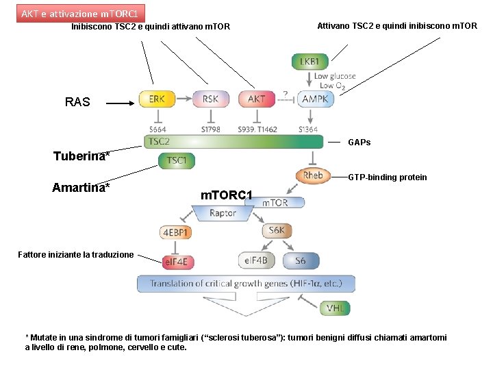
AKT e attivazione m. TORC 1 Inibiscono TSC 2 e quindi attivano m. TOR Attivano TSC 2 e quindi inibiscono m. TOR RAS GAPs Tuberina* Amartina* GTP-binding protein m. TORC 1 Fattore iniziante la traduzione * Mutate in una sindrome di tumori famigliari (“sclerosi tuberosa”): tumori benigni diffusi chiamati amartomi a livello di rene, polmone, cervello e cute.
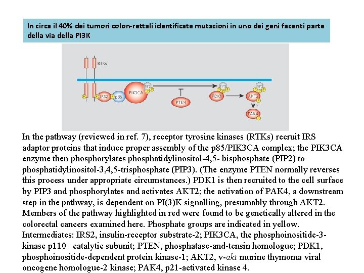
In circa il 40% dei tumori colon-rettali identificate mutazioni in uno dei geni facenti parte della via della PI 3 K In the pathway (reviewed in ref. 7), receptor tyrosine kinases (RTKs) recruit IRS adaptor proteins that induce proper assembly of the p 85/PIK 3 CA complex; the PIK 3 CA enzyme then phosphorylates phosphatidylinositol-4, 5 - bisphosphate (PIP 2) to phosphatidylinositol-3, 4, 5 -trisphosphate (PIP 3). (The enzyme PTEN normally reverses this process under appropriate circumstances. ) PDK 1 is then recruited to the cell surface by PIP 3 and phosphorylates and activates AKT 2; the activation of PAK 4, a downstream step in the pathway, is dependent on PI(3)K signalling, presumably through AKT 2. Members of the pathway highlighted in red were found to be genetically altered in the colorectal cancers examined here. Phosphate groups are indicated in yellow. Intermediates: IRS 2, insulin-receptor substrate-2; PIK 3 CA, the phosphoinositide-3 kinase p 110 catalytic subunit; PTEN, phosphatase-and-tensin homologue; PDK 1, phosphoinositide-dependent protein kinase-1; AKT 2, v-akt murine thymoma viral oncogene homologue-2 kinase; PAK 4, p 21 -activated kinase 4.
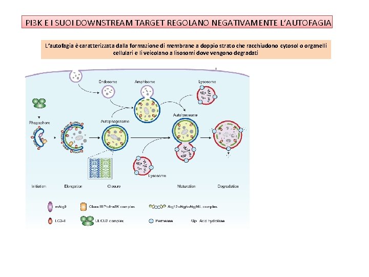
PI 3 K E I SUOI DOWNSTREAM TARGET REGOLANO NEGATIVAMENTE L’AUTOFAGIA L’autofagia è caratterizzata dalla formazione di membrane a doppio strato che racchiudono cytosol o organelli cellulari e li veicolano a lisosomi dove vengono degradati
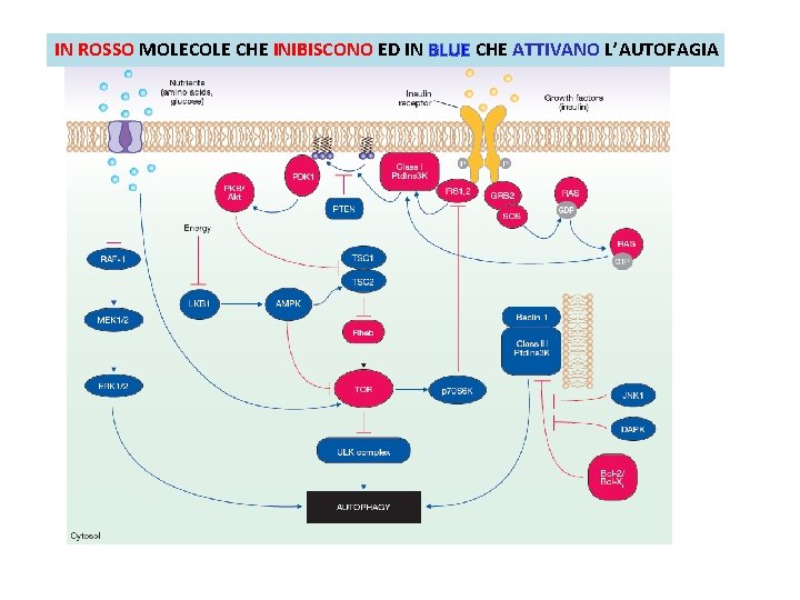
IN ROSSO MOLECOLE CHE INIBISCONO ED IN BLUE CHE ATTIVANO L’AUTOFAGIA
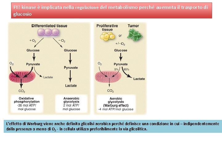
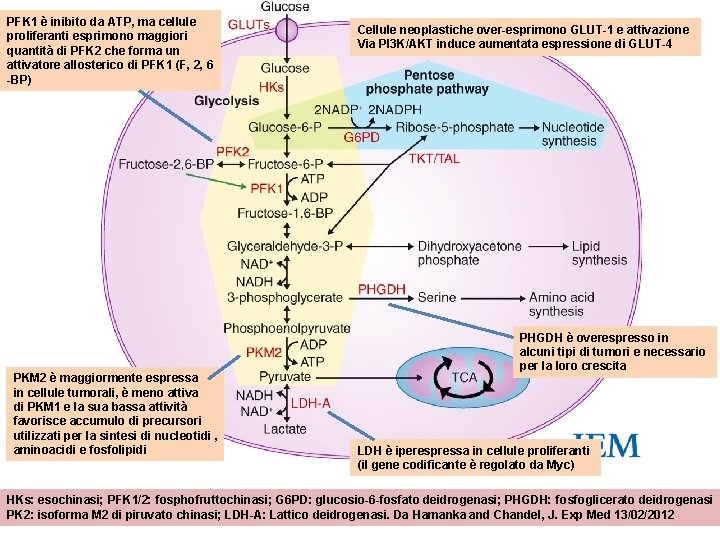
PI 3 kinase è implicata nella regolazione del metabolismo perché auemnta il trasporto di glucosio L’effetto di Warburg viene anche definito glicolisi aerobica perché definisce una condizione in cui – indipendentemente dalla presenza o meno di O 2 - la cellula utilizza preferibilmente la via glicolitica.
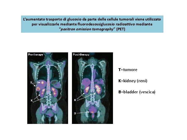
L’aumentato trasporto di glucosio da parte delle cellule tumorali viene utilizzato per visualizzarle mediante fluorodesossiglucosio radioattivo mediante “positron emission tomography” (PET) T=tumore K=kidney (reni) B=bladder (vescica)
PFK 1 è inibito da ATP, ma cellule proliferanti esprimono maggiori quantità di PFK 2 che forma un attivatore allosterico di PFK 1 (F, 2, 6 -BP) PKM 2 è maggiormente espressa in cellule tumorali, è meno attiva di PKM 1 e la sua bassa attività favorisce accumulo di precursori utilizzati per la sintesi di nucleotidi , aminoacidi e fosfolipidi Cellule neoplastiche over-esprimono GLUT-1 e attivazione Via PI 3 K/AKT induce aumentata espressione di GLUT-4 PHGDH è overespresso in alcuni tipi di tumori e necessario per la loro crescita LDH è iperespressa in cellule proliferanti (il gene codificante è regolato da Myc) HKs: esochinasi; PFK 1/2: fosphofruttochinasi; G 6 PD: glucosio-6 -fosfato deidrogenasi; PHGDH: fosfoglicerato deidrogenasi PK 2: isoforma M 2 di piruvato chinasi; LDH-A: Lattico deidrogenasi. Da Hamanka and Chandel, J. Exp Med 13/02/2012
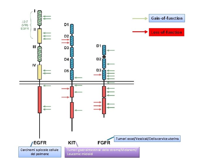
Gain-of-function Loss-of-function Tumori ossei/Vescicali/Della cervice uterina Carcinomi a piccole cellule del polmone Tumori gastrointestinali dello stroma/Melanomi/ Leucemie mieloidi
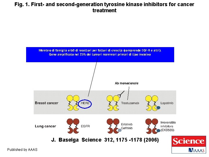
Fig. 1. First- and second-generation tyrosine kinase inhibitors for cancer treatment Membro di famiglia erb. B di recettori per fattori di crescita (comprende EGF-R e altri). Gene amplificato nel 25% dei tumori mammari primari di tipo invasivo Sindrome ipereosinofilica Ab monoclonale J. Baselga Science 312, 1175 -1178 (2006) Published by AAAS
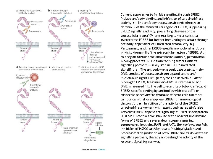
Current approaches to inhibit signalling through ERBB 2 include antibody binding and inhibition of tyrosine kinase activity. a | The antibody trastuzumab binds directly to domain IV of the extracellular region of ERBB 2, suppressing ERBB 2 signalling activity, preventing cleavage of the extracellular domain 74 and marking tumour cells that overexpress ERBB 2 for further immunological attack through antibody-dependent cell-mediated cytotoxicity. b | Pertuzumab, another ERBB 2 -specific monoclonal antibody, binds to domain II of the extracellular region of ERBB 2. As this region contains the dimerization domain, pertuzumab binding prevents ERBB 2 from forming dimers with its signalling partners — a key step in ERBB 2 -mediated signalling. c | The antibody–drug conjugate trastuzumab– DM 1 consists of trastuzumab conjugated to the antimicrotubule agent DM 1 (a maytansine derivative). After binding to ERBB 2, trastuzumab–DM 1 is internalized and DM 1 is released into the cell to exert its cytotoxic effects. d | ERBB 2 -specific binding by antibodies with bispecific or trispecific selectivity for cytotoxic effector cells can mark tumour cells that overexpress ERBB 2 for immunological destruction. e | Inhibition of the activity of the ERBB 2 tyrosine kinase domain with agents such as lapatinib also prevents ERBB 2 -dependent signalling. f | Heat shock protein 90 (HSP 90) controls the stability of the nascent and mature forms of ERBB 2 and several downstream signalling components, including RAF 1 and AKT 1 (for reviews, see Refs. Inhibition of HSP 90 activity results in ubiquitylation and proteasomal degradation of both ERBB 2 and its downstream signalling partners, thereby abrogating the activity of the relevant signalling pathway.