Overview of Tubular Reabsorption Note Most of the

Overview of Tubular: Reabsorption Note: Most of the essential materials (Glucose, Amino Acids, Vitamins, Pro-) are reabsorbed immediately! Secretion Note: Urea and Uric Acid are both reabsorbed and secreted. Each tubule region has specific substances that are secreted. Excretion By the end of the Collecting Ducts, filtrate is no longer modified and it becomes Urine, stored in the bladder.
 – Water")
Proximal Convoluted Tubule • Active Reabsorption – Nutrients (glucose, amino acids, vitamins) – Water and Ions (K+, Na+, Cl-, Ca 2+, Mg 2+, HCO 3 -…) – Small plasma proteins ~70% of Filtrate is – Some urea and uric acid reabsorbed in PCT Question: How are these Reabsorbed?

Na+ is Actively Reabsorbed: Reabsorption of Na+ : First – simple diffusion: Then – 1 o active transport:

Na+ linked 2 o Active Transport Symport with: – Glucose – Amino acids – Ions (e. g. , Ca 2+)

Passive Transport of: Water - as Na+ pumped out, H 2 O follows by osmosis. Urea - as other solutes leave lumen, the [urea] is higher than ECF, thus diffuses into ECF. Note: About 50% of the Urea filtered is Reabsorbed. Urea is also Secreted into the Loop of Henle and plays a major role in maintaining the Osmotic (Salinity) Gradient in the medulla.

Transcytosis of Proteins: Small proteins can get into filtrate, reabsorbed via vesicular transport. Glucose, Amino Acids, some vitamins are reabsorbed via protein carrier transporters

Protein Carrier Transporter Characteristics – Specificity: glucose, fructose, tyrosine, valine, etc. , all have own carriers. – Competition: maltose instead of glucose – takes a seat, but not transported. – Saturation (# of carriers): limited # of carriers to transport solutes back into body. A substance can exceed renal threshold, e. g. , glucosuria.

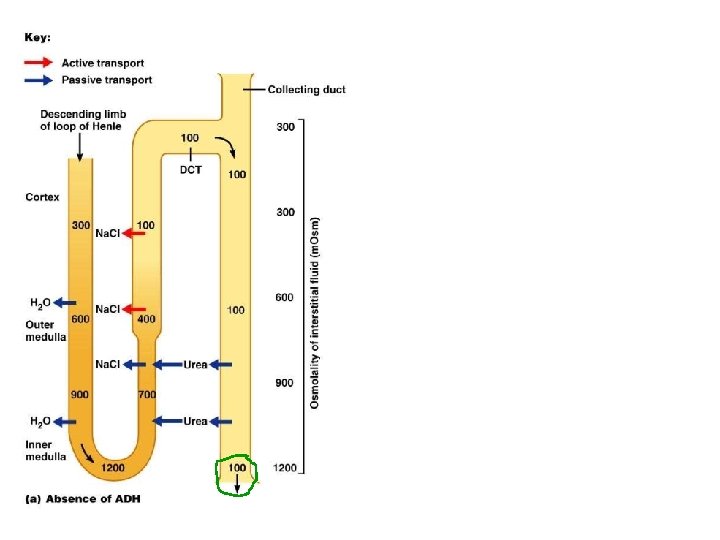
H 2 O Reabsorption – Loop of Henle • Much of Na+ already transported out of filtrate. • Osmolarity of ECF gets higher (counter current). • More H 2 O drawn out of filtrate deeper into medulla. • Filtrate becomes Very concentrated at hair pin loop! • Ascending thick segment of Loop impermeable to H 2 O – thus now filtrate gets more and more dilute. *Collecting duct also a key site for H 2 O reabsorption (via ADH).

Countercurrent Multiplier System The hair pin Loop of Henle creates the steep osmotic gradient in the renal medulla – as the concentrations of urea and Na. Cl multiply deep in the medulla. The adjacent loops have fluid flow in opposite (countercurrent) directions. Countercurrent Exchanger Is the vasa recta arrangement which can reabsorb both H 20 and Na. Cl yet can still maintain the osmotic gradient. Ascending thick segment impermeable to H 2 O! ECF Osmotic Gradient 1200

Filtrate Modification: Changes is Osmolarity

Secretion – DCT a key site. • Active Transport into nephron tubules + + + e. g. , K , H and NH 4 • Fine-tuning - eliminate unwanted items. For example, creatinine* is secreted at the PCT. • Filtrate in tubules is destined to be urine unless reabsorbed in collecting ducts.

Final Modification: Collecting Ducts • Reabsorption of Na+ • Reabsorption of H 2 O • Under Endocrine Control – ADH (vasopressin) • After passing through the collecting duct, the filtrate is no longer modified, now called urine.

Autoregulation of Renal System Renin-Angiotensin-Aldosterone
 ______ (activated) ________ (active) Kidneys ____________ (_______)")
Liver Lungs Kidneys Adrenal Cortex ______________________ (inactive) ______ (activated) ________ (active) Kidneys ____________ (_______) Na+ _______ Vasoconstriction Reabsorption of H 2 O Thirst Stimulation H 2 O _______
 Renin Angiotensinogen (inactive) Angiotensin")
Liver Lungs Kidneys Adrenal Cortex Aldosterone Angiotensin Converting Enzyme (ACE) Renin Angiotensinogen (inactive) Angiotensin I (activated) Angiotensin II (active) Kidneys Anti Diuretic Hormone (ADH) (Vasopressin) Na+ retention Vasoconstriction Reabsorption of H 2 O Thirst Stimulation H 2 O retention

Substances Filtered and Reabsorbed by the Kidney per 24 Hours Water Substance Proteins Chlorine Sodium Bicarbonate Glucose Urea Potassium Uric acid Creatinine 180 L 179 L 1 L Amount Filtered (grams) Amount Reabsorbed (grams) Amount in Urine (grams) 10– 20 0 630 625 5 540 537 3 300 299. 7 0. 3 180 0 53 28 25 28 24 4 8. 5 7. 7 0. 8 1. 4 0 1. 4
 Blood Plasma Filtrate Urine Volume 5. 0 L Rate")
Comparison of Fluids Substance (parameter) Blood Plasma Filtrate Urine Volume 5. 0 L Rate 1 -2 L/day p. H 7. 35 – 7. 45 Osmolarity 295 – 310 m. Os. M Cells No Na+, K+ Yes Glucose Yes No Large Proteins No Small Proteins Yes Urea Yes Creatinine Yes

Micturition is coordinated between the central, autonomic, and somatic nervous systems. Brain centers involved are the pontine micturition center and cerebral cortex.
. Stretch (mechano-) receptors detect bladder wall tension")
Micturition Reflex Voiding urine mostly Para (ANS). Stretch (mechano-) receptors detect bladder wall tension and send signals that ascend via the spinal cord and project into up to the pons where the micturition center is located. Cerebral integration (conscious decision to urinate) activates neurons of the pontine micturition center, excites sacral neurons (preganglionic) activating Para pelvic nerves (S 2 -4) a releasing ACh which binds to M 3 receptors on detrusor muscle causing contraction and increased pressure. The Pons also inhibits Sym stimulation to internal urethral sphincter causing relaxation. Combined with conscious relaxation of the external urethral sphincter (cerebrum), this allows voiding of urine.

Renal Failure When kidney function is disrupted to the point they are unable to perform their normal regulatory and excretory functions sufficient to maintain homeostasis. Acute – sudden onset with rapid reduction in urine formation (less than 500 ml/day minimum being excreted). Chronic – slow, progressive, insidious loss of renal function. Up to 75% of function can be lost before detected.

Normal Healthy Kidney

Polycystic kidneys
.")
Enlarged Polycystic kidneys (16 to 18 pounds combined).
 leading to a decrease")
Acute Kidney Failure Pre-renal: decreased renal perfusion (often from hypovolemia) leading to a decrease in GFR; heart failure; liver cirrhosis. Reversible. Intra-renal: intrinsic kidney damage; most common due to ischemic or nephrotoxic injury (nephritis). Also medications can cause damage. Post-renal: extrinsic/intrinsic obstruction (e. g. , kidney stone) of the urinary collection system. BUN = blood urea nitrogen. Normal adult blood range 6 to 20 mg/d. L (1. 8 to 7. 1 mmol/L). BUN an indication of renal health. Blood tests also check amount of creatinine or a BUN-to-creatinine ratio.

Variety of Causes of Renal Failure: 1. Infectious organisms. - Blood borne microbes - UTI’s 2. Toxic agents. - lead, arsenic, pesticides, additives, medications - long-term exposure to high aspirin doses 3. Inflammatory immune response (allergic). - glomerulonephritis, sepsis - e. g. , after strep throat (streptoccocus)

Variety of Causes: 4. Obstruction of urine flow. - Kidney stone (calcium oxalate, uric acid crystals) - Tumors - Enlarged prostate gland All create back pressure, decreasing GFR 5. Insufficient renal blood flow. - 2 o to heart failure - Hemorrhage (e. g. shock) - Atherosclerosis Leads to inadequate Filtration pressure

Potential Ramifications: 1. Uremic Toxicity - Caused by retention of toxins/waste products in blood. 2. Metabolic Acidosis - From inability of kidneys to secrete H+. 3. Potassium (K+) Retention - Inability to secrete K+ (effects RMP).

4. Na+, Ca 2+ and phosphate and Imbalances - Inability of kidneys to regulate ion reabsorption and secretion. 5. Loss of plasma proteins - Result of increased leakiness of glomerulus. 6. Anemia - Inadequate erythropoiten production. 7. Depressed immune system - Increased toxic waste and acidic conditions.

Possible Treatments for Renal Failure: Stop or Treat the Cause Dialysis Kidney Transplant
- Slides: 30