
Overview of Heart Failure Fourth year medical students
- Slides: 58
Overview of Heart Failure Fourth year medical students Tareq Yousef Goussous, M. D. , FACC Interventional Cardiologis
A 72 -year-old-male patient with a pmhs of HTN presents with dyspnea on exertion and orthopnea for the past 5 months that got worse in the last week. On P/E: BP 150/80 mm Hg, HR 110 bpm, S 3 & S 4, 4/6 SEM in the 2 RICS, bilateral crackles & bilateral LLE. CXR: pulmonary vascular congestion.
A 28 -year-old-male patient presents with dyspnea on exertion and fatigue of several months duration. P/E: BP 140/40 mm Hg, HR 90 bpm & 3/6 diastolic murmur at the left lower ICS.
A 60 -year-old-male patient who sustained an acute MI 2 weeks ago presented with acute dyspnea, orthopnea and PNDs of 1 day duration. On P/E: BP 100/60 mm Hg, HR 110 bpm, S 3 and bilateral crackles.
A 65 -year-old-female patient presented with acute dyspnea on exertion, feeling of suffocation and frothy sputum. P/E: BP 190/100 mm Hg, HR 115 bpm, & bilateral crackles all over the chest.
Introduction Heart failure (HF) is a complex clinical syndrome that can result from any structural or functional cardiac disorder that impairs the ability of the ventricle to fill with or eject blood. It is characterized by specific symptoms, such as dyspnea and fatigue, and signs, such as fluid retention. There are many ways to assess cardiac function.
However, there is no diagnostic test for HF, since it is largely a clinical diagnosis that is based upon a careful history and physical examination.
Systolic heart failure. Diastolic heart failure, or heart failure with preserved ejection fraction. (relaxation & filling).
Systolic dysfunction: ischemia/MI, DCMP, chronic AI/MR, Diastolic dysfunction: HCMP, AS, HTN, RCMP, ischemia. High output HF: A-V fistula, Paget’s, sepsis, Beriberi, anemia, thyrotoxicosis. Pericardial diseases: (usually right-sided HF): tamponade and constriction.
Classification of HF severity: The classification system that is most commonly used to quantify the degree of functional limitation imposed by HF is one first developed by the New York Heart Association (NYHA). This system assigns patients to one of four functional classes, depending on the degree of effort needed to elicit symptoms:
Class I — symptoms of HF only at activity levels that would limit normal individuals. Class II — symptoms of HF with ordinary exertion. Class III — symptoms of HF with less than ordinary exertion. Class IV — symptoms of HF at rest.
Stages in the development of HF: There are several stages in the evolution of HF, as outlined by the American College of Cardiology/American Heart Association (ACC/AHA) guidelines: Stage A: High risk for HF, without structural heart disease or symptoms. Stage B: Heart disease with asymptomatic left ventricular dysfunction. Stage C: Prior or current symptoms of HF. Stage D: Refractory end stage HF.
Etiology: There are two basic pathophysiologic mechanisms that cause reduced cardiac output and HF: systolic dysfunction and diastolic dysfunction. Systolic and diastolic dysfunction each may be due to a variety of etiologies. Effective management is often dependent upon establishing the correct etiologic.
Systolic dysfunction: The most common causes of systolic dysfunction are: Coronary (ischemic) heart disease. Idiopathic dilated cardiomyopathy (DCM). Hypertension. Valvular disease.
Diastolic dysfunction: Diastolic dysfunction can be induced by many of the same conditions that lead to systolic dysfunction. The most common causes are: Hypertension. Ischemic heart disease. Hypertrophic obstructive cardiomyopathy. Restrictive cardiomyopathy.
Clinical assessment: The approach to the patient with HF or cardiomyopathy includes the history and physical examination, and diagnostic tests to establish the diagnosis, assess acuity, severity and etiology.
History: Symptoms of HF include those due to excess fluid accumulation (dyspnea, edema, hepatic congestion, and ascites) and those due to a reduction in cardiac output (fatigue, weakness) that is most pronounced with exertion.
The history and other findings may be helpful in identifying the etiology of HF. As examples: Classic exertional angina usually indicates ischemic heart disease. Acute HF after an antecedent flu-like illness suggests viral myocarditis. Long-standing hypertension or alcohol use suggests hypertensive or alcoholic cardiomyopathy.
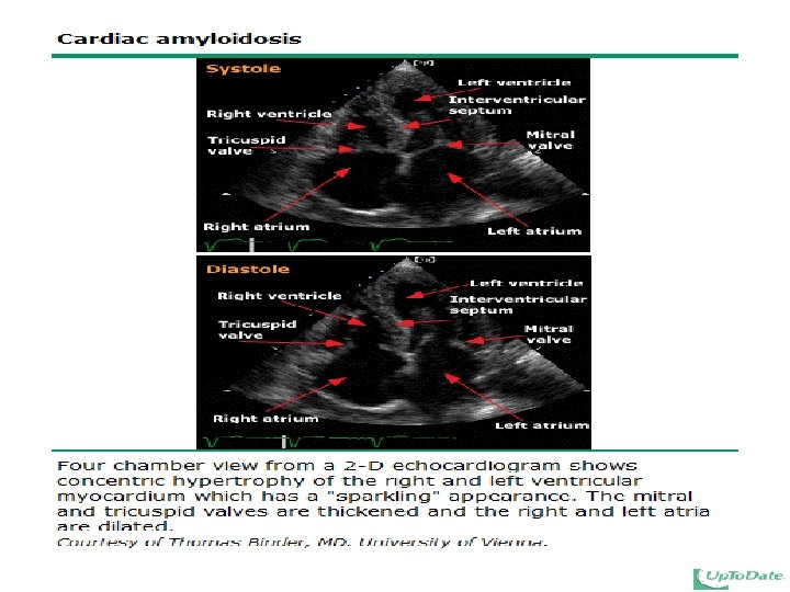
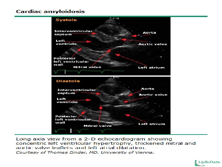
Primary valvular dysfunction should be considered in a patient with a history of murmurs. A diagnosis of amyloidosis should be strongly considered in patients who have a family history of unexplained cardiomyopathy or amyloidosis, low voltage on EKG, left ventricular hypertrophy (especially without hypertension), and a history of heavy proteinuria.
HF may be provoked or worsened by drugs, including antiarrhythmic agents such as disopyramide and flecainide; calcium channel blockers, particularly verapamil; beta blockers; and nonsteroidal antiinflammatory drugs (NSAIDs).
Acute pulmonary edema occurring during, or shortly after, infusion of blood products suggests transfusional volume overload.
Physical examination: There are three major manifestations of volume overload in patients with HF: pulmonary congestion. peripheral edema. elevated jugular venous pressure.
Pulmonary congestion is more prominent in acute or subacute disease. Peripheral edema is manifested by swelling of the legs (which is more prominent when the patient is upright), ascites, hepatomegaly, and splenomegaly. Hepatojugular reflux.
Manual compression of the right upper quadrant to increase venous return may elevate jugular venous pressure above the transient 1 to 3 cm elevations seen in normal individuals. This sign is known as the hepatojugular reflux.
Elevated jugular venous pressure is usually present if peripheral edema is due to HF, since it is the high intracapillary pressure that is responsible for fluid movement into the interstitium. With the patient sitting at 45º jugular venous pressure can be estimated from the height above the left atrium of venous pulsations in the internal jugular vein.
Pulsus alternans — Pulsus alternans, if present, is virtually pathognomonic of severe left ventricular failure. This phenomenon is characterized by evenly spaced alternating strong and weak peripheral pulses. It is best appreciated by applying light pressure on the peripheral arterial pulse
Precordial palpation — Ventricular chamber size can be estimated by precordial palpation. An apical impulse that is laterally displaced past the midclavicular line is usually indicative of left ventricular enlargement.
Heart sounds — An S 3 gallop is associated with left atrial pressures exceeding 20 mm. Hg, increased left ventricular end-diastolic pressures (>15 mm. Hg) and elevated serum brain natriuretic peptide concentrations.
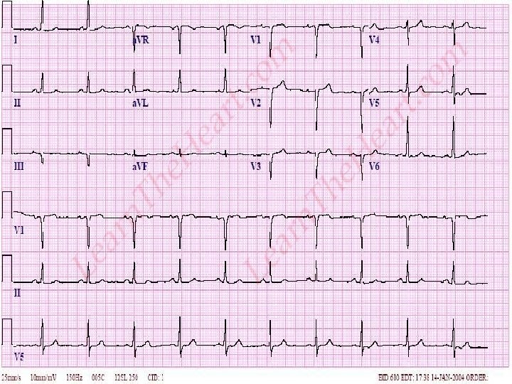
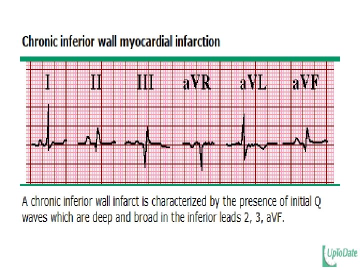
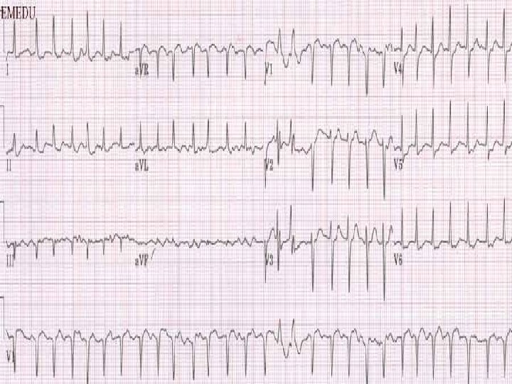
Initial tests: Electrocardiogram: Potentially diagnostic findings on ECG include the following: Evidence of ischemic heart disease including evidence of prior or acute myocardial infarction or ischemia.
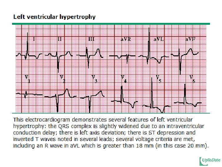
Left ventricular hypertrophy due to hypertension. Low limb lead voltage on the surface ECG with a pseudoinfarction pattern (loss of precordial R wave progression in leads V 1 -V 6) can suggest an infiltrative process such as amyloidosis.
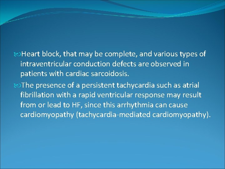
Heart block, that may be complete, and various types of intraventricular conduction defects are observed in patients with cardiac sarcoidosis. The presence of a persistent tachycardia such as atrial fibrillation with a rapid ventricular response may result from or lead to HF, since this arrhythmia can cause cardiomyopathy (tachycardia-mediated cardiomyopathy).
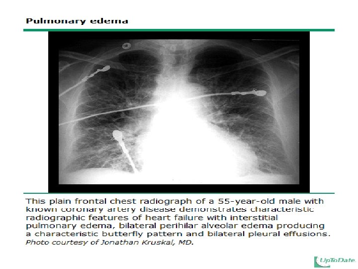
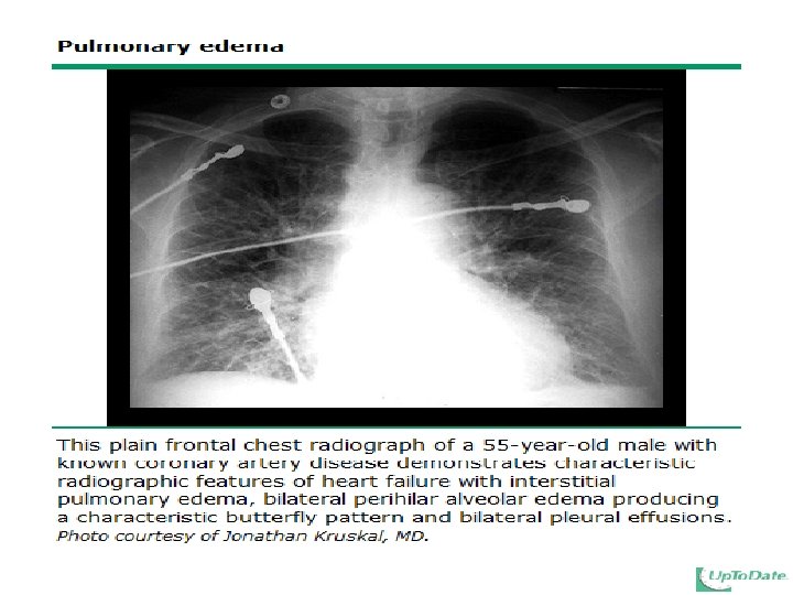
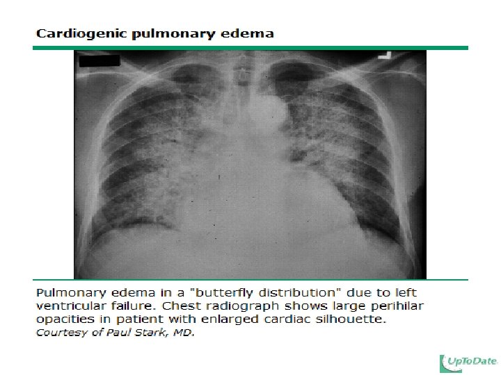
Chest x-ray — A chest x-ray is generally indicated to evaluate pulmonary edema, cardiopulmonary structural abnormalities and other potential causes of dyspnea.
Initial blood tests — Recommended initial blood tests for patients with symptoms and signs of HF include: CBC. Serum electrolytes, Creatinine & urea. LFT. FBS.
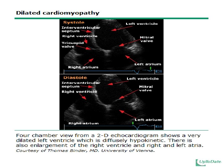
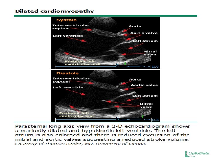
Echocardiography: Echocardiography should be performed in all patients with new onset HF and can provide important information about ventricular size and function. For example, patients with idiopathic dilated cardiomyopathy typically have both left and right ventricular enlargement (four chamber dilatation) with decreased left systolic ventricular function.
Treatment Chronic compensated heart failure. Acute decompensated heart failure.
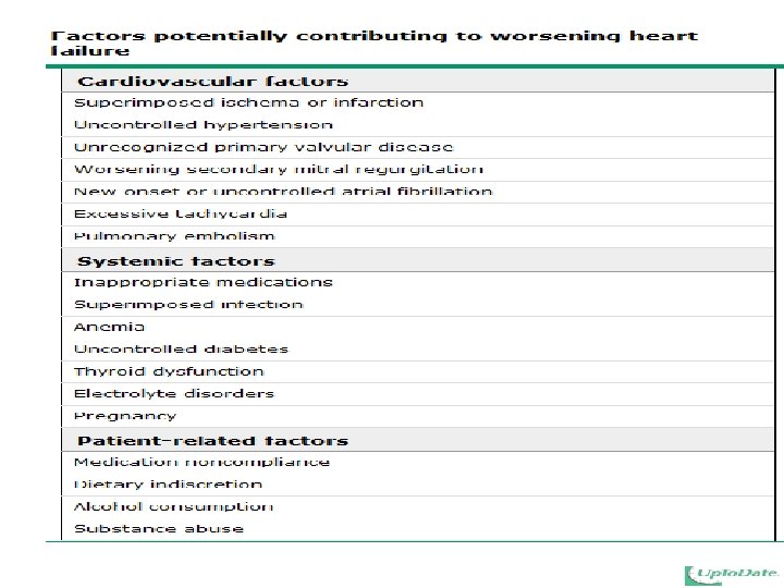
General principles: The management of HF begins with an accurate assessment of the etiology and severity of the disease. This is followed by a therapeutic regimen aimed at the following factors: Correction of systemic factors (eg, thyroid dysfunction, infection, uncontrolled diabetes). Lifestyle modification:
Cessation of smoking. Restriction of alcohol consumption. Salt restriction to approximately 2 to 3 g (or less) of sodium per day to minimize fluid accumulation. Weight reduction in obese subjects with goal of being within 10 percent of ideal body weight. Daily weight monitoring to detect fluid accumulation before it becomes symptomatic.
Review of drugs that may contribute to HF (eg, nonsteroidal antiinflammatory drugs, antiarrhythmic drugs, calcium channel blockers, thiazolidinediones). Pneumococcal vaccination and annual influenza vaccination. Treatment of the cause of the heart disease. Pharmacologic therapy directed at relieving symptoms, slowing the progression of the HF, and improving patient survival.
Pharmacologic therapy: The goals of pharmacologic therapy are to improve symptoms, slow or reverse deterioration in myocardial function, and reduce mortality. Additional pharmacologic therapy is directed at the prevention of arrhythmias and embolic events and the treatment of anemia and other possible exacerbating factors.
A number of drugs are recommended in HF for symptom relief and improvement in outcome: Improvement in symptoms can be achieved by digoxin, diuretics, beta blockers, angiotensin converting enzyme (ACE) inhibitors, and angiotensin II receptor blockers (ARBs). Prolongation of patient survival has been documented with ACE inhibitors, beta blockers, ARBs, hydralazine/nitrates, and aldosterone antagonists.
Order of therapy — the following sequence of drugs is recommended in the typical patient, with allowance for variations depending upon clinical response: Loop diuretics are introduced first for fluid control in patients in overt HF. The goal is relief of signs or symptoms of volume overload, such as dyspnea and peripheral edema.
ACE inhibitors, or if not tolerated, angiotensin II receptor blockers (ARBs) are typically initiated during or after the optimization of diuretic therapy. These drugs are usually started at low doses and then titrated to goals based upon trial data. Beta blockers are initiated after the patient is stable on ACE inhibitors, again beginning at low doses with titration to trial goals as tolerated.
The following drugs should be given to selected patients in the absence of a contraindication: The addition of an aldosterone antagonist (spironolactone or, if not tolerated, eplerenone) to improve survival in patients who can be monitored for preserved renal function and a normal plasma potassium concentration and have NYHA functional class II HF and a LVEF ≤ 30% ; or NYHA functional class III to IV HF and an LVEF <35%.
Angiotensin II receptor blockers (ARBs) as an alternative to ACE inhibitors in patients who cannot tolerate these drugs. The addition of the combination of hydralazine and a nitrate for patients (particularly blacks) with a reduced LVEF who have persistent symptoms despite therapy with an ACE inhibitor and beta blocker.
Digoxin to reduce hospitalization for HF or for patients with concomitant atrial fibrillation, for rate control.