Overview of Gastroenteritis is inflammation of the lining


- Slides: 53

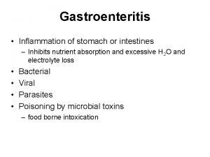
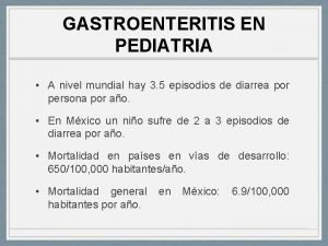
Overview of Gastroenteritis is inflammation of the lining of the stomach and small and large intestines. Most cases are infectious, although gastroenteritis may occur after ingestion of drugs and chemical toxins (eg, metals, plant substances). Acquisition may be foodborne, waterborne, or via person-to-person spread. In the US, an estimated 1 in 6 people contracts foodborne illness each year. Symptoms include anorexia, nausea, vomiting, diarrhea, and abdominal discomfort. Diagnosis is clinical or by stool culture, although PCR and immunoassays are increasingly used. Treatment is symptomatic, although some parasitic and some bacterial infections require specific anti-infective therapy. Gastroenteritis is usually uncomfortable but self-limited. Electrolyte and fluid loss is usually little more than an inconvenience to an otherwise healthy adult but can be grave for people who are very young, elderly, or immunocompromised or who have serious concomitant illnesses. Worldwide, an estimated 1. 5 million children die each year from infectious gastroenteritis; although high, this number represents one half to one quarter of previous mortality. Improvements in water sanitation in many parts of the world and the appropriate use of oral rehydration therapy for infants with diarrhea are likely responsible for this decrease.

Etiology Infectious gastroenteritis may be caused by viruses, bacteria, or parasites. Many specific organisms are discussed further in the Infectious Diseases section. Viral gastroenteritis The viruses most commonly implicated are • Norovirus • Rotavirus Viruses are the most common cause of gastroenteritis in the US. They infect enterocytes in the villous epithelium of the small bowel. The result is transudation of fluid and salts into the intestinal lumen; sometimes, malabsorption of carbohydrates worsens symptoms by causing osmotic diarrhea. Diarrhea is watery. Inflammatory diarrhea (dysentery), with fecal WBCs and RBCs or gross blood, is uncommon. Four categories of viruses cause most gastroenteritis: norovirus and rotavirus cause the majority of viral gastroenteritis, followed by astrovirus and enteric adenovirus. Norovirus infects people of all ages. Since the introduction of rotavirus vaccines, norovirus has become the most common cause of acute gastroenteritis in the US, including in children. Infections occur year-round, but 80% occur from November to April. Norovirus is now the principal cause of sporadic and epidemic viral gastroenteritis in all age groups; however, the peak age is between 6 mo and 18 mo. Large waterborne and foodborne outbreaks occur. Person-to-person transmission also occurs because the virus is highly contagious. This virus causes most cases of gastroenteritis epidemics on cruise ships and in nursing homes. Incubation is 24 to 48 h. Rotavirus is the most common cause of sporadic, severe, dehydrating diarrhea in young children worldwide (peak incidence, 3 to 15 mo). Its incidence has decreased by about 80% in the US since the introduction of routine rotavirus immunization. Rotavirus is highly contagious; most infections occur by the fecal-oral route. Adults may be infected after close contact with an infected infant. The illness in adults is generally mild. Incubation is 1 to 3 days. In temperate climates, most infections occur in the winter. Each year in the US, a wave of rotavirus illness begins in the Southwest in November and ends in the Northeast in March. Astrovirus can infect people of all ages but usually infects infants and young children. Infection is most common in winter. Transmission is by the fecal-oral route. Incubation is 3 to 4 days. Adenoviruses are the 4 th most common cause of childhood viral gastroenteritis. Infections occur year-round, with a slight increase in summer. Children < 2 yr are primarily affected. Transmission is by the fecal-oral route. Incubation is 3 to 10 days. In immunocompromised patients, additional viruses (eg, cytomegalovirus, enterovirus) can cause gastroenteritis.

Symptoms and Signs The character and severity of symptoms of gastroenteritis vary. Generally, onset is sudden, with anorexia, nausea, vomiting, abdominal cramps, and diarrhea (with or without blood and mucus). Malaise, myalgias, and prostration may occur. The abdomen may be distended and mildly tender; in severe cases, muscle guarding may be present. Gas-distended intestinal loops may be palpable. Hyperactive bowel sounds are present on auscultation even without diarrhea (an important differential feature from paralytic ileus, in which bowel sounds are absent or decreased). Persistent vomiting and diarrhea can result in intravascular fluid depletion with hypotension and tachycardia. In severe cases, shock, with vascular collapse and oliguric renal failure, occurs. If vomiting is the main cause of fluid loss, metabolic alkalosis with hypochloremia can occur. If diarrhea is more prominent, acidosis is more likely. Both vomiting and diarrhea can cause hypokalemia. Hyponatremia may develop, particularly if hypotonic fluids are used in replacement therapy. Viral gastroenteritis In viral infections, watery diarrhea is the most common symptom; stools rarely contain mucus or blood.

Rotavirus gastroenteritis in infants and young children may last 5 to 7 days. Vomiting occurs in 90% of patients, and fever > 39° C (>102. 2° F) occurs in about 30%. Norovirus typically causes acute onset of vomiting, abdominal cramps, and diarrhea, with symptoms lasting only 1 to 2 days. In children, vomiting is more prominent than diarrhea, whereas in adults, diarrhea usually predominates. Patients may also have fever, headache, and myalgias. The hallmark of adenovirus gastroenteritis is diarrhea lasting 1 to 2 wk. Affected infants and children may have mild vomiting that typically starts 1 to 2 days after the onset of diarrhea. Low-grade fever occurs in about 50% of patients. Respiratory symptoms may be present. Symptoms are generally mild but can last longer than with other viral causes of gastroenteritis. Astrovirus causes a syndrome similar to mild rotavirus infection.

Diagnosis • Clinical evaluation • Stool testing in select cases Other GI disorders that cause similar symptoms (eg, appendicitis, cholecystitis, ulcerative colitis) must be excluded (see also evaluation of diarrhea). Findings suggestive of gastroenteritis include copious, watery diarrhea; ingestion of potentially contaminated food (particularly during a known outbreak), untreated surface water, or a known GI irritant; recent travel; or contact with certain animals or similarly ill people. E. coli O 157: H 7–induced diarrhea is notorious for appearing to be a hemorrhagic rather than an infectious process, manifesting as GI bleeding with little or no stool. Hemolytic-uremic syndrome may follow as evidenced by renal failure and hemolytic anemia. Recent oral antibiotic use (within 3 mo) must raise suspicion for C. difficile infection. However, about one fourth of patients with community-associated C. difficile infection do not have a history of recent antibiotic use. Stool testing is guided by clinical findings and the organisms that are suspected based on patient history and epidemiologic factors (eg, immunosuppression, exposure to a known outbreak, recent travel, recent antibiotic use). Cases are typically stratified into • Acute watery diarrhea • Subacute or chronic watery diarrhea • Acute inflammatory diarrhea Multiplex PCR platforms that can identify causative organisms in each of these categories are being used more often. However, this testing is expensive, and because the categories are distinguishable clinically, se it is usually more cost-effective to test for specific microorganisms depending on the type and duration of diarrhea. Acute watery diarrhea is probably viral and testing is not indicated unless the diarrhea persists. Although rotavirus and enteric adenovirus infections can be diagnosed using commercially available rapid assays that detect viral antigen in the stool, these assays are rarely indicated. Subacute and chronic watery diarrhea require testing for parasitic causes, typically with microscopic stool examination for ova and parasites. Fecal antigen tests are available for Giardia, Cryptosporidia, and Entamoeba histolytica. Acute inflammatory diarrhea without gross blood can be recognized by the presence of WBCs on stool examination. Patients should have stool culture for typical enteric pathogens (eg, Salmonella, Shigella, Campylobacter, E. coli). Acute inflammatory diarrhea with gross blood should also prompt testing specifically for E. coli O 157: H 7, as should nonbloody diarrhea during a known outbreak. Specific cultures must be requested because this organism is not detected on standard stool culture media. Alternatively, a rapid enzyme assay for the detection of Shiga toxin in stool can be done; a positive test indicates infection with E. coli O 157: H 7 or one of the other serotypes of enterohemorrhagic E. coli. (Note: Shigella species in the US do not produce Shiga toxin. ) However, a rapid enzyme assay is not as sensitive as culture. PCR is used to detect Shiga toxin in some centers. Adults with grossly bloody diarrhea should usually have sigmoidoscopy with cultures and biopsy. Appearance of the colonic mucosa may help diagnose amebic dysentery, shigellosis, and E. coli O 157: H 7 infection, although ulcerative colitis may cause similar lesions. Patients with a history of recent antibiotic use or other risk factors for C. difficile infection (eg, inflammatory bowel disease, use of proton pump inhibitors) should have a stool assay for C. difficiletoxin, but testing should also be done in patients with significant illness even when these risk factors are not present because about 25% of cases of C. difficile infection currently occur in people without identified risk factors. Historically, enzyme immunoassays for toxins A and B were used to diagnose C. difficile infection. However, nucleic acid amplification tests targeting one of the C. difficile toxin genes or their regulator have been shown to have higher sensitivity and are now the diagnostic tests of choice. General tests Serum electrolytes, BUN, and creatinine should be obtained to evaluate hydration and acid-base status in patients who appear seriously ill. CBC is nonspecific, although eosinophilia may indicate parasitic infection. Renal function tests and CBC should be done about a week after the start of symptoms in patients with E. coli O 157: H 7 to detect early-onset hemolytic-uremic syndrome. It is unclear whether this testing is necessary in patients with non–E. coli O 157: H 7 Shiga toxin infection

. Treatment • Oral or IV rehydration • Consideration of antidiarrheal agents if C. difficile or E. coli O 157: H 7 infection is not suspected • Antibiotics only in select cases Supportive treatment is all that is needed for most patients. Bed rest with convenient access to a toilet or bedpan is desirable. Oral glucoseelectrolyte solutions, broth, or bouillon may prevent dehydration or treat mild dehydration. Even if vomiting, the patient should take frequent small sips of such fluids; vomiting may abate with volume replacement. For patients with E. coli O 157: H 7 infection, rehydration with isotonic IV fluids may attenuate the severity of any renal injury should hemolytic-uremic syndrome develop. Children may become dehydrated more quickly and should be given an appropriate rehydration solution. Carbonated beverages and sports drinks lack the correct ratio of glucose to sodium and thus are not appropriate, particularly for children < 5 yr. If the child is breastfed, breastfeeding should continue. If vomiting is protracted or if severe dehydration is prominent, IV replacement of volume and electrolytes is necessary. When the patient can tolerate fluids without vomiting and the appetite has begun to return, food may be gradually restarted. There is no demonstrated benefit from restriction to bland food (eg, cereal, gelatin, bananas, toast). Some patients have temporary lactose intolerance. Antidiarrheal agents are safe for patients > 2 yr with watery diarrhea (as shown by heme-negative stool). However, antidiarrheals may cause deterioration of patients with C. difficile or E. coli O 157: H 7 infection and thus should not be given to any patient with recent antibiotic use or heme-positive stool, pending specific diagnosis. Effective antidiarrheals include loperamide 4 mg po initially, followed by 2 mg po for each subsequent episode of diarrhea (maximum of 6 doses/day or 16 mg/day), or diphenoxylate 2. 5 to 5 mg tid or qid in tablet or liquid form. For children, loperamide is used. The dose for children 13 to 21 kg is 1 mg after the first loose stool then 1 mg after each subsequent loose stool (maximum dose is 3 mg/day); for children 21 to 28 kg, 2 mg after the first loose stool then 1 mg after each subsequent loose stool (maximum dose is 4 mg/day); and for children 27 to 43 kg, up to age 12, 2 mg after the first loose stool followed by 1 mg after each subsequent loose stool (maximum dose is 6 mg/day). If vomiting is severe and a surgical condition has been excluded, an antiemetic may be beneficial. Drugs useful in adults include prochlorperazine 5 to 10 mg IV tid or qid, or 25 mg per rectum bid and promethazine 12. 5 to 25 mg IM tid or qid, or 25 to 50 mg per rectum qid. These drugs are usually avoided in children because of lack of demonstrated efficacy and the high incidence of dystonic reactions. Ondansetron is safe and effective in decreasing nausea and vomiting in children and in adults, including those with gastroenteritis, and is available as a standard tablet, oral disintegrating pill, or IV formulation. The dose for adults is 4 or 8 mg po or IV tid. For children, the IV dose is 0. 15 or 0. 3 mg/kg (maximum 16 mg) and the oral dose for children 8 to 15 kg is 2 mg, for children > 15 to 30 kg, 4 mg, and for children > 30 kg, 8 mg. A single dose of ondansetron is usually adequate for children, but if needed the dose may be repeated q 8 h for 2 more doses; children still vomiting after 24 h require reevaluation. Although probiotics appear to briefly shorten the duration of diarrhea, there is insufficient evidence that they affect major clinical outcomes (eg, decrease the need for IV hydration and/or hospitalization) to support their routine use in the treatment or prevention of infectious diarrhea.

Prevention Two live-attenuated oral rotavirus vaccines are available that are safe and effective against the majority of strains responsible for disease. Rotavirus immunization is part of the recommended infant vaccination schedule. Prevention of infection is complicated by the frequency of asymptomatic infection and the ease with which many agents, particularly viruses, are transmitted from person to person. In general, proper procedures for handling and preparing food must be followed. Travelers should avoid potentially contaminated food and drink. To prevent recreational waterborne infections, people should not swim if they have diarrhea. Infants and toddlers should have frequent diaper checks and should be changed in a bathroom and not near the water. Swimmers should avoid swallowing water when they swim. Infants and other immunocompromised people are particularly predisposed to developing severe cases of salmonellosis and should not be exposed to reptiles, birds, or amphibians, which commonly carry Salmonella. Breastfeeding affords some protection to neonates and infants. Caregivers should wash their hands thoroughly with soap and water after changing diapers, and diaper-changing areas should be disinfected with a freshly prepared solution of 1: 64 household bleach (¼ cup diluted in 1 gallon of water). Children with diarrhea should be excluded from child care facilities for the duration of symptoms. Children infected with enterohemorrhagic E. coli or Shigella should also have two negative stool tests before readmission to the facility.

Hepatic infections At least 5 specific viruses (hepatitis A, B, C, D, and E viruses) can cause hepatitis; each causes a specific type of hepatitis. Hepatitis D virus can infect only when hepatitis B is present. Transmission is from person to person by contact with infected blood or body secretions or by the fecal-oral route for hepatitis A and E. Other viruses can affect the liver as part of their disease process. Common examples are cytomegalovirus, Epstein-Barr virus, and yellow fever virus. Less common examples are echovirus, coxsackievirus, and herpes simplex, rubeola, rubella, and varicella viruses.

Principal Syndromes Prevalence and Distribution Specific Therapy Specific Prevention* Hepatitis A (acute) Widespread, often epidemic None γ-Globulin, vaccine Hepatitis B (acute and chronic) Widespread Interferon, other antivirals, including nucleoside analogs (eg, entecavir) and nucleotide analogs (eg, tenofovirdisoproxil fumarate) Screening for hepatitis B surface antigen Vaccine, γ- or hyperimmune globulin Hepatitis C (acute and chronic) Widespread Interferon, ribavirin Screening for hepatitis C Hepatitis D (delta) Endemic pockets in several countries Interferon None Vaccine (not available in US) Parenteral drug users at relatively high risk Can infect only in the presence of hepatitis B Hepatitis E Outbreaks Developing world Severe during pregnancy *Nonspecific precautions (eg, avoidance of body fluids of infected patients, aseptic precautions, routine hygiene measures) are also recommended.

Less common causes of hepatitis include other viral infections Disease or Organism Manifestations Viruses Cytomegalovirus In neonates: Hepatomegaly, jaundice, congenital defects In adults: Mononucleosis-like illness with hepatitis; may occur posttransfusion Epstein-Barr Infectious mononucleosis Clinical hepatitis with jaundice in 5– 10%; subclinical liver involvement in 90– 95% Acute hepatitis sometimes severe in young adults Yellow fever Jaundice, systemic toxicity, bleeding Liver necrosis with little inflammatory reaction Other Hepatic infection occasionally due to echovirus or coxsackievirus infections, varicella, herpes simplex, rubella, or rubeola

Overview of Acute Viral Hepatitis Acute viral hepatitis is diffuse liver inflammation caused by specific hepatotropic viruses that have diverse modes of transmission and epidemiologies. A nonspecific viral prodrome is followed by anorexia, nausea, and often fever or right upper quadrant pain. Jaundice often develops, typically as other symptoms begin to resolve. Most cases resolve spontaneously, but some progress to chronic hepatitis. Occasionally, acute viral hepatitis progresses to acute liver failure (indicating fulminant hepatitis). Diagnosis is by liver function tests and serologic tests to identify the virus. Good hygiene and universal precautions can prevent acute viral hepatitis. Depending on the specific virus, preexposure and postexposure prophylaxis may be possible using vaccines or serum globulins. Treatment is usually supportive. Acute viral hepatitis is a common, worldwide disease that has different causes; each type shares clinical, biochemical, and morphologic features. Liver infections caused by nonhepatitis viruses (eg, Epstein-Barr virus, yellow fever virus, cytomegalovirus) generally are not termed acute viral hepatitis.

Characteristic Hepatitis A Virus Hepatitis B Virus Hepatitis C Virus Hepatitis D Virus Hepatitis E Virus Nucleic acid RNA DNA RNA * RNA Serologic diagnosis Ig. M anti-HA HBs. Ag Anti-HCV Anti-HDV Anti-HEV Major transmission Fecal-oral Blood Needle Water Incubation period (days) 15– 45 40– 180 20– 120 30– 180 14– 60 Epidemics Yes No No No Yes Chronicity No Yes Yes No Liver cancer No Yes Yes No *Incomplete RNA; requires presence of hepatitis B virus for replication. Anti-HCV = antibody to hepatitis C virus; anti-HDV = antibody to hepatitis D virus; anti-HEV = antibody to hepatitis E virus; HBs. Ag = hepatitis B surface antigen; Ig. M anti-HAV = Ig. M antibody to hepatitis A virus.

Symptoms and Signs Some manifestations of acute hepatitis are virus-specific (see discussions of individual hepatitis viruses), but in general, acute infection tends to develop in predictable phases: • Incubation period: The virus multiplies and spreads without causing symptoms. • Prodromal (pre-icteric) phase: Nonspecific symptoms occur; they include profound anorexia, malaise, nausea and vomiting, a newly developed distaste for cigarettes (in smokers), and often fever or right upper quadrant abdominal pain. Urticaria and arthralgias occasionally occur, especially in HBV infection. • Icteric phase: After 3 to 10 days, the urine darkens, followed by jaundice. Systemic symptoms often regress, and patients feel better despite worsening jaundice. The liver is usually enlarged and tender, but the edge of the liver remains soft and smooth. Mild splenomegaly occurs in 15 to 20% of patients. Jaundice usually peaks within 1 to 2 wk. • Recovery phase: During this 2 - to 4 -wk period, jaundice fades. Appetite usually returns after the first week of symptoms. Acute viral hepatitis usually resolves spontaneously 4 to 8 wk after symptom onset. Anicteric hepatitis (hepatitis without jaundice) occurs more often than icteric hepatitis in patients with HCV infection and in children with HAV infection. It typically manifests as a minor flu-like illness. Recrudescent hepatitis occurs in a few patients and is characterized by recurrent manifestations during the recovery phase. Manifestations of cholestasis may develop during the icteric phase (called cholestatic hepatitis) but usually resolve. When they persist, they cause prolonged jaundice, elevated alkaline phosphatase, and pruritus, despite general regression of inflammation.

Diagnosis • Liver function tests (AST and ALT elevated out of proportion to alkaline phosphatase, usually with hyperbilirubinemia) • Viral serologic testing • PT/INR measurement Initial diagnosis of acute viral hepatitis Acute hepatitis must first be differentiated from other disorders that cause similar symptoms. In the prodromal phase, hepatitis mimics various nonspecific viral illnesses and is difficult to diagnose. Anicteric patients suspected of having hepatitis based on risk factors are tested initially with nonspecific liver function tests, including aminotransferases, bilirubin, and alkaline phosphatase. Usually, acute hepatitis is suspected only during the icteric phase. Thus, acute hepatitis should be differentiated from other disorders causing jaundice. Acute hepatitis can usually be differentiated from other causes of jaundice by • Its marked elevations of AST and ALT (typically ≥ 400 IU/L) ALT is typically higher than AST, but absolute levels correlate poorly with clinical severity. Values increase early in the prodromal phase, peak before jaundice is maximal, and fall slowly during the recovery phase. Urinary bilirubin usually precedes jaundice. Hyperbilirubinemia in acute viral hepatitis varies in severity, and fractionation has no clinical value. Alkaline phosphatase is usually only moderately elevated; marked elevation suggests extrahepatic cholestasis and prompts imaging tests (eg, ultrasonography). Liver biopsy is usually not needed unless the diagnosis is uncertain. If laboratory results suggest acute hepatitis, particularly if ALT and AST are>1000 IU/L, PT/INR is measured. Manifestations of portosystemic encephalopathy, bleeding diathesis, or prolongation of INR suggest acute liver failure, indicating fulminant hepatitis. If acute hepatitis is suspected, efforts are next directed toward identifying its cause. A history of exposure may provide the only clue of drug-induced or toxic hepatitis. The history should also elicit risk factors for viral hepatitis. Prodromal sore throat and diffuse adenopathy suggest infectious mononucleosis rather than viral hepatitis. Alcoholic hepatitis is suggested by a history of drinking, more gradual onset of symptoms, and presence of vascular spiders or signs of chronic alcohol use or chronic liver disease; aminotransferase levels rarely exceed 300 IU/L, even in severe cases. Also, unlike in viral hepatitis, AST is typically higher than ALT, although this difference by itself does not reliably differentiate the two. In uncertain cases, liver biopsy usually distinguishes alcoholic from viral hepatitis.

Simplified diagnostic approach to possible acute viral hepatitis. Anti-HCV = antibody to hepatitis C virus; HBs. Ag = hepatitis B surface antigen; Ig. M anti-HAV = Ig. M antibody to hepatitis A virus.

Serology In patients with findings suggesting acute viral hepatitis, the following studies are done to screen for hepatitis viruses A, B, and C: • Ig. M antibody to HAV (Ig. M anti-HAV) • Hepatitis B surface antigen (HBs. Ag) • Ig. M antibody to hepatitis B core (Ig. M anti-HBc) • Antibody to HCV (anti-HCV) If any are positive, further serologic testing may be necessary to differentiate acute from past or chronic infection. If serology suggests hepatitis B, testing for hepatitis B e antigen (HBe. Ag) and antibody to hepatitis B e antigen (anti-HBe) is usually done to help determine the prognosis and to guide antiviral therapy. If serologically confirmed HBV infection is severe, anti-HDV is measured. If the patient has recently traveled to an endemic area, Ig. M antibody to HEV (Ig. M anti-HEV) should be measured if the test is available. Biopsy is usually unnecessary but, if done, usually reveals similar histopathology regardless of the specific virus: • Patchy cell dropout • Acidophilic hepatocellular necrosis • Mononuclear inflammatory infiltrate • Histologic evidence of regeneration • Preservation of the reticulin framework HBV infection can occasionally be diagnosed based on the presence of ground-glass hepatocytes (caused by HBs. Ag-packed cytoplasm) and using special immunologic stains for the viral components. However, these findings are unusual in acute HBV infection and are much more common in chronic HBV infection. HCV causation can sometimes be inferred from subtle morphologic clues. Liver biopsy may help predict prognosis in acute hepatitis but is rarely done solely for this purpose. Complete histologic recovery occurs unless extensive necrosis bridges entire acini (bridging necrosis). Most patients with bridging necrosis recover fully. However, some cases progress to chronic hepatitis.

Treatment • Supportive care No treatments attenuate acute viral hepatitis. Alcohol should be avoided because it can increase liver damage. Restrictions on diet or activity, including commonly prescribed rest, have no scientific basis. Most patients may safely return to work after jaundice resolves, even if AST or ALT levels are slightly elevated. For cholestatic hepatitis, cholestyramine 8 g po once/day or bid can relieve itching. Viral hepatitis should be reported to the local or state health department. Prevention Because treatments have limited efficacy, prevention of viral hepatitis is very important. General measures Good personal hygiene helps prevent transmission, particularly fecal-oral transmission, as occurs with HAV and HEV. Blood and other body fluids (eg, saliva, semen) of patients with acute HBV and HCV infection and stool of patients with HAV infection are considered infectious. Barrier protection is recommended, but isolation of patients does little to prevent spread of HAV and is of no value in HBV or HCV infection. Posttransfusion infection is minimized by avoiding unnecessary transfusions and screening all donors for HBs. Ag and anti. HCV. Screening has decreased the incidence of posttransfusion hepatitis, probably to about 1/100, 000 units of blood component transfused.

Immunoprophylaxis can involve active immunization using vaccines and passive immunization. Vaccines for hepatitis A and hepatitis B and hepatitis Eare available. Standard immune globulin prevents or decreases the severity of HAV infection and should be given to family members and close contacts of patients. Hepatitis B immune globulin (HBIG) probably does not prevent infection but prevents or attenuates clinical illness. No product exists for immunoprophylaxis of HCV or HDV. However, prevention of HBV infection prevents HDV infection. The propensity of HCV for changing its genome hampers vaccine development. Key Points • Transmission is the fecal-oral route for hepatitis A and parenterally or via blood for hepatitis B and C. • Hepatitis B and C, unlike hepatitis A, predispose to chronic hepatitis and liver cancer. • Patients with acute viral hepatitis may be anicteric or even asymptomatic. • Do viral serologic testing (Ig. M anti-HAV, HBs. Ag, anti-HCV) if clinical findings are consistent with acute viral hepatitis and AST and ALT are elevated out of proportion to alkaline phosphatase. • Treat patients supportively. • Routine vaccination for hepatitis A and B is recommended for all children and adults at high risk.

Fulminant Hepatitis Fulminant hepatitis is a rare syndrome of massive necrosis of liver parenchyma and a decrease in liver size (acute yellow atrophy) that usually occurs after infection with certain hepatitis viruses, exposure to toxic agents, or drug-induced injury. Hepatitis B virus is sometimes responsible for fulminant hepatitis, and up to 50% of cases of fulminant hepatitis B involve hepatitis D virus coinfection. Fulminant hepatitis with hepatitis A virus is rare but may be more likely in people with preexisting liver disorders. The role of hepatitis C virus remains uncertain. Patients rapidly deteriorate because portosystemic encephalopathy develops, progressing to coma and cerebral edema over a period of several days to several weeks. Coagulopathy commonly results from liver failure or disseminated intravascular coagulation, and functional renal failure (hepatorenal syndrome) may develop. Increasing PT or INR, portosystemic encephalopathy, and particularly renal failure are ominous. Diagnosis • Clinical evaluation • Liver function tests • PT/INR measurement Fulminant hepatitis should be suspected if patients are acutely ill with new-onset jaundice, rapid changes in mental status, or unexplained bleeding or if patients with known liver disease rapidly deteriorate. Laboratory tests to confirm the diagnosis of fulminant hepatitis include liver function tests and PT/INR. Laboratory tests for acute hepatitis A, B, and C viruses, as well as some other viruses (eg, cytomegalovirus, Epstein-Barr virus, herpes simplex virus), are done to determine whether a virus is the cause. Treatment • Oral nucleoside or nucleotide analogs • Liver transplantation Meticulous nursing care and aggressive treatment of complications improve the outcome. If fulminant hepatitis results from hepatitis B, treatment with oral nucleoside or nucleotide analogs can increase the likelihood of survival. However, emergency liver transplantation provides the best hope for survival. Survival in adults is uncommon without transplantation; children tend to do better. Patients who survive usually recover fully.

Overview of Chronic Hepatitis Chronic hepatitis is hepatitis that lasts > 6 mo. Common causes include hepatitis B and C viruses, autoimmune liver disease (autoimmune hepatitis), steatohepatitis (nonalcoholic steatohepatitis or alcoholic hepatitis), and some drugs. Many patients have no history of acute hepatitis, and the first indication is discovery of asymptomatic aminotransferase elevations. Some patients present with cirrhosis or its complications (eg, portal hypertension). Biopsy is necessary to confirm the diagnosis and to grade and stage the disease. Treatment is directed toward complications and the underlying condition (eg, corticosteroids for autoimmune hepatitis, antiviral therapy for viral hepatitis). Liver transplantation is often indicated for decompensated cirrhosis. Hepatitis lasting > 6 mo is generally defined as chronic, although this duration is arbitrary. Etiology Common causes The most common causes are • Hepatitis B virus • Hepatitis C virus • Nonalcoholic steatohepatitis (NASH) • Alcoholic hepatitis • Idiopathic (probably autoimmune) Hepatitis B virus (HBV) and hepatitis C virus (HCV) are frequent causes of chronic hepatitis; 5 to 10% of cases of HBV infection, with or without hepatitis D virus (HDV) coinfection, and about 75% of cases of HCV infection become chronic. Rates are higher for HBV infection in children (eg, up to 90% of infected neonates and 30 to 50% of young children). Although the mechanism of chronicity is uncertain, liver injury is mostly determined by the patient’s immune reaction to the infection. Rarely, hepatitis E virus genotype 3 has been implicated in chronic hepatitis. Hepatitis A virus does not cause chronic hepatitis. Other causes of chronic hepatitis include nonalcoholic steatohepatitis (NASH) and alcoholic hepatitis. NASH develops most often in patients with at least one of the following risk factors: • Obesity • Dyslipidemia • Glucose intolerance Alcoholic hepatitis (a combination of fatty liver, diffuse liver inflammation, and liver necrosis) results from excess consumption.

Symptoms and Signs Clinical features of chronic hepatitis vary widely. About one third of cases develop after acute hepatitis, but most develop insidiously de novo. Many patients are asymptomatic, especially in chronic HCV infection. However, malaise, anorexia, and fatigue are common, sometimes with low-grade fever and nonspecific upper abdominal discomfort. Jaundice is usually absent. Often, particularly with HCV, the first findings are • Signs of chronic liver disease (eg, splenomegaly, spider nevi, palmar erythema) • Complications of cirrhosis (eg, portal hypertension, ascites, encephalopathy) A few patients with chronic hepatitis develop manifestations of cholestasis (eg, jaundice, pruritus, pale stools, steatorrhea). In autoimmune hepatitis, especially in young women, manifestations may involve virtually any body system and can include acne, amenorrhea, arthralgia, ulcerative colitis, pulmonary fibrosis, thyroiditis, nephritis, and hemolytic anemia. Chronic HCV is occasionally associated with lichen planus, mucocutaneous vasculitis, glomerulonephritis, porphyria cutanea tarda, and, perhaps, non-Hodgkin B-cell lymphoma. About 1% of patients develop symptomatic cryoglobulinemia with fatigue, myalgias, arthralgias, neuropathy, glomerulonephritis, and rashes (urticaria, purpura, leukocytoclastic vasculitis); asymptomatic cryoglobulinemia is more common.

Diagnosis • Liver function test results compatible with hepatitis • Viral serologic tests • Possibly autoantibodies, immunoglobulins, alpha-1 antitrypsin level, and other tests • Usually biopsy • Serum albumin, platelet count, and PT/INR The diagnosis is suspected in patients with any of the following: • Suggestive symptoms and signs • Incidentally noted elevations in aminotransferase levels • Previously diagnosed acute hepatitis In addition, to identify asymptomatic patients, the Centers for Disease Control and Prevention (CDC) recommends testing all people born between 1945 and 1965 once for hepatitis C. Liver function tests are needed if not previously done and include serum ALT, AST, alkaline phosphatase, and bilirubin. Aminotransferase elevations are the most characteristic laboratory abnormalities. Although levels can vary, they are typically 100 to 500 IU/L. ALT is usually higher than AST. Aminotransferase levels can be normal during chronic hepatitis if the disease is quiescent, particularly with HCV. Alkaline phosphatase is usually normal or only slightly elevated but is occasionally markedly high. Bilirubin is usually normal unless the disease is severe or advanced. However, abnormalities in these laboratory tests are not specific and can result from other disorders, such as

Other laboratory tests If laboratory results are compatible with hepatitis, viral serologic tests are done to exclude HBV and HCV. Unless these tests indicate viral etiology, further testing is required. The next tests done include • Autoantibodies (antinuclear antibody, anti–smooth muscle antibody, antimitochondrial antibody, liver-kidney microsomal antibody) • Immunoglobulins • Thyroid tests (thyroid-stimulating hormone) • Tests for celiac disease (tissue transglutaminase antibody) • Alpha-1 antitrypsin level • Iron and ferritin levels and total iron-binding capacity Children and young adults are screened for Wilson disease by measuring the ceruloplasmin level. Marked elevations in serum immunoglobulins suggest chronic autoimmune hepatitis but are not conclusive. Autoimmune hepatitis is normally diagnosed based on the presence of antinuclear (ANA), anti–smooth muscle (ASMA), or anti-liver/kidney microsomal type 1 (anti-LKM 1) antibodies at titers of 1: 80 (in adults) or 1: 20 (in children). Antimitochondrial antibodies are occasionally present in patients with autoimmune hepatitis. Clinical Calculator: Autoimmune Hepatitis Diagnostic Criteria Serum albumin, platelet count, and PT should be measured to determine severity; low serum albumin, a low platelet count, or prolonged PT may suggest cirrhosis and even portal hypertension.

Biopsy Unlike in acute hepatitis, biopsy is necessary. Mild cases may have only minor hepatocellular necrosis and inflammatory cell infiltration, usually in portal regions, with normal acinar architecture and little or no fibrosis. Such cases rarely develop into clinically important liver disease or cirrhosis. In more severe cases, biopsy typically shows periportal necrosis with mononuclear cell infiltrates (piecemeal necrosis) accompanied by variable periportal fibrosis and bile duct proliferation. The acinar architecture may be distorted by zones of collapse and fibrosis, and frank cirrhosis sometimes coexists with signs of ongoing hepatitis. Biopsy is also used to grade and stage the disease. In most cases, the specific cause of chronic hepatitis cannot be discerned via biopsy alone, although cases caused by HBV can be distinguished by the presence of ground-glass hepatocytes and special stains for HBV components. Autoimmune cases usually have a more pronounced infiltration by lymphocytes and plasma cells. In patients with histologic but not serologic criteria for chronic autoimmune hepatitis, variant autoimmune hepatitis is diagnosed; many have overlap syndromes. Screening for complications If symptoms or signs of cryoglobulinemia develop during chronic hepatitis, particularly with HCV, cryoglobulin levels and rheumatoid factor should be measured; high levels of rheumatoid factor and low levels of complement suggest cryoglobulinemia. Patients with chronic HBV infection should be screened every 6 mo for hepatocellular cancer with ultrasonography and serum alpha-fetoprotein measurement, although the cost-effectiveness of this practice is debated. Patients with chronic HCV infection should be similarly screened only if advanced fibrosis or cirrhosis is present.

Prognosis is highly variable. Chronic hepatitis caused by a drug often regresses completely when the causative drug is withdrawn. Without treatment, cases caused by HBV can resolve (uncommon), progress rapidly, or progress slowly to cirrhosis over decades. Resolution often begins with a transient increase in disease severity and results in seroconversion from hepatitis B e antigen (HBe. Ag) to antibody to hepatitis B e antigen (anti-HBe). Coinfection with HDV causes the most severe form of chronic HBV infection; without treatment, cirrhosis develops in up to 70% of patients. Untreated chronic hepatitis due to HCV causes cirrhosis in 20 to 30% of patients, although development may take decades and varies because it is often related to a patient's other risk factors for chronic liver disease, including alcohol use and obesity. Chronic autoimmune hepatitis usually responds to therapy but sometimes causes progressive fibrosis and eventual cirrhosis. Chronic HBV infection increases the risk of hepatocellular cancer. The risk is also increased in chronic HCV infection, but only if cirrhosis or advanced fibrosis has developed.

Treatment • Supportive care • Treatment of cause (eg, corticosteroids for autoimmune hepatitis, antivirals for HBV and HCV infection) There are specific antiviral treatments for chronic hepatitis B (eg, entecavir and tenofovir as first-line therapies) and antiviral treatments for chronic hepatitis C (eg, interferon-free regimens of direct-acting antivirals). General treatment Treatment goals for chronic hepatitis include treating the cause and managing complications (eg, ascites, encephalopathy) if cirrhosis and portal hypertension have developed. Drugs that cause hepatitis should be stopped. Underlying disorders, such as Wilson disease, should be treated. In chronic hepatitis due to HBV, prophylaxis (including immunoprophylaxis) for contacts of patients may be helpful. No vaccination is available for contacts of patients with HCV infection. Corticosteroids and immunosuppressants should be avoided in chronic hepatitis B and C because these drugs enhance viral replication. If patients with chronic hepatitis B require treatment with corticosteroids, immunosuppressive therapies, or cytotoxic chemotherapy for other disorders, they should be treated with antiviral drugs at the same time to prevent a flare-up of acute hepatitis B or acute liver failure due to hepatitis B. A similar situation with hepatitis C being activated or causing acute liver failure has not been described.

Key Points • Chronic hepatitis is usually not preceded by acute hepatitis and is often asymptomatic. • If liver function test results (eg, unexplained elevations in aminotransferase levels) are compatible with chronic hepatitis, do serologic tests for hepatitis B and C. • If serologic results are negative, do tests (eg, autoantibodies, immunoglobulins, alpha-1 antitrypsin level) for other forms of hepatitis. • Do a liver biopsy to confirm the diagnosis and assess the severity of chronic hepatitis. • Consider entecavir and tenofovir as first-line therapies for chronic hepatitis B. • Treat chronic hepatitis C of all genotypes with interferon-free regimens of direct-acting antivirals. • Treat autoimmune hepatitis with corticosteroids and transition to maintenance treatment with azathioprine or mycophenolate mofetil.

Hepatitis A, Acute Hepatitis A is caused by an enterically transmitted RNA virus that, in older children and adults, causes typical symptoms of viral hepatitis, including anorexia, malaise, and jaundice. Young children may be asymptomatic. Fulminant hepatitis and death are rare. Chronic hepatitis does not occur. Diagnosis is by antibody testing. Treatment is supportive. Vaccination and previous infection are protective. Hepatitis A virus (HAV) is a single-stranded RNA picornavirus. It is the most common cause of acute viral hepatitis and is particularly common among children and young adults. In some countries, > 75% of adults have been exposed to HAV. In the US, an estimated 3000 cases occur annually—a decrease from 25, 000 to 35, 000 annual cases before the hepatitis A vaccine became available in 1995. HAV spreads primarily by fecal-oral contact and thus may occur in areas of poor hygiene. Waterborne and foodborne epidemics occur, especially in developing countries. Eating contaminated raw shellfish is sometimes responsible. Sporadic cases are also common, usually as a result of person-to-person contact. Fecal shedding of the virus occurs before symptoms develop and usually ceases a few days after symptoms begin; thus, infectivity often has already ceased when hepatitis becomes clinically evident. HAV has no known chronic carrier state and does not cause chronic hepatitis or cirrhosis.

Symptoms and Signs In children < 6 yr, 70% of hepatitis A infections are asymptomatic, and in children with symptoms, jaundice is rare. In contrast, most older children and adults have typical manifestations of viral hepatitis, including anorexia, malaise, fever, nausea, and vomiting; jaundice occurs in over 70%. Manifestations typically resolve after about 2 mo, but in some patients, symptoms continue or recur for up to 6 mo. Recovery from acute hepatitis A is usually complete. Fulminant hepatitis rarely occurs. Diagnosis • Serologic testing In the initial diagnosis of acute hepatitis, viral hepatitis should be differentiated from other disorders causing jaundice. If acute viral hepatitis is suspected, the following tests are done to screen for hepatitis viruses A, B, and C: • Ig. M antibody to HAV (Ig. M anti-HAV) • Hepatitis B surface antigen (HBs. Ag) • Ig. M antibody to hepatitis B core (Ig. M anti-HBc) • Antibody to hepatitis C virus (anti-HCV) If the Ig. M anti-HAV test is positive, acute hepatitis A is diagnosed. The Ig. G antibody to HAV (Ig. G anti-HAV) test is done to help distinguish acute from prior infection. A positive Ig. G anti-HAV test suggests prior HAV infection or acquired immunity. There is no further testing for hepatitis A. HAV is present in serum only during acute infection and cannot be detected by clinically available tests. Ig. M antibody typically develops early in the infection and peaks about 1 to 2 wk after the development of jaundice. It diminishes within several weeks, followed by the development of protective Ig. G antibody (Ig. G anti-HAV), which

Hepatitis A Serology Marker Acute HAV Infection Prior HAV Infection* Ig. M anti-HAV + − Ig. G anti-HAV − + *HAV does not cause chronic hepatitis. HAV = hepatitis A virus; Ig. M anti-HAV = Ig. M antibody to HAV.

Treatment • Supportive care No treatments attenuate acute viral hepatitis, including hepatitis A. Alcohol should be avoided because it can increase liver damage. Restrictions on diet or activity, including commonly prescribed rest, have no scientific basis. Most patients may safely return to work after jaundice resolves, even if AST or ALT levels are slightly elevated. For cholestatic hepatitis, cholestyramine 8 g po once/day or bid can relieve itching. Viral hepatitis should be reported to the local or state health department. Prevention Good personal hygiene helps prevent fecal-oral transmission of hepatitis A. Barrier protection is recommended, but isolation of patients does little to prevent spread of HAV. Spills and contaminated surfaces in the home of patients can be cleaned with dilute household bleach. Vaccination The hepatitis A vaccine is recommended for all children beginning at age 1 yr, with a 2 nd dose 6 to 18 mo after the first. Preexposure HAV vaccination should be provided for • Travelers to high or intermediate HAV endemicity • Diagnostic laboratory workers • Men who have sex with men • People who use injection or noninjection illicit drugs • People with chronic liver disorders (including chronic hepatitis C) because they have an increased risk of developing fulminant hepatitis due to HAV • People who receive clotting factor concentrates • People who anticipate close contact with an international adoptee during the first 60 days after arrival from a country with high or intermediate HAV endemicity Preexposure HAV prophylaxis can be considered for day-care center employees and for military personnel. Several vaccines against HAV are available, each with different doses and schedules; they are safe, provide protection within about 4 wk, and provide prolonged protection (probably for > 20 yr). Previously, travelers were advised to get the hepatitis A vaccine ≥ 2 wk before travel; those leaving in < 2 wk should also be given standard immune globulin. Current evidence suggests immune globulin is necessary only for older travelers and travelers with chronic liver disease or another chronic disorder.

Postexposure prophylaxis should be given to family members and close contacts of patients with hepatitis A. For healthy, unvaccinated patients aged 1 to 40 yr, a single dose of hepatitis A vaccine is given. For other patients, particularly those > 75 yr, those with chronic liver disease, and immunocompromised patients, standard immune globulin (formerly immune serum globulin) prevents or decreases the severity of hepatitis A. A dose 0. 02 m. L/kg IM is generally recommended, but some experts advise 0. 06 m. L/kg (3 to 5 m. L for adults). It can be given up to 2 wk after exposure, but the earlier, the better. Key Points • Hepatitis A virus is the most common cause of acute viral hepatitis; it is spread by the fecal-oral route. • Children < 6 yr may be asymptomatic; older children and adults have anorexia, malaise, and jaundice. • Fulminant hepatitis is rare, and chronic hepatitis, cirrhosis, and cancer do not occur. • Treat supportively. • Routine vaccination beginning at age 1 is recommended for all. • Vaccinate people at risk (eg, travelers to endemic areas, laboratory workers), and provide postexposure prophylaxis with standard immune globulin or, for some, vaccination.

Hepatitis B, Acute Hepatitis B is caused by a DNA virus that is often parenterally transmitted. It causes typical symptoms of viral hepatitis, including anorexia, malaise, and jaundice. Fulminant hepatitis and death may occur. Chronic infection can lead to cirrhosis and/or hepatocellular carcinoma. Diagnosis is by serologic testing. Treatment is supportive. Vaccination is protective and postexposure use of hepatitis B immune globulin may prevent or attenuate clinical disease. Hepatitis B virus (HBV) is the most thoroughly characterized and complex hepatitis virus. The infective particle consists of a viral core plus an outer surface coat. The core contains circular double-stranded DNA and DNA polymerase, and it replicates within the nuclei of infected hepatocytes. A surface coat is added in the cytoplasm and, for unknown reasons, is produced in great excess. HBV is the 2 nd most common cause of acute viral hepatitis. Prior unrecognized infection is common but is much less widespread than that with hepatitis A virus. HBV, for unknown reasons, is sometimes associated with several primarily extrahepatic disorders, including polyarteritis nodosa, other connective tissue diseases, membranous glomerulonephritis, and essential mixed cryoglobulinemia. The pathogenic role of HBV in these disorders is unclear, but autoimmune mechanisms are suggested.

Transmission of hepatitis B HBV is often transmitted parenterally, typically by contaminated blood or blood products. Routine screening of donor blood for hepatitis B surface antigen (HBs. Ag) has nearly eliminated the previously common posttransfusion transmission, but transmission through needles shared by drug users remains common. Risk of HBV is increased for patients in renal dialysis and oncology units and for hospital personnel in contact with blood. Infants born to infected mothers have a 70 to 90% risk of acquiring hepatitis B during delivery (neonatal hepatitis B virus infection) unless they are treated with hepatitis B immune globulin (HBIG) and are vaccinated immediately after delivery. Earlier transplacental transmission can occur but is rare. The virus may be spread through mucosal contact with other body fluids (eg, between sex partners, both heterosexual and homosexual; in closed institutions, such as mental health institutions and prisons), but infectivity is far lower than that of hepatitis A virus, and the means of transmission is often unknown. The role of insect bites in transmission is unclear. Many cases of acute hepatitis B occur sporadically without a known source. Chronic HBV carriers provide a worldwide reservoir of infection. Prevalence varies widely according to several factors, including geography (eg, < 0. 5% in North America and northern Europe, > 10% in some regions of the Far East and Africa).

Symptoms and Signs Hepatitis B infection causes a wide spectrum of liver diseases, from a subclinical carrier state to severe hepatitis or acute liver failure (fulminant hepatitis), particularly in the elderly, in whom mortality can reach 10 to 15%. Most patients have typical manifestations of viral hepatitis, including anorexia, malaise, fever, nausea, and vomiting, followed by jaundice. Symptoms persist from a few weeks up to 6 mo. Five to 10% of all patients with HBV develop chronic hepatitis B or become inactive carriers. The younger the age when acute infection occurs, the higher the risk of developing chronic infection: • For infants: 90% • For children aged 1 to 5 yr: 25 to 50% • Adults: About 5% Cirrhosis can develop. Hepatocellular carcinoma can ultimately develop in chronic HBV infection, even without being preceded by cirrhosis.

Diagnosis • Serologic testing In the initial diagnosis of acute hepatitis, viral hepatitis should be differentiated from other disorders causing jaundice. If acute viral hepatitis is suspected, the following tests are done to screen for hepatitis viruses A, B, and C: • Ig. M antibody to HAV (Ig. M anti-HAV) • Hepatitis B surface antigen (HBs. Ag) • Ig. M antibody to hepatitis B core (Ig. M anti-HBc) • Antibody to hepatitis C virus (anti-HCV) If any of the hepatitis B tests are positive, further serologic testing may be necessary to differentiate acute from past or chronic infection. If serology suggests hepatitis B, testing for hepatitis B e antigen (HBe. Ag) and antibody to hepatitis B e antigen (anti-HBe) is usually done to help determine the prognosis and to guide antiviral therapy. If serologically confirmed HBV infection is severe, antibody to hepatitis D virus (anti-HDV) is measured. Hepatitis B has at least 3 distinct antigen-antibody systems that can be tested: • HBs. Ag • Hepatitis B core antigen (HBc. Ag) • HBe. Ag HBs. Ag characteristically appears during the incubation period, usually 1 to 6 wk before clinical or biochemical illness develops, and implies infectivity of the blood. It disappears during convalescence. However, HBs. Ag is occasionally transient. The corresponding protective antibody (anti-HBs) appears weeks or months later, after clinical recovery, and usually persists for life; thus, its detection indicates past HBV infection and relative immunity. In 5 to 10% of patients, HBs. Ag persists and antibodies do not develop; these patients become asymptomatic carriers of the virus or develop chronic hepatitis. HBc. Ag reflects the viral core. It is detectable in infected liver cells but not in serum except by special techniques. Antibody to HBc. Ag (anti-HBc) usually appears at the onset of clinical illness; thereafter, titers gradually diminish, usually over years or life. Its presence with anti-HBs indicates recovery from previous HBV infection. Anti-HBc is also present in chronic HBs. Ag carriers, who do not mount an anti-HBs response. In acute infection, anti-HBc is mainly of the Ig. M class, whereas in chronic infection, Ig. G anti-HBc predominates. Ig. M anti-HBc is a sensitive marker of acute HBV infection and occasionally is the only marker of recent infection, reflecting a window between disappearance of HBs. Ag and appearance of anti-HBs. HBe. Ag is a protein derived from the viral core (not to be confused with hepatitis E virus). Present only in HBs. Ag-positive serum, HBe. Ag tends to suggest more active viral replication and greater infectivity. In contrast, presence of the corresponding antibody (anti. HBe) suggests lower infectivity. Thus, e antigen markers are more helpful in prognosis than in diagnosis. Chronic liver disease develops more often among patients with HBe. Ag and less often among patients with anti-HBe. HBV-DNA can be detected in the serum of patients with active HBV infection.

Hepatitis B Serology* Marker Acute HBV Infection Chronic HBV Infection Prior HBV Infection† HBs. Ag + + − Anti-HBs − − +‡ Ig. M anti-HBc + − − Ig. G anti-HBc − + ± HBe. Ag ± ± − Anti-HBe − ± ± HBV-DNA + + − *Antibody to hepatitis D virus (anti-HDV) levels should be measured if serologic tests confirm HBV and infection is severe. †Patients have had HBV infection and recovered. ‡Anti-HBs is also seen as the sole serologic marker after HBV vaccination. Anti-HBc = antibody to hepatitis B core; anti-HBe = antibody to HBe. Ag; anti-HBs = antibody to HBs. Ag; HBe. Ag =hepatitis B e antigen; HBs. Ag = hepatitis B surface antigen; HBV = hepatitis B virus.

Other tests Liver function tests are needed if not previously done; they include serum ALT, AST, alkaline phosphatase, and bilirubin. Other tests should be done to evaluate disease severity; they include serum albumin, platelet count, and PT/INR. Treatment • Supportive care • For fulminant hepatitis B, antiviral drugs and liver transplantation No treatments attenuate acute viral hepatitis, including hepatitis B. Alcohol should be avoided because it can increase liver damage. Restrictions on diet or activity, including commonly prescribed rest, have no scientific basis. If fulminant hepatitis occurs, treatment with oral nucleoside or nucleotide analogs can increase the likelihood of survival. However, emergency liver transplantation provides the best hope for survival. Survival in adults is uncommon without transplantation; children tend to do better. Most patients may safely return to work after jaundice resolves, even if AST or ALT levels are slightly elevated. For cholestatic hepatitis, cholestyramine 8 g po once/day or bid can relieve itching. Viral hepatitis should be reported to the local or state health department. Prevention Patients should be advised to avoid high-risk behavior (eg, sharing needles to inject drugs, having multiple sex partners). Blood and other body fluids (eg, saliva, semen) are considered infectious. Spills should be cleaned up using dilute bleach. Barrier protection is recommended, but isolation of patients is of no value. Posttransfusion infection is minimized by avoiding unnecessary transfusions and screening all donors for HBs. Ag and anti-HCV. Screening has decreased the incidence of posttransfusion hepatitis, probably to about 1/100, 000 units of blood component transfused.

Vaccination Hepatitis B vaccinationin endemic areas has dramatically reduced local prevalence. Preexposure immunization has long been recommended for people at high risk. However, selective vaccination of high-risk groups in the US and other nonendemic areas has not substantially decreased the incidence of HBV infection; thus, vaccination is now recommended for all US residents < 18 beginning at birth. Universal worldwide vaccination is desirable but is too expensive to be feasible. Adults at high risk of HBV infection should be screened and vaccinated if they are not already immune or infected. These high-risk groups include • Men who have sex with men • People with a sexually transmitted disease • People who have had > 1 sex partner during the previous 6 mo • Health care and public safety workers potentially exposed to blood or other infectious body fluids • People who have diabetes and are < 60 yr (or ≥ 60 yr if their risk of acquiring HBV is considered increased) • People with end-stage renal disease, HIV, or chronic liver disease • Household contacts and sex partners of people who are HBs. Ag-positive • Clients and staff members of institutions and nonresidential day care facilities for people with developmental disabilities • People in correctional facilities or facilities providing drug abuse treatment and prevention services • International travelers to regions with high or intermediate HBV endemicity Two recombinant vaccines are available; both are safe, even during pregnancy. Three IM deltoid injections are given: at baseline, at 1 mo, and at 6 mo. Children are given lower doses, and immunosuppressed patients and patients receiving hemodialysis are given higher doses. After vaccination, levels of anti-HBs remain protective for 5 yr in 80 to 90% of immunocompetent recipients and for 10 yr in 60 to 80%. Booster doses of vaccine are recommended for patients receiving hemodialysis and immunosuppressed patients whose anti-HBs is < 10 m. IU/m. L.

Postexposure prophylaxis Hepatitis B postexposure immunoprophylaxis combines vaccination with hepatitis B immune globulin (HBIG), a product with high titers of anti-HBs. HBIG probably does not prevent infection but prevents or attenuates clinical illness. For infants born to HBs. Ag-positive mothers, an initial dose of vaccine plus 0. 5 m. L of HBIG is given IM in the thigh immediately after birth. For anyone having sexual contact with an HBs. Ag-positive person or percutaneous or mucous membrane exposure to HBs. Ag-positive blood, 0. 06 m. L/kg of HBIG is given IM within days, along with vaccine. Any previously vaccinated patient sustaining a percutaneous HBs. Ag-positive exposure is tested for anti-HBs; if titers are < 10 m. IU/m. L, a booster dose of vaccine is given. Key Points • Hepatitis B is often transmitted by parenteral contact with contaminated blood but can result from mucosal contact with other body fluids. • Infants born to mothers with hepatitis B have a 70 to 90% risk of acquiring infection during delivery unless the infants are treated with hepatitis B immune globulin (HBIG) and are vaccinated after delivery. • Chronic infection develops in 5 to 10% of patients with acute hepatitis B and often leads to cirrhosis and/or hepatocellular carcinoma. • Diagnose by testing for hepatitis B surface antigen and other serologic markers. • Treat supportively. • Routine vaccination beginning at birth is recommended for all. • Postexposure prophylaxis consists of HBIG and vaccine; HBIG probably does not prevent infection but may

Hepatitis B, Chronic Hepatitis B is a common cause of chronic hepatitis. Patients may be asymptomatic or have nonspecific manifestations such as fatigue and malaise. Without treatment, cirrhosis often develops; risk of hepatocellular carcinoma is increased. Antiviral drugs may help, but liver transplantation may become necessary. Hepatitis lasting > 6 mo is generally defined as chronic hepatitis, although this duration is arbitrary. Acute hepatitis B becomes chronic in about 5 to 10% of patients overall. However, the younger the age when acute infection occurs, the higher the risk of developing chronic infection: • For infants: 90% • For children aged 1 to 5 yr: 25 to 50% • Adults: About 5% The Centers for Disease Control and Prevention (CDC) estimates that 700, 000 to 1. 4 million people in the US and about 240 million people worldwide have chronic hepatitis B infection. Without treatment, chronic hepatitis B can resolve (uncommon), progress rapidly, or progress slowly to cirrhosis over decades. Resolution often begins with a transient increase in disease severity and results in seroconversion from hepatitis B e antigen (HBe. Ag) to antibody to hepatitis B e antigen (anti-HBe). Coinfection with hepatitis D virus (HDV) causes the most severe form of chronic HBV infection; without treatment, cirrhosis develops in up to 70% of patients. Chronic HBV infection increases the risk of hepatocellular cancer.

Symptoms and Signs Symptoms vary depending on the degree of underlying liver damage. Many patients, particularly children, are asymptomatic. However, malaise, anorexia, and fatigue are common, sometimes with low-grade fever and nonspecific upper abdominal discomfort. Jaundice is usually absent. Often, the first findings are • Signs of chronic liver disease or portal hypertension (eg, splenomegaly, spider nevi, palmar erythema) • Complications of cirrhosis (eg, portal hypertension, ascites, encephalopathy) A few patients with chronic hepatitis develop manifestations of cholestasis (eg, jaundice, pruritus, pale stools, steatorrhea). Extrahepatic manifestations may include polyarteritis nodosa and glomerular disease. Diagnosis • Serologic testing • Liver biopsy The diagnosis of chronic hepatitis B is suspected in patients with suggestive symptoms and signs, incidentally noted elevations in aminotransferase levels, or previously diagnosed acute hepatitis. Diagnosis is confirmed by finding positive hepatitis B surface antigen (HBs. Ag) and Ig. G anti-HBc and negative Ig. M antibody to HBc. Ag (anti-HBc) and by measuring hepatitis B virus DNA (quantitative HBV-DNA).

Hepatitis B Serology* Marker Acute HBV Infection Chronic HBV Infection Prior HBV Infection† HBs. Ag + + − Anti-HBs − − +‡ Ig. M anti-HBc + − − Ig. G anti-HBc − + ± HBe. Ag ± ± − Anti-HBe − ± ± HBV-DNA + + − *Antibody to hepatitis D virus (anti-HDV) levels should be measured if serologic tests confirm HBV and infection is severe. †Patients have had HBV infection and recovered. ‡Anti-HBs is also seen as the sole serologic marker after HBV vaccination. Anti-HBc = antibody to hepatitis B core; anti-HBe = antibody to HBe. Ag; anti-HBs = antibody to HBs. Ag; HBe. Ag =hepatitis B e antigen; HBs. Ag = hepatitis B surface antigen; HBV = hepatitis B virus.

If chronic hepatitis B is confirmed, testing for hepatitis B e antigen (HBe. Ag) and antibody to hepatitis B e antigen (anti-HBe) is usually done to help determine the prognosis and to guide antiviral therapy. If serologically confirmed HBV infection is severe, antibody to hepatitis D virus (anti-HDV) is measured. Quantitative HBV-DNA tests are also used before and during treatment to assess response. Biopsy is typically done to evaluate the extent of liver damage and to exclude other causes of liver disease. Liver biopsy is most useful in cases that do not meet clear-cut guidelines for treatment. Other tests Liver function tests are needed if not previously done; they include serum ALT, AST, alkaline phosphatase, and bilirubin. Other tests should be done to evaluate disease severity; they include serum albumin, platelet count, and PT/INR. Patients should also be tested for HIV and hepatitis C infection because transmission of these infections is similar. If symptoms or signs of cryoglobulinemia develop during chronic hepatitis, cryoglobulin levels and rheumatoid factor should be measured; high levels of rheumatoid factor and low levels of complement suggest cryoglobulinemia. Screening for complications Patients with chronic HBV infection should be screened every 6 mo for hepatocellular cancer with ultrasonography and serum alpha-fetoprotein measurement, although the cost-effectiveness of this practice is debated

Treatment • Antiviral drugs • Sometimes liver transplantation Antiviral treatment is indicated for patients with • Elevated aminotransferase levels • Clinical or biopsy evidence of progressive disease • Both of the above The goal is to eliminate HBV-DNA (1). Treatment can occasionally cause loss of hepatitis B e antigen (HBe. Ag), or, even more rarely, loss of hepatitis B surface antigen (HBs. Ag). However, the majority of patients treated for chronic hepatitis B must be treated indefinitely; thus, treatment may be very expensive. Stopping treatment prematurely can lead to relapse, which may be severe. However, treatment may be stopped if one of the following occurs: • HBe. Ag converts to antibody to HBe. Ag (anti-HBe). • Tests for HBs. Ag become negative. Drug resistance is also a concern. Seven antiviral drugs—entecavir, adefovir, lamivudine, interferon alfa (INF-alpha), pegylated INF-alpha (peginterferonalpha), telbivudine, and tenofovir—are available

Comparison of Drugs Commonly Used to Treat Chronic Viral Hepatitis B Effect INF-alpha (% of patients) Serum HBVDNA becomes undetectable Seroconversion from HBe. Ag to anti-Hbe occurs ALT normalizes Histologic improvement occurs HBs. Ag becomes undetectable (at 1 yr) Resistance develops PEG IFN-alpha Lamivudine Adefovir Entecavir Telbivudine Tenofovir 37% 30– 42% 44% 21% 60% 76% 18% 29– 36% 16– 21% 12% 21– 22% 21% 23% NA 34– 52% 38% 41– 75% 49– 56% 48% 53% 68– 81% 72% 77% 65% 68% 74% 8% 3% < 1% 0% 2– 3% 0% 3% None At 1 yr: ~14– 32% At 5 yr: ~60– 70% At 1 yr: 0% At 5 yr: 29% At 1 yr: 0% At 6 yr: 1. 2% At 1 yr: 5% At 2 yr: 25% After 6 yr: 0 Anti-Hbe = antibody to HBe. Ag; HBe. Ag = hepatitis B e antigen; HBs. Ag = hepatitis B surface antigen; HBV = hepatitis B virus; INF alpha = interferon alfa; PEG IFN-alpha = pegylated INF-alpha.

Key Points • Acute hepatitis B becomes chronic in about 5 to 10% of patients overall; risk is highest at a young age (90% for infants, 25 to 50% for children aged 1 to 5 yr, and about 5% for adults). • The CDC estimates about 240 million people worldwide have chronic hepatitis B infection. • Symptoms vary depending on the degree of underlying liver damage. • Antiviral drugs can improve liver function test results and liver histology and delay progression to cirrhosis but may need to be taken indefinitely; drug resistance is a concern. • Liver transplantation may be required in patients with decompensated cirrhosis due to hepatitis B.

Hepatitis C, Acute Hepatitis C is caused by an RNA virus that is often parenterally transmitted. It sometimes causes typical symptoms of viral hepatitis, including anorexia, malaise, and jaundice but may be asymptomatic. Fulminant hepatitis and death rarely occur. Chronic hepatitis develops in about 75% and can lead to cirrhosis and rarely hepatocellular carcinoma. Diagnosis is by serologic testing. Treatment is supportive. No vaccine is available. In the US, about 2000 cases of acute hepatitis C infection are reported annually. However, because many cases are not recognized or not reported, the CDC estimates that the actual number of new infections is close to 30, 000 annually. Hepatitis C virus (HCV) is a single-stranded RNA flavivirus that causes acute viral hepatitis and is a common cause of chronic viral hepatitis. Six major HCV subtypes exist with varying amino acid sequences (genotypes); these subtypes vary geographically and in virulence and response to therapy. HCV can also alter its amino acid pattern over time in an infected person, producing quasispecies. HCV infection sometimes occurs simultaneously with specific systemic disorders, including the following: • Essential mixed cryoglobulinemia • Porphyria cutanea tarda (about 60 to 80% of porphyria patients have HCV infection, but only a few patients infected with HCV develop porphyria) • Glomerulonephritis The mechanisms are uncertain. In addition, up to 20% of patients with alcoholic liver disease harbor HCV. The reasons for this high association are unclear because concomitant alcohol and drug use accounts for only a portion of cases. In these patients, HCV and alcohol act synergistically to worsen liver inflammation and fibrosis.

Transmission of hepatitis C Infection is most commonly transmitted through blood, primarily when parenteral drug users share needles, but also through tattoos or body piercing. Sexual transmission and vertical transmission from mother to infant are relatively rare. Transmission through blood transfusion has become very rare since the advent of screening tests for donated blood. Some sporadic cases occur in patients without apparent risk factors. HCV prevalence varies with geography and other risk factors. Symptoms and Signs Hepatitis C may be asymptomatic during the acute infection. Its severity often fluctuates, sometimes with recrudescent hepatitis and roller-coaster aminotransferase levels for many years or even decades. Fulminant hepatitis is extremely rare. HCV has the highest rate of chronicity (about 75%). The resultant chronic hepatitis C is usually asymptomatic or benign but progresses to cirrhosis in 20 to 30% of patients; cirrhosis often takes decades to appear. Hepatocellular carcinoma can result from HCV-induced cirrhosis but results only rarely from chronic infection without cirrhosis (unlike in hepatitis B).

Diagnosis • Serologic testing In the initial diagnosis of acute hepatitis, viral hepatitis should be differentiated from other disorders causing jaundice. If acute viral hepatitis is suspected, the following tests are done to screen for hepatitis viruses A, B, and C: • Ig. M antibody to hepatitis A virus (Ig. M anti-HAV) • Hepatitis B surface antigen (HBs. Ag) • Ig. M antibody to hepatitis B core (Ig. M anti-HBc) • Antibody to HCV (anti-HCV) If the anti-HCV test is positive, HCV-RNA is measured to distinguish active from past hepatitis C infection. In hepatitis C, serum anti-HCV represents chronic, past, or acute infection; the antibody is not protective. In unclear cases, HCV-RNA is measured. Anti-HCV usually appears within 2 wk of acute infection but is sometimes delayed; however, HCV-RNA is positive sooner.

Hepatitis C Serology Marker Acute HCV Infection Chronic HCV Infection Prior HCV Infection* Anti-HCV + + + HCV-RNA + + − *Patients have had HCV infection and spontaneously recovered or been successfully treated. Anti-HCV = antibody to HCV; HCV = hepatitis C virus.

Other tests Liver function tests are needed if not previously done; they include serum ALT, AST, alkaline phosphatase, and bilirubin. Other tests should be done to evaluate disease severity; they include serum albumin, platelet count, and PT/INR. Treatment • Supportive care No treatments attenuate acute viral hepatitis, including hepatitis C. There a number of new, highly effective direct-acting antiviral drugs for chronic hepatitis C that may decrease the likelihood of developing chronic infection. However, the regimens are very expensive and have not been studied in acute infection; current recommendations are to follow patients for 6 mo to allow spontaneous clearance and then treat those who have persistent viremia (ie, chronic hepatitis C). Alcohol should be avoided because it can increase liver damage. Restrictions on diet or activity, including commonly prescribed rest, have no scientific basis. Most patients may safely return to work after jaundice resolves, even if AST or ALT levels are slightly elevated. For cholestatic hepatitis, cholestyramine 8 g po once/day or bid can relieve itching. Viral hepatitis should be reported to the local or state health department.

Prevention Patients should be advised to avoid high-risk behavior (eg, sharing needles to inject drugs, getting tattoos and body piercings). Blood and other body fluids (eg, saliva, semen) are considered infectious. Risk of infection after a single needlestick exposure is about 1. 8%. Barrier protection is recommended, but isolation of patients is of no value in preventing acute hepatitis C. Risk of transmission from HCV-infected medical personnel appears to be low, and there are no CDC recommendations to restrict health care workers with hepatitis C infection. Posttransfusion infection is minimized by avoiding unnecessary transfusions and screening all donors for HBs. Ag and anti-HCV. Screening has decreased the incidence of posttransfusion hepatitis, probably to about 1/100, 000 units of blood component transfused. No product exists for immunoprophylaxis of HCV. The propensity of HCV for changing its genome hampers vaccine development. Key Points • Hepatitis C is usually transmitted by parenteral contact with contaminated blood; transmission from mucosal contact with other body fluids and perinatal transmission from infected mothers are rare. • About 75% of patients with acute hepatitis C develop chronic hepatitis C, which leads to cirrhosis in 20 to 30%; some patients with cirrhosis develop hepatocellular carcinoma. • Diagnose by testing for antibody to HCV and other serologic markers. • Treat supportively. • There is no vaccine for hepatitis C.
 Shigellosis causative agent
 Akutni gastroenteritis
 Viral gastroenteritis
 Rotatest
 Gastroenteritis suffix
 Akutni gastroenteritis
 Acute gastroenteritis
 Gastroenteritis bacterial
 Elements of a medical term are the
 Gastroenteritis at a university in texas
 Gastroenteritis adalah
 Thickening of uterine lining
 A sac shaped like an upside down pear with a thick lining
 Dot and dab drywall
 Full sectional view examples
 Moist lining
 Concrete lined canal
 Soft palate epithelium
 Simple squamous epithelium is found lining the
 Types of underlining
 Section view engineering drawing
 A watched pot never boils origin
 Sources of wholesome water
 Price lining definition
 Pyloric glands histology
 Soft palate lining epithelium
 Cheminée lining
 Price lining
 Respiratory system histology
 Sectional view drawing
 Price lining
 Ampulla uterus
 Sectional views
 Edlp pricing
 Windpipe cells
 Tư thế ngồi viết
 Ví dụ về giọng cùng tên
 Thẻ vin
 Thơ thất ngôn tứ tuyệt đường luật
 Hát lên người ơi
 Hươu thường đẻ mỗi lứa mấy con
 Từ ngữ thể hiện lòng nhân hậu
 Diễn thế sinh thái là
 Vẽ hình chiếu vuông góc của vật thể sau
 Phép trừ bù
 Tỉ lệ cơ thể trẻ em
 Lời thề hippocrates
 đại từ thay thế
 Vẽ hình chiếu đứng bằng cạnh của vật thể
 Quá trình desamine hóa có thể tạo ra
 Các môn thể thao bắt đầu bằng từ đua
 Cong thức tính động năng
 Thế nào là mạng điện lắp đặt kiểu nổi
 Hát kết hợp bộ gõ cơ thể
