
Ovarian structures and function Structures on the Ovaries



- Slides: 40
Ovarian structures and function
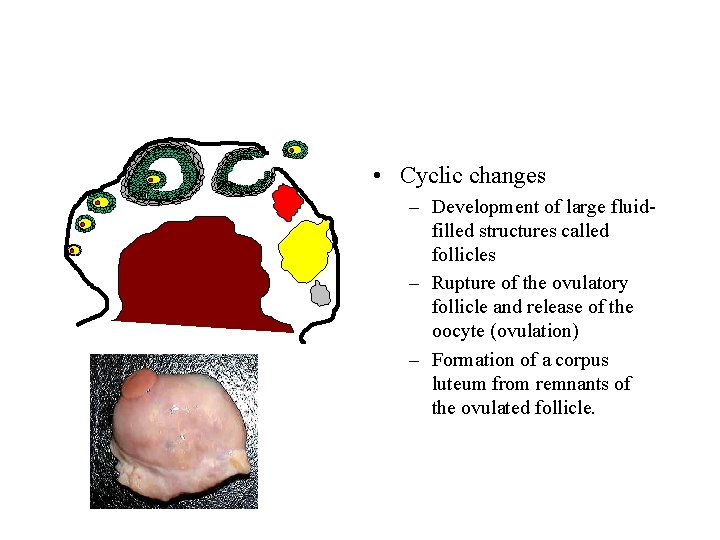
Structures on the Ovaries and Their Function • Ovaries – Characterized as an organ of constant change • A series of dynamic changes in a very predictable manner during the reproductive cycle.
• Cyclic changes – Development of large fluidfilled structures called follicles – Rupture of the ovulatory follicle and release of the oocyte (ovulation) – Formation of a corpus luteum from remnants of the ovulated follicle.
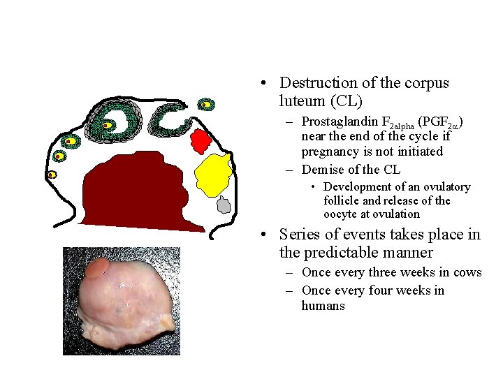
• Destruction of the corpus luteum (CL) – Prostaglandin F 2 alpha (PGF 2 a) near the end of the cycle if pregnancy is not initiated – Demise of the CL • Development of an ovulatory follicle and release of the oocyte at ovulation • Series of events takes place in the predictable manner – Once every three weeks in cows – Once every four weeks in humans
Association between cyclic changes in ovarian structures and hormone production • Production of two steroid hormones (Estradiol and Progesterone) – Key regulators of the reproductive cycle, reproductive behavior, and maintenance of pregnancy
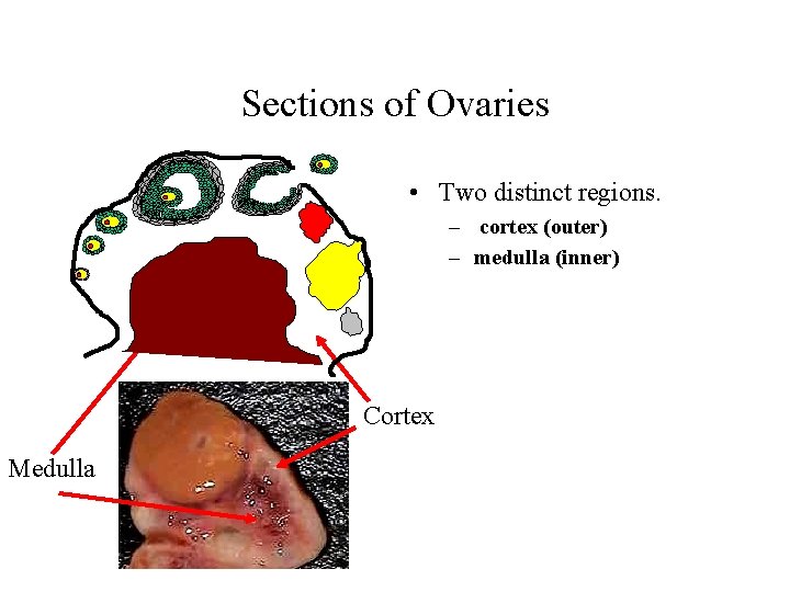
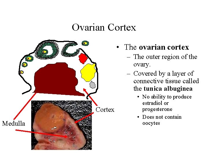
Sections of Ovaries • Two distinct regions. – cortex (outer) – medulla (inner) Cortex Medulla
Ovarian Cortex • The ovarian cortex – The outer region of the ovary. – Covered by a layer of connective tissue called the tunica albuginea Cortex Medulla • No ability to produce estradiol or progesterone • Does not contain oocytes
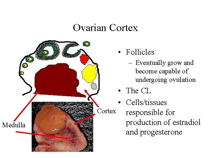
Ovarian Cortex • Follicles – Eventually grow and become capable of undergoing ovulation Medulla • The CL • Cells/tissues Cortex responsible for production of estradiol and progesterone
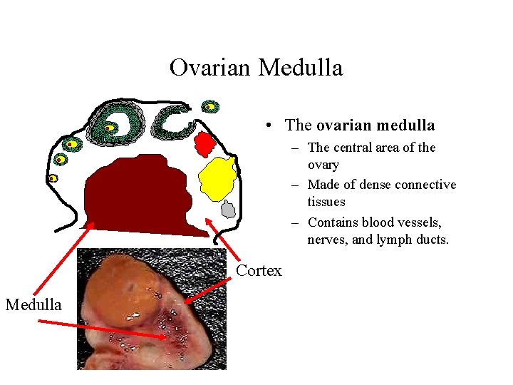
Ovarian Medulla • The ovarian medulla – The central area of the ovary – Made of dense connective tissues – Contains blood vessels, nerves, and lymph ducts. Cortex Medulla
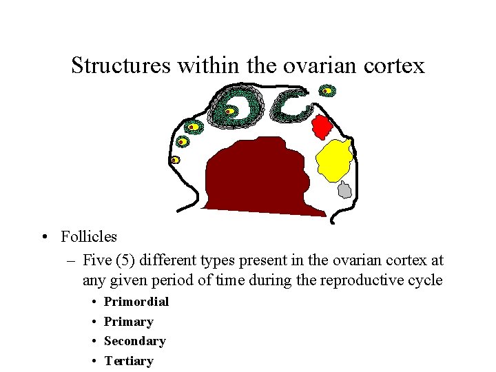
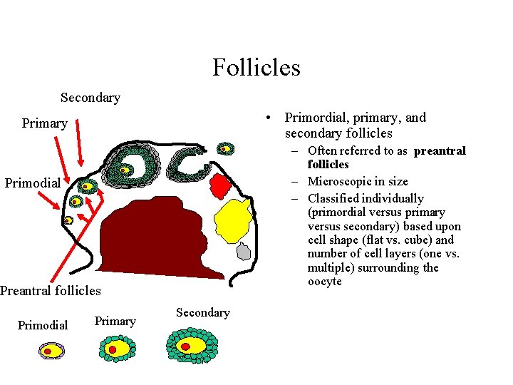
Structures within the ovarian cortex • Follicles – Five (5) different types present in the ovarian cortex at any given period of time during the reproductive cycle • • Primordial Primary Secondary Tertiary
Follicles Secondary • Primordial, primary, and secondary follicles Primary – Often referred to as preantral follicles – Microscopic in size – Classified individually (primordial versus primary versus secondary) based upon cell shape (flat vs. cube) and number of cell layers (one vs. multiple) surrounding the oocyte Primodial Preantral follicles Primodial Primary Secondary
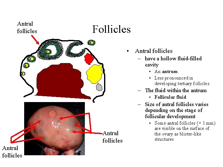
Antral follicles Follicles • Antral follicles – have a hollow fluid-filled cavity • An antrum. • Less pronounced in developing tertiary follicles. – The fluid within the antrum • Follicular fluid – Size of antral follicles varies depending on the stage of follicular development Antral follicles • Some antral follicles (> 1 mm) are visible on the surface of the ovary as blister-like structures
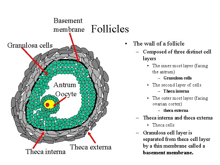
Basement membrane Follicles • The wall of a follicle Granulosa cells – Composed of three distinct cell layers • The inner most layer (facing the antrum) – Granulosa cells Antrum Oocyte • The second layer of cells – Theca interna • The outer most layer (facing ovarian cortex) – theca externa – Theca interna and theca externa • Theca cells Theca interna Theca externa – Granulosa cell layer is separated from theca cell layer by a thin membrane called a basement membrane.
Follicles • Both theca and granulosa cells are involved in production of estradiol – Theca cells produce androgens – Granulosa cells convert androgens to estradiol • Granulosa cells – Nurse cells by producing • Numerous materials essential for development of the oocyte
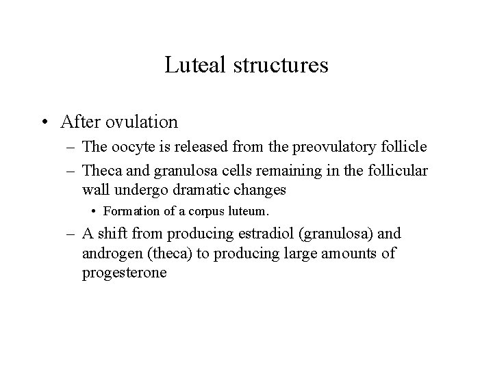
Luteal structures • After ovulation – The oocyte is released from the preovulatory follicle – Theca and granulosa cells remaining in the follicular wall undergo dramatic changes • Formation of a corpus luteum. – A shift from producing estradiol (granulosa) androgen (theca) to producing large amounts of progesterone
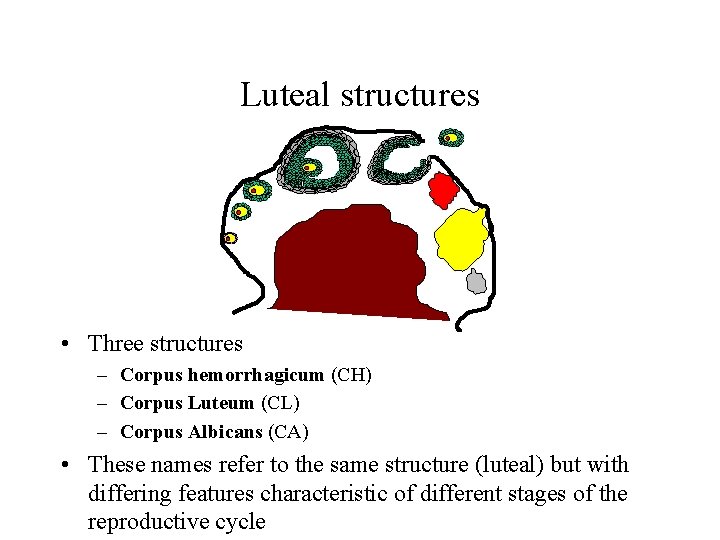
Luteal structures • Three structures – Corpus hemorrhagicum (CH) – Corpus Luteum (CL) – Corpus Albicans (CA) • These names refer to the same structure (luteal) but with differing features characteristic of different stages of the reproductive cycle
Luteal structures • The corpus hemorrhagicum CH (bloody body) – During the early part of the luteal phase of the reproductive cycle – Appears red • Small blood vessels within the follicle rupture during ovulation • Collapse of follicular wall into many folds after leakage of follicular fluid into many folds CH Developing CH Early CH
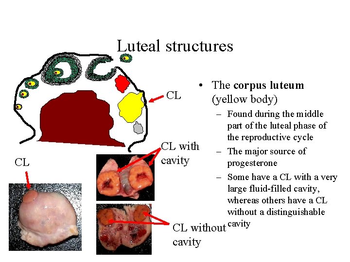
Luteal structures CL CL • The corpus luteum (yellow body) – Found during the middle part of the luteal phase of the reproductive cycle CL with – The major source of cavity progesterone – Some have a CL with a very large fluid-filled cavity, whereas others have a CL without a distinguishable CL without cavity
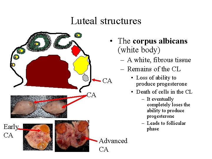
Luteal structures • The corpus albicans (white body) – A white, fibrous tissue – Remains of the CL CA CA Early CA • Loss of ability to produce progesterone • Death of cells in the CL – It eventually completely loses the ability to produce progesterone – Leads to follicular phase Advanced CA
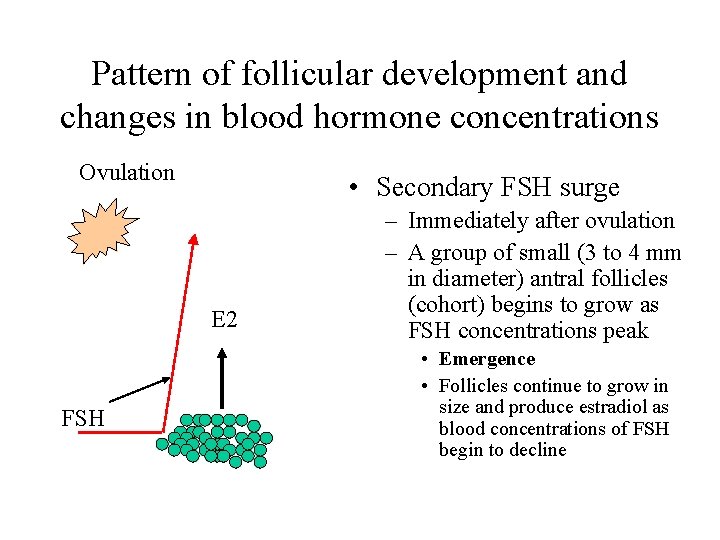
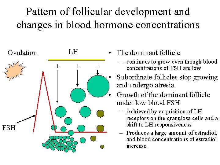
Pattern of follicular development and changes in blood hormone concentrations Ovulation • Secondary FSH surge E 2 FSH – Immediately after ovulation – A group of small (3 to 4 mm in diameter) antral follicles (cohort) begins to grow as FSH concentrations peak • Emergence • Follicles continue to grow in size and produce estradiol as blood concentrations of FSH begin to decline
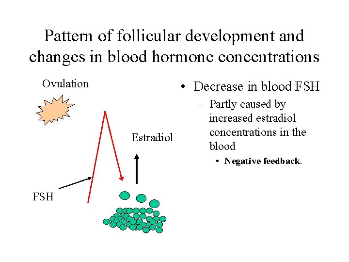
Pattern of follicular development and changes in blood hormone concentrations Ovulation • Decrease in blood FSH Estradiol – Partly caused by increased estradiol concentrations in the blood • Negative feedback. FSH
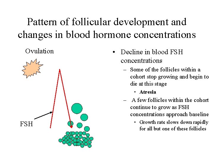
Pattern of follicular development and changes in blood hormone concentrations Ovulation • Decline in blood FSH concentrations – Some of the follicles within a cohort stop growing and begin to die at this stage • Atresia – A few follicles within the cohort continue to grow as FSH concentrations approach baseline FSH • Growth rate slows down rapidly for all but one of these follicles
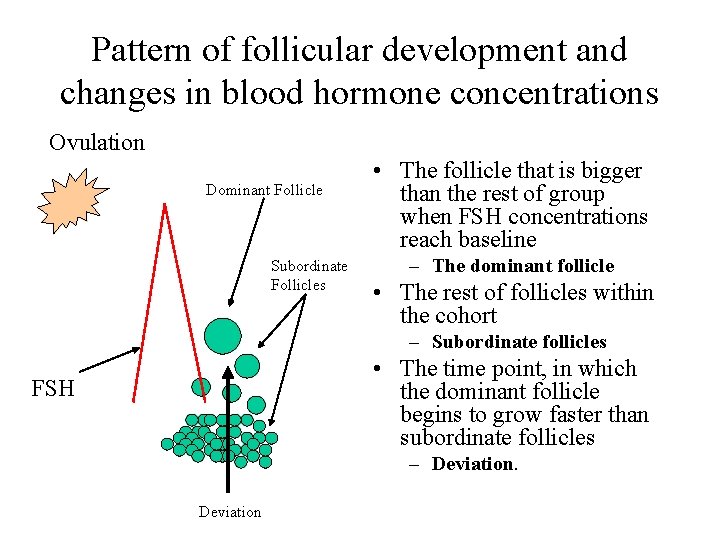
Pattern of follicular development and changes in blood hormone concentrations Ovulation Dominant Follicle Subordinate Follicles • The follicle that is bigger than the rest of group when FSH concentrations reach baseline – The dominant follicle • The rest of follicles within the cohort – Subordinate follicles • The time point, in which the dominant follicle begins to grow faster than subordinate follicles FSH – Deviation
Pattern of follicular development and changes in blood hormone concentrations LH Ovulation + + • The dominant follicle + – continues to grow even though blood concentrations of FSH are low • Subordinate follicles stop growing and undergo atresia • Growth of the dominant follicle under low blood FSH – Achieved by acquisition of LH receptors on the granulosa cells and a shift to LH responsiveness – Produces a large amount of estradiol, and blood concentrations of estradiol increase.
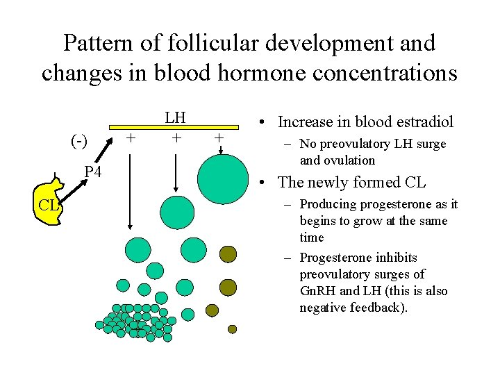
Pattern of follicular development and changes in blood hormone concentrations (-) P 4 CL + LH + + • Increase in blood estradiol – No preovulatory LH surge and ovulation • The newly formed CL – Producing progesterone as it begins to grow at the same time – Progesterone inhibits preovulatory surges of Gn. RH and LH (this is also negative feedback).
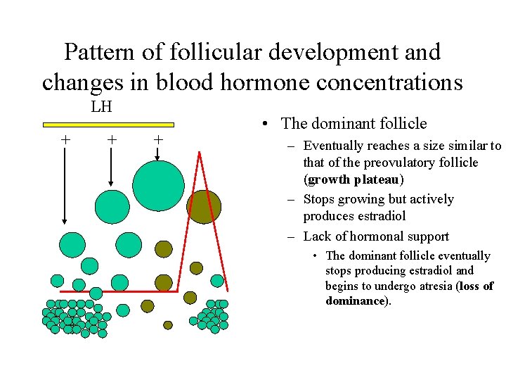
Pattern of follicular development and changes in blood hormone concentrations LH + + + • The dominant follicle – Eventually reaches a size similar to that of the preovulatory follicle (growth plateau) – Stops growing but actively produces estradiol – Lack of hormonal support • The dominant follicle eventually stops producing estradiol and begins to undergo atresia (loss of dominance).
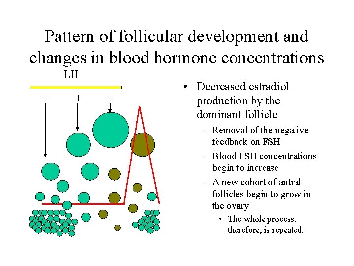
Pattern of follicular development and changes in blood hormone concentrations LH + + + • Decreased estradiol production by the dominant follicle – Removal of the negative feedback on FSH – Blood FSH concentrations begin to increase – A new cohort of antral follicles begin to grow in the ovary • The whole process, therefore, is repeated.
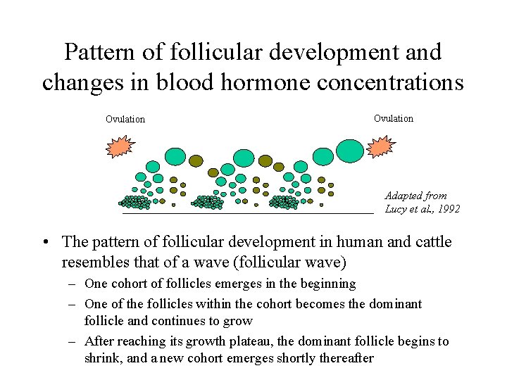
Pattern of follicular development and changes in blood hormone concentrations Ovulation CL CH Ovul ation Ovulation C Ovul ation Adapted from Lucy et al. , 1992 • The pattern of follicular development in human and cattle resembles that of a wave (follicular wave) – One cohort of follicles emerges in the beginning – One of the follicles within the cohort becomes the dominant follicle and continues to grow – After reaching its growth plateau, the dominant follicle begins to shrink, and a new cohort emerges shortly thereafter
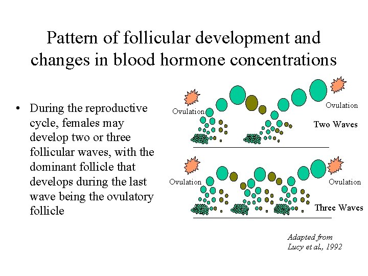
Pattern of follicular development and changes in blood hormone concentrations CL • During the reproductive cycle, females may develop two or three follicular waves, with the dominant follicle that develops during the last wave being the ovulatory follicle Ovulation O v ul at io n Ovulation Ov ula tio n CH Ovulation Two Waves CH Ovulation Ov ula tio. Waves Three n Adapted from Lucy et al. , 1992
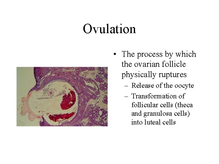
Ovulation • The process by which the ovarian follicle physically ruptures – Release of the oocyte – Transformation of follicular cells (theca and granulosa cells) into luteal cells
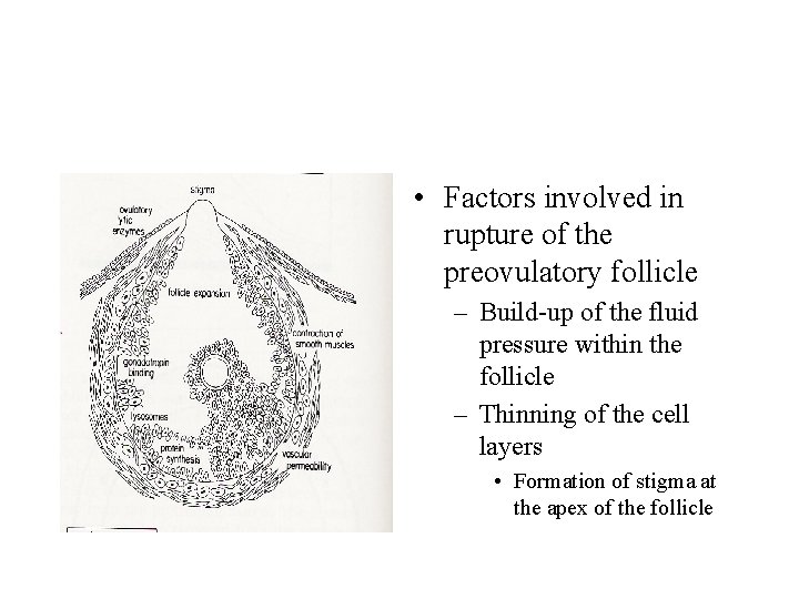
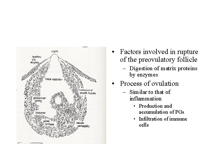
• Factors involved in rupture of the preovulatory follicle – Build-up of the fluid pressure within the follicle – Thinning of the cell layers • Formation of stigma at the apex of the follicle
• Factors involved in rupture of the preovulatory follicle – Digestion of matrix proteins by enzymes • Process of ovulation – Similar to that of inflammation • Production and accumulation of PGs • Infiltration of immune cells
Regulation of luteal function • Luteal cells – Small (around 25 % of total cells in the CL) • Very small contribution to basal production of progesterone • Responds to LH and produce progesterone (5 to 20 X above the basal level) – Large (around 10 % of total cells in the CL) • Very high • No significant response to LH
• Hormonal factors – LH • Extremely crucial during development • May not be necessary during the middle of luteal phase – Estradiol • Only in some species (i. e. rabbits) – Prolactin • During early stage of pregnancy in rats and mice
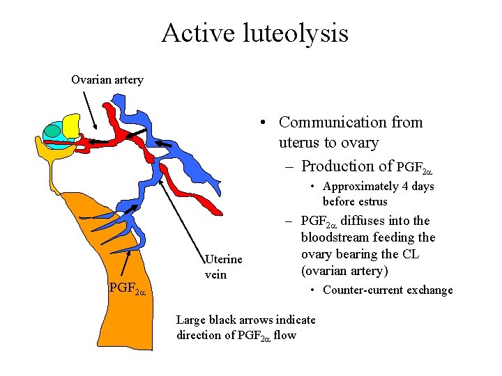
Luteolysis • Death of luteal structure – Formation of CA • Active or passive – Active • Uterine secretion of luteolytic agent – PGF 2 a – Passive • Loss of luteotropic agent
Active luteolysis Ovarian artery • Communication from uterus to ovary – Production of PGF 2 a • Approximately 4 days before estrus PGF 2 a Uterine vein – PGF 2 a diffuses into the bloodstream feeding the ovary bearing the CL (ovarian artery) • Counter-current exchange Large black arrows indicate direction of PGF 2 a flow
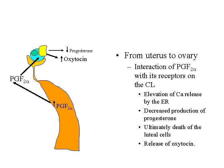
Progesterone Oxytocin • From uterus to ovary – Interaction of PGF 2 a with its receptors on the CL PGF 2 a • Elevation of Ca release by the ER • Decreased production of progesterone • Ultimately death of the luteal cells • Release of oxytocin.
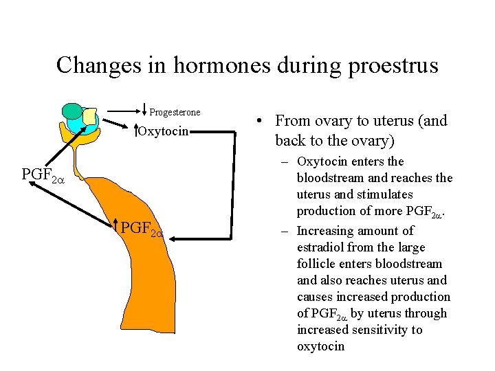
Changes in hormones during proestrus Progesterone Oxytocin PGF 2 a • From ovary to uterus (and back to the ovary) – Oxytocin enters the bloodstream and reaches the uterus and stimulates production of more PGF 2 a. – Increasing amount of estradiol from the large follicle enters bloodstream and also reaches uterus and causes increased production of PGF 2 a by uterus through increased sensitivity to oxytocin
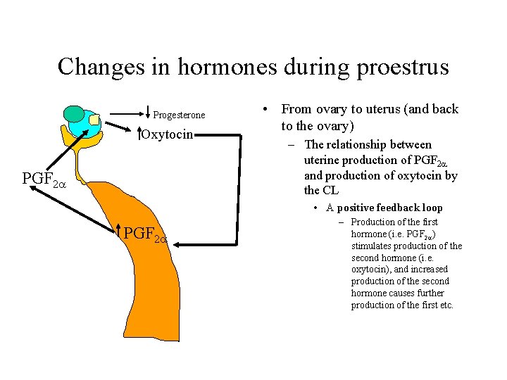
Changes in hormones during proestrus Progesterone Oxytocin PGF 2 a • From ovary to uterus (and back to the ovary) – The relationship between uterine production of PGF 2 a and production of oxytocin by the CL • A positive feedback loop PGF 2 a – Production of the first hormone (i. e. PGF 2 a) stimulates production of the second hormone (i. e. oxytocin), and increased production of the second hormone causes further production of the first etc.
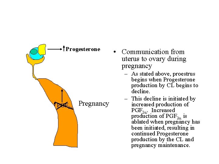
Progesterone PGF 2 a Pregnancy • Communication from uterus to ovary during pregnancy – As stated above, proestrus begins when Progesterone production by CL begins to decline. – This decline is initiated by increased production of PGF 2 a. Increased production of PGF 2 a is ablated when pregnancy has been initiated, resulting in continued Progesterone production by the CL and pregnancy maintenance.