Otitis Media Felasten Abed The ear consists of

Otitis Media Felasten Abed

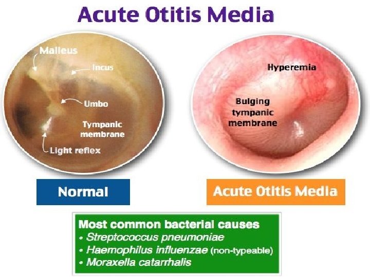
� The ear consists of three major parts: the outer ear, the middle ear, and the inner ear. The outer ear includes the pinna—the visible part of the ear—and the ear canal. The outer ear extends to the tympanic membrane or eardrum, which separates the outer ear from the middle ear. � The middle ear is an air-filled space that is located behind the eardrum. The middle ear contains three tiny bones, the malleus, incus, and stapes, which transmit sound from the eardrum to the inner ear. � The inner ear contains the hearing and balance organs. The cochlea contains the hearing organ which converts sound into electrical signals which are associated with the origin of impulses carried by nerves to the brain where their meanings are appreciated
 : is a short-lived (usually 1– 5 days)")
• Acute otitis media (AOM) : is a short-lived (usually 1– 5 days) infection of the middle ear. If it is viral it may last as little as a day or so but it can persist, causing pus to accumulate under pressure behind the eardrum, which may perforate Before the eardrum perforates, AOM is intensely painful. It mainly occurs in children. Recurrent otitis media (ROM) refers to repeated such episodes, typically more than three in a 6 -month period. • Otitis media with eff usion (OME) : is also common in children. Fluid – often thick sticky ‘glue’ – accumulates in the middle ear behind an intact drum. Because some fluid in the middle ear is normal for up to several weeks after an episode of AOM, the term OME requires that the fluid be persistent for at least 3 months. • Chronic otitis media : This implies that the eardrum has perforated, the perforation has failed to heal and there is ongoing infection. Th e term chronic suppurative otitis media (CSOM) is often used to emphasize the tendency for ears with longstanding perforations to become infected and discharge
 Duration • 2)Nature of fluid/discharge :")
Classification of otitis media according to: • 1) Duration • 2)Nature of fluid/discharge : suppurative and nonsuppurative • 3)Otitis media with effusion and Aero-otitis media • 4)Causative organism –viral /bacterial
 Acute otitis media (AOM):")
Clssification of OM according to the duratoin of illness 1) Acute otitis media (AOM): Rapid onset of symptoms < 3 weeks 2)Subacute otitis media : Symptoms lasting for 3 weeks to 3 months 3) Chronic otitis media : Symptoms lasting for 3 months or longer
 is the second most common disease of childhood. o")
Epidemiology o Otitis media (OM) is the second most common disease of childhood. o Most children will experience some form of acute otits media during their lifetime , 3 out 4 kids have had at least one ear infection by the time they reach 3 years of age. o The peak age of incidence is 6 months to 3 years old and it is rarely seen above 5 years of age. o Otitis media occurs more in the winter than summer months as it is usually associated with a cold. o It can occur in adults but this is unusual.

Risk factor of otitis media : o Boys are more likely than girls to develop otitis media. o Children with older siblings at school or nursery are exposed to infections that may be brought home. o Children who suffer with many colds or respiratory infections are more likely to develop OME. o Parent's smoking is thought to be associated with an increase in both acute and chronic otitis media as it is believed to increase S. pneumonia colonization.
 ET shorter, wider, more horizontal 2) Upper")
Why OM more common in childhood? 1) ET shorter, wider, more horizontal 2) Upper Resp tract infection is more 3) Adenoid obst 4)regurgitation of milk + vomitus
 • Acute otitis media is common and frequently bilateral. Most")
Acute otitis media (AOM) • Acute otitis media is common and frequently bilateral. Most children will develop one or more episodes typically before they are 2 years old. • It can follow an acute upper respiratory tract infection and may be viral or bacterial. A viral infection is short-lived (1 or 2 days) and often accompanied by some general features of an upper respiratory infection, e. g. pharyngitis and a runny nose.

pathogenesis • Acute otitis media is an infection of the mucous membrane of the whole middle-ear Eustachian tube, tympanic cavity, attic, aditus, mastoid antrum and air cells. • Th e bacteria responsible for acute otitis media are: Streptococcus pneumoniae 35% • Haemophilus influenzae 25% • Moraxella catarrhalis 15% • Group A streptococci and Staphylococcus aureus may also be responsible.

• Th e sequence of events in acute otitis media is as follows: 1) Organisms invade the mucous membrane causing inflammation, oedema, exudate and later pus. 2) Oedema closes the Eustachian tube, preventing aeration and drainage. 3) Pressure from the pus rises, causing the drum to bulge and perforate. 4) Most cases resolve completely. A small number cause complications or persistent perforation.
 Common cold 2) Acute tonsillitis 3)")
Causes of AOM : • More common 1) Common cold 2) Acute tonsillitis 3) Influenza 4) Coryza of measles, scarlet fever, whooping cough • Less common 1) Sinusitis 2) Haemotympanum 3) Trauma to the tympanic membrane 4) Barotrauma (air flight) 5) Diving 6) Temporal bone fracture
 : may be slight in a mild case, but")
Symptoms : • Earache (otalgia) : may be slight in a mild case, but more usually it is throbbing and severe. Th e child may cry and scream inconsolably until the ear perforates, the pain is relieved and peace is restored. • Deafness : is always present in acute otitis media but if the infection is unilateral this can go unnoticed. It is conductive in nature and may be accompanied by tinnitus. In an adult deafness or tinnitus may be the first complaint. • Discharge : Pressure builds up in the middle ear and the drum ruptures. Th e child gets immediate pain relief but the parents notice a sticky discharge, often purulent. The perforation formed in this way usually heals.
 Pyrexia : The child is flushed and ill. Th e")
• Signs 1) Pyrexia : The child is flushed and ill. Th e temperature may be as high as 40 °C. 2) Tenderness: There is usually some tenderness to pressure on the mastoid bone. 3)Mucoid (sticky) discharge from an ear must mean that there is a perforation of the tympanic membrane. Th ere are no mucous glands in the external canal. Otoscopy and interpreting the findings can be difficult in a fractious child.

The tympanic membrane varies in appearance according to the stage of the infection: 1. 2. 3. 4. 5. Loss of lustre and break-up of the light reflex. Congestion of the small vessels around the periphery and along the handle of the malleus. Redness and fullness of the drum; the malleus handle becomes more vertical. Bulging, with loss of landmarks. Purple colour. Outer layer may desquamate, causing bloodstained serous discharge. Early necrosis may be recognized, heralding imminent perforation. Perforation with otorrhoea, which will often be blood-stained. Profuse and mucoid at first, later becoming thick and yellow

Investigations • Usually no investigation is required. • Culture of discharge from ear may be indicated in chronic or recurrent perforation. • Audiometry should be performed if chronic hearing loss is suspected; however, not during acute infection. So diagnosis simply by detect an active infection in the middle ear is to look in the child’s ear with an otoscope, a light instrument that allows the physician to examine the outer ear and the eardrum. Inflammation of the eardrum indicates infection.
 2) 3) § The treatment depends on the stage reached")
Treatment § § 1) 2) 3) § The treatment depends on the stage reached by the infection. The following stages may be considered: Early bulging discharging. Most cases of AOM improve spontaneously. Cases that require treatment may be managed with antibiotics and analgesics or with observation alone. § The recommendations offer more rigorous diagnostic criteria to reduce unnecessary antibiotic use.

For early stage : 1. Antibiotics o Penicillin remains the drug of choice in most cases, and ideally should be given initially by injection followed by oral medication. o In children under 5 years, when Haemophilus influenzae is likely to be present, amoxycillin will be more effective, and should always be considered if there is not a rapid response to penicillin. o Co-amoxiclav is useful in Moraxella infections. o Be guided by sensitivity reports from the laboratory. 2. Analgesics o Simple analgesics, such as aspirin or paracetamol, should suffice. o Avoid the use of aspirin in children because of the risk of Reye’s syndrome

3. Nasal vasoconstrictors § The role of 0. 5% ephedrine nasal drops is traditional but its value is uncertain in the presence of acute inflammation of the middle ear. 4. Ear drops § Ear drops are of no value in acute otitis media with an intact drum. Especially illogical is the use of drops containing local anaesthetics, which can have no effect on the middle-ear mucosa yet may cause a sensitivity reaction in the meatal skin.

In bulging stage: § Myringotomy is necessary when bulging of the tympanic membrane persists, despite adequate antibiotic therapy. § It should be carried out under general anaesthesia in theatre and a large incision in the membrane should be made to allow the ear to drain. Pus should be sent for bacteriological assessment. § Following myringotomy, the ear will discharge and the outer meatus should be dry-mopped regularly.

In discharge : § Discharging—nature’s myringotomy § If the ear is already discharging when the patient is first seen, a swab should be sent for culture of the organism. Antibiotic therapy should be started but modified if necessary when the result of the sensitivities is known. § Regular aural toilet will be necessary. Further management Acute otitis media is not cured until the hearing and the appearance of the membrane have returned to normal. This can take several weeks and a persistent effusion of fluid in the middle ear is especially common in children. If there is no resolution suspect: 1) the nose, sinuses or nasopharynx; infection may be present; 2) low-grade infection in the mastoid cells. 3) the choice or dose of antibiotic

Recurrent Otitis Media o Some children are susceptible to repeated attacks of AOM. o There may be an underlying immunological deficit such as Ig. A deficiency or hypogammaglobulinaemia that will need to be investigated. o Long-term treatment with half-dose cotrimoxazole may be beneficial. o If the attacks persist, grommet insertion Tympanostomy tube may prevent further attacks but may result in purulent discharge.

Tympanostomy tube indication o people who have three or more episodes of acute otitis media in 6 months. o or four or more in a year, with at least one episode or more attacks in the preceding 6 months complication : o most common otorrhea Grommet insertion

COMPLICATIONS Otologic complications : o TM perforation, o chronic suppurative OM, o cholesteatoma, o ossicular necrosis o chronic otorrhoea Intracranial complications o meningitis, o extradural abscess, o brain abscess Other complications include: o mastoiditis o labyrinthitis osigmoid sinus thrombophlebitis.

CLINICAL PRACTICE POINTS • • Eardrops are of no value in acute otitis media with an intact drum. • • Adequate analgesia is essential. • • If antibiotics are withheld, make sure you can review the child after 24 h. • • Passive smoking predisposes children to otitis media.

Otitis Media with Effusion abla sultan qudah
, or ‘glue ear’, The condition is due")
INTRODUCTION • Otitis media with effusion (OME), or ‘glue ear’, The condition is due to the accumulation of non-infected fluid, either serous or viscous, within the middle-ear cleft, resulting in conductive deafness. • It is commonest in small children (6 months- 3 years) and those of primary school age and may cause significant deafness. • It occurs in adults, usually as a serous effusion and may rarely be a sign of nasopharyngeal malignancy. • self-limited , takes 4 -6 weeks to resolve

CAUSES OF OME • 1 - Nasopharyngeal obstruction, e. g. large adenoids(mc cause in children) or tumour resulting in Eustachian tube dysfunction. • 2 - untreated Acute otitis media, , will often give rise to a spontaneous perforation and drainage of the middle ear.

Cont. • 3 - Allergic rhinitis, often missed in children, will predispose to middle-ear effusions. • 4 - Parental smoking has been shown to predispose to OME in children. • 5 - OME is commoner in winter months. • 6 - In many cases of secretory otitis media, no cause is apparent.

SYMPTOMS § Not all children with OME have symptoms , painless § Symptoms are often mild or minimal. They can vary based on the child’s age. § It may be responsible for developmental and educational impairment (delayed speech development) , and if untreated may result in permanent middle-ear changes. § Deafness may be the only symptom. § Discomfort in the ear —rarely severe. § Occasionally, tinnitus or unsteadiness.

SIGNS • 1 Fluid in the middle ear —a variable appearance that may be difficult to recognize. 2 Dull appearance with radial vessels visible on the tympanic membrane and handle of the malleus. • 3 Retraction of the tympanic membrane. • 4 Yellow/orange tinge to tympanic membrane. •

Cont. • 5 Dark blue or grey colour of tympanic membrane. • 6 Hair lines or bubbles —rarely seen. • 7 Tuning fork tests show conductive deafness, i. e. bone conduction > air conduction.

DIAGNOSIS • 1 - Pneumatic otoscopy: Primary diagnostic • • Ø Ø method. Using an otoscope. The doctor will be looking for: Air bubbles on the tympanic membrane surface. A tympanic membrane that appear dull instead of smooth and shiny. Visible fluid behind the tympanic membrane. A tympanic membrane that does not move when a small amount of air is blown into it.

CONT. • 2 - Tympanometry: Ø Flat, type B tympanogram.

CONT. • 3 - Audiometry: Ø Unilateral conductive hearing loss.

TREATMENT >> In children • 1 Many cases will resolve spontaneously, and the child should usually be observed for 3 months before embarking on surgery. • 2 The use of antihistamines and mucolytics is of no proven benefit. Antibiotic therapy may help in the short term. Surgery is indicated if hearing loss persists for 3 months or if there is recurring pain. • 3 Surgical treatment.

Adenoidectomy • It has been shown that adenoidectomy is beneficial in the long-term resolution of OME. • The maximum benefit occurs between the ages of 4 and 8 years.

Myringotomy and grommet insertion • Under general anaesthetic, the tympanic membrane is incised anteroinferiorly. • The glue is aspirated and a grommet inserted into the incision. • The function of the grommet is to ventilate the middle ear and not to drain the fluid.

TREATMENT >> In adults • Examination of the nasopharynx to exclude tumour is essential, especially if the effusion is unilateral. • Under the same anaesthetic, a grommet may be inserted. • Secretory otitis media in adults not due to tumour usually follows a cold. Resolution is usually spontaneous, but may take up to 6 weeks.

Complications and Long-Term Effects of OME • If OME is associated with frequent ear infections, more complications can occur. • These can include: Ø Acute ear infections. Ø Cysts in the middle ear. Ø Eardrum scarring. Ø Damage to the ear that causes hearing loss. Ø Affected speech or language delay.

THANK YOU! REFERENCE: LECTURE NOTES ON Diseases of the Ear, Nose and Throat , 9 th ed 2002
- Slides: 42