Osteoporosis Strategies for Screening and Intervention March 2


- Slides: 66

Osteoporosis Strategies for Screening and Intervention March 2, 2005 Steven R. Brown, MD Family Physician, Whiteriver Indian Health Service Roy Teramoto, MD, MPH Maternal Child Health Consultant Phoenix Area Indian Health Service

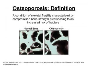
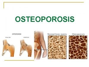
Definition A skeletal disorder characterized by compromised bone strength predisposing to an increased risk of fracture. NIH Consensus Statement 2000

Diagnostic Criteria Bone mineral density (BMD) 2. 5 standard deviations below the mean bone mineral density of healthy, young, white women (T-score of -2. 5) or n Fracture in the absence of significant trauma in a postmenopausal woman n World Health Organization working group 1992

Diagnostic Criteria Osteopenia – BMD 1 to 2. 5 standard deviations below the mean (T-score -1. 0 to -2. 5) n Unclear clinical value – wide range of fracture risk found among women in this category n

Gold Standard Central DEXA scan for bone mineral density, BUT true “gold standard” is fracture/fracture rates for public health purposes.

Epidemiology Rates vary among racial and ethnic groups n Data is best established for Caucasian females n Limited data available regarding osteoporosis and fracture rates in American Indian and Alaska Natives n Data suggests that they are at least as great a problem as for the general population n

Epidemiology National Osteoporosis Risk Assessment (NORA) n n Study of peripheral osteoporosis screening 1708 “Native American” women – risk of fracture over life of the study was the same as that of “white” women

Epidemiology Small study in the Sac and Fox Nation of Oklahoma Lower BMD in postmenopausal women as measured by central DEXA than that reported for “white” women The effect of aging on bone mineral metabolism and bone mass in Native American Women, J Am Geriatr Soci 1998; 46: 1418 -22

Hip Fracture Risk by Race/Ethnicity Relative Hazard (95% Confidence interval) White 1. 00 (referant) n African American 0. 54 (0. 41 -0. 72) n Native American 0. 89 (0. 59 -1. 34) n Hispanic 0. 91 (0. 72 -1. 15) n Asian 0. 41 (0. 21 -1. 79) n

Epidemiology Review of hip fractures at Alaska Native Medical Center Higher fracture rates than reported for white US women during the periods of 1979 -1989 and 1996 -1999 Incidence of hip fracture in Alaska Inuit people, Alaska Med; 2001, 43: 2 -5

Risk Factors for Osteoporosis (AI/AN) n Low calcium intake n Sedentary lifestyle n Issues with body mass index n Smoking Osteoporosis in Native Americans, IHS Provider 2002; 94: 94 -101

Clinical Risk Factors for Hip Fracture n Skeletal History of maternal hip fracture (RR=2. 0) n Age, per 5 year (RR=1. 5) n Current cortisone use (RR=1. 57) n Current smoking (RR=1. 14 - 2. 1) n Low Body Mass Index (BMI >30 has RR 0. 16 of having osteoporosis) n Race (African-American RR= 0. 54) n

Skeletal status by age

Clinical Risk Factors for Hip Fracture Use of anticonvulsant drugs (RR=2. 8) n Serious long-term conditions thought to increase fracture risk such as hyperthyroidism or malabsorption n

Clinical Risk Factors for Hip Fracture n Non-skeletal Poor vision (RR=1. 5) n Inability to rise from chair (RR=2. 1) n Benzodiazapine use (RR=1. 6) n Fall in the previous year (RR=1. 6) n Self-rated health status of fair/poor (RR=1. 79) n

The Osteoporosis Pyramid 1

Risk Factors for Falls Risk Factor Mean RR-OR Muscle weakness 4. 4 History of falls 3. 0 Gait deficit 2. 9 Balance deficit 2. 9 Use assistive device 2. 6 Visual deficit 2. 5 From: Guidelines for the Prevention of Falls in Older Persons, Journal of the American Geriatric Society 49: 664 -672, 2001

Approach to Older Persons as Part of Routine Care (Not Presenting After A Fall) 1. 2. All older persons who are under the care of a health professional (or their caregivers) should be asked at least once a year about falls. All older persons who report a single fall should be observed as they stand up from a chair without using their arms, walk several paces, and return (i. e. , the “Get Up and Go Test”). Those demonstrating no difficulty or unsteadiness need no further assessment. From: Guidelines for the Prevention of Falls in Older Persons, Journal of the American Geriatric Society 49: 664 -672, 2001

Approach to Older Persons as Part of Routine Care (Not Presenting After A Fall) 3. Persons who have difficulty or demonstrate unsteadiness performing this test require further assessment. From: Guidelines for the Prevention of Falls in Older Persons, Journal of the American Geriatric Society 49: 664 -672, 2001

Approach to Older Persons Presenting with One or More Falls or Have Abnormalities of Gait and/or Balance, or Who Report Recurrent Falls Older persons who should have a fall evaluation performed: n Present for medical attention because of a fall n Report recurrent falls in the past year n Demonstrate abnormalities of gait and/or balance.

Approach to Older Persons Presenting with One or More Falls or Have Abnormalities of Gait and/or Balance, or Who Report Recurrent Falls Fall Evaluation Should be performed by a clinician with appropriate skills and experience n May necessitate referral to a specialist (e. g. , geriatrician). n

Approach to Older Persons Presenting with One or More Falls or Have Abnormalities of Gait and/or Balance, or Who Report Recurrent Falls Fall evaluation n Assessment that includes the following: n n n A history of fall circumstances Medications Acute or chronic medical problems Mobility levels Examination of vision Examination of gait, balance and lower extremity joint function

Approach to Older Persons Presenting with One or More Falls or Have Abnormalities of Gait and/or Balance, or Who Report Recurrent Falls n Examination of basic neurological function n Mental status n Muscle strength n Lower extremity peripheral nerves n Proprioception n Reflexes n Tests of cortical, extrapyramidal, and cerebellar functions

Approach to Older Persons Presenting with One or More Falls or Have Abnormalities of Gait and/or Balance, or Who Report Recurrent Falls n Assessment of basic cardiovascular status including heart rate and rhythm n Postural pulse and blood pressure n If appropriate, heart rate and blood pressure responses to carotid sinus stimulation.

Approaches Demonstrating Benefit Muscle strengthening and balance retraining n Professional home hazard assessment and modification n Stopping or reducing psychotropic medication n NEJM 348: 42 -9, 2003; NEJM 331: 821 -7, 1994 Note: Optimal duration or intensity of these approaches have not been defined

Hip Protectors/Pads Hip protectors reduce the risk of hip fracture for elderly individuals who live in nursing homes and residential care facilities, as well as those in supported living at home n The generalization of the results beyond this high risk population is unknown n

Hip Protectors/Pads One study found that 41 patients would need to be offered treatment with a hip protector to prevent one hip fracture over the course of one year (NEJM 343: 1506 -13; 2000) n Hip protectors may be a reasonable and costeffective option for patients at high risk of falls. n

Hip Protectors/Pads Randomized controlled trial among women over 70 living in the community found that hip protectors offered no significant reduction in the risk of hip fracture (Birks -Osteoporos Int 2004, Mar 3) n Compliance rates were quite low (about 30%) n Need for more acceptable, easy-to-use devices that can protect fragile bones n


The Third Level of the Pyramid: Medical Interventions n Bisphonates n n n n Alendronate Risedronate Calcium + Vitamin D Calcitonin Estrogen Selective estrogen receptor modulator (Raloxifene) Teriparatide (recombinant human parathyroid hormone)

POEM vs. DOE n POEM: Patient Oriented Evidence that Matters n n Hip and Vertebral Fractures DOE: Disease Oriented Evidence n Bone Density

Intervention: Alendronate In postmenopausal women with osteoporosis does alendronate compared to placebo reduce hip and vertebral fractures? n Answer: Good evidence based on randomized, controlled trials… n

Treatment of Osteoporosis: The Fracture Intervention Trial 2 (FIT)

FIT: Hip fracture Population: 3658 white women age 55 -80 with osteoporosis (femoral-neck T-score <-2. 5 or existing vertebral fracture. ) * = achieved statistical significance

FIT: Vertebral Fracture Population: 3658 white women age 55 -80 with osteoporosis (femoral-neck T-score <-2. 5 or existing vertebral fracture. ) * = achieved statistical significance

FIT: Number Needed to Treat (NNT)

Alendronate: Harms n n n “Esophagitis, esophageal ulcers and esophageal erosions, occasionally with bleeding and rarely followed by esophageal stricture or perforation, have been reported in patients receiving treatment with alendronate sodium. In some cases these have been severe and required hospitalization. ” 3 One RCT found that when taken correctly, there was no significant difference in esophagitis with alendronate versus placebo 4 From the SG: “appears to be safe and effective for up to 10 years. ”

Interventions n n n Calcium and Vitamin D In mobile elderly women in nursing homes does Calcium and Vit D compared to placebo reduce hip fracture? Yes, quite impressively. The NNT at 3 years to prevent one hip fracture in this population is 25. NNT at 18 months to prevent one hip fracture is 50.


Ref 5

Key Question #1 in the analytic framework from the USPSTF 5 n n n Does screening for osteoporosis using risk factor assessment or bone density testing reduce fractures? USPSTF answer: “We identified no studies about the effectiveness of screening in reducing osteoporotic fractures. ” Therefore: “Recommendations about screening need to rely on evidence that risk factor assessment or bone density testing can adequately identify women who could ultimately benefit from treatment. ”

Risk Factor Screening Osteoporosis Risk Assessment Instrument (ORAI)6 is one of two validated risk factor assessment tools given a “good” quality rating by the USPSTF 5 n 95 % sensitive and 41 % specific for detecting hip or lumbar spine BMD T-score <-2. 5. n

Risk Factor Screening n ORAI scoring 6

Radiologic Screening Methods n DXA (dual energy x-ray absorptiometry) n n n Peripheral Densitometry (DXA or SXA) n n Distal radius Calcaneus Finger Quantitative Computed Tomography n n Hip Spine Quantitative Ultrasound n n Calcaneus Tibia

“Looking to the future…” 1

Radiologic Screening Methods n 1996 BMJ meta-analysis 7 showed all measuring sites had similar predictive abilities for fracture. n RR 1. 5 (CI 1. 4 -1. 6) for 1 SD decrease in BMD for age (Z-score) except: n Measurement at spine for vertebral fractures: RR 2. 3 (CI 1. 9 -2. 8) n Measurement at hip for hip fractures: RR 2. 6 (CI 2. 03. 5)

Screening Methods n n n As stated by the USPSTF: “the probability of receiving a diagnosis of osteoporosis depends on the choice of test and site. ” USPSTF: “the likelihood of receiving a diagnosis of osteoporosis also depends on the number of sites tested. ” National Osteoporosis Risk Assessment 8: n n the probability of having osteoporosis varied from 3. 4% (heel ultrasound) to 13. 5% (finger DXA) depending on site tested and method of testing. Odds of osteoporosis by site tested: Heel ultrasound OR 0. 79, DXA at forearm or finger OR 2. 86, 4. 82.

Screening Methods Again from the USPSTF: “DXA is considered the gold standard because it is the most extensively validated test against fracture outcomes. ” n Randomized, controlled trials, such as FIT, use femoral neck DXA to identify patients with osteoporosis that might benefit from therapy. n

Number needed…

Number needed to screen 5

Screening Methods: Ultrasound n n n EPIDOS 9: 1996 prospective study of 5662 elderly women with 115 hip fractures in average follow-up of two years Patients had calcaneal ultrasound (Lunar Corp. ) and femoral DXA Relative risk of hip fracture for 1 SD reduction n in ultrasound attenuation at heel 2. 0 (CI 1. 6 -2. 4) in hip BMD 1. 9 (CI 1. 6 -2. 4) Author conclusions: Calcaneal ultrasound “predicts the risk of hip fracture as efficiently as DXA. ”

Screening Methods: Ultrasound n n The 1997 Study of Osteoporotic Fractures Research Group 10 followed 6189 women over 65 for an average of 2 years 54 hip fractures Calcaneal quantitative ultrasound (Walker-Sonix) and hip and calcaneal DXA were performed Relative risk of hip fracture for 1 SD reduction n In heel ultrasound attenuation 2. 0 (1. 5 -2. 7) In hip BMD 2. 6 (1. 9 -3. 8) Author conclusion: “the strength for the association between BUA and fracture is similar to that observed with bone mineral density. ”

An ultrasound study in primary 11 care n n 200 women age 60 -69 in British general practice had DXA and heel ultrasound (Sahara) and risk factor screening Ultrasound T-score <1. 7 SD (lowest quartile) compared to a “gold standard” of osteoporosis on DXA yielded n n sensitivity of 71% and specificity of 83% PPV of 45% and NPV of 94% 16. 3 % of patients had osteoporosis Including risk factor screening n n Sensitivity 90%, specificity 38% PPV 22%, NPV 95%

A Public Health Approach to a Fracture Prevention Program at Your Service Unit n Why? n n n It will help your patients “live long and live well. ” October 2004: “The intent of this Surgeon General’s Report is to serve as a catalyst for the development of a public health approach to promoting bone health. ” The Surgeon General urges: “implement a comprehensive, systems-based approach to promoting bone health. ” 2002 -2011 Decade of Bone and Joint Healthy People 2010 n n “reduce the number of individuals with osteoporosis and hip fractures…” HEDIS, GPRA

Outcomes and Cost/Effort Designing an approach at your Service Unit


Designing a Fracture Prevention Approach at your Service Unit n Avoid pitfalls No screening program n Not identifying and treating the highest risk individuals n Not appropriately utilizing cost/effort n Focusing on unproven technologies if DEXA is available n Remember the pyramid n

Fracture Prevention Program: Avoiding Pitfalls n. Pitfall #1: program No screening

Fracture Prevention Program: Avoiding Pitfalls n Pitfall #2: Not identifying and treating the highestrisk individuals n n Consider secondary osteoporosis (e. g. ESRD, rheumatoid arthritis, steroid use) Treat patients with fragility fractures (the “sentinel event”) n n Only 22 -24% of patients with fractures treated in two recent studies A recent controlled trial showed improved treatment after fracture with faxed reminders to physicians (still only 40%). Do not delay treatment for imaging Screen women over 75 and frail elderly first

Fracture Prevention Program: Avoiding Pitfalls n Pitfall #3: Not appropriately utilizing cost and effort Do not prioritize screening young, well-nourished, otherwise healthy, women. n Initiate population screening no earlier than 65 years of age n

Number needed to screen 5

Fracture Prevention Program: Avoiding Pitfalls n Pitfall #4: Focusing on unproven technologies if DEXA is available

Fracture Prevention Program: Avoiding Pitfalls n Pitfall #5: Remember the Pyramid! (and don’t invert it. ) n Use Calcium n NNT in some populations 25

“When so many hours have been spent in convincing myself that I am right, is there not some reason to fear I may be wrong? ” - Jane Austen

References 1 2 3 4 5 6 7 8 US Dept of HHS. Bone health and osteoporosis: A Report of the Surgeon General. Rockville, MD: HHS, Office of the Surgeon General, 2004. Black DM, et al. Fracture risk reduction with alendronate in women with osteoporosis: The fracture intervention trial. J Clin Endocrinol Metab 2000; 85: 4118 -4124. Mosby’s Drug Consult 2003, accessed on mdconsult. com 12/1/03 Bruyere O, et al. Fracture prevention in postmenopausal women. Clinical Evidence 2003; 9. BMJ Publishing Group. Nelson HD, et al. Screening for postmenopausal osteoporosis: a review of the evidence for the USPSTF. Ann Intern Med 2002; 137: 529 -41. Cadarette SM, et al. Development and validation of the Osteoporosis Risk Assessment Instrument to facilitate selection of women for bone densitometry. CMAJ 2000; 162(9): 12891294. Marshall D, Johnell O, Wedel H. Meta-analysis of how well measures of bone mineral density predict occurrence of osteoporotic fractures. BMJ 1996; 312: 125459. Siris ES, et al. Identification and fracture outcomes of of undiagnosed low bone mineral density in postmenopausal women. Results from the National Osteoporosis Risk Assessment. JAMA 2001; 286: 2815 -2822.

References 9 10 11 Hans D, et al. Ultrasonographic heel measurements to predict hip fracture in elderly women: the EPIDOS prospective study. Lancet 1996; 348: 511 -514. Bauer DC, et al. Broadband ultrasound attenuation predicts fractures strongly and independently of densitometry in older women. Arch Intern Med 1997; 157: 629 -634. Hodson J, Marsh J. Quantitative ultrasound and risk factor enquiry as predictors of postmenopausal osteoporosis: comparative study in primary care. BMJ 2003; 326: 1250 -1251.
 March march dabrowski
 Tbi intervention strategies
 Comprehensive intervention in od
 Culturally appropriate intervention strategies
 Group intervention strategies
 Pes statement for osteoporosis
 Osteoporosis t score
 Is osteoporosis hypokinetic or hyperkinetic
 Osteoporosis defination
 Nursing diagnosis for osteoporosis
 Woc atrial fibrilasi
 Is osteoporosis additive or destructive
 Royal osteoporosis society leaflets
 Osteoporosis
 Eva decroli
 Alendronate mechanism
 Tbs osteoporosis
 Defined
 Bendaggio
 Whole muscle
 Fspos vägledning för kontinuitetshantering
 Novell typiska drag
 Tack för att ni lyssnade bild
 Returpilarna
 Shingelfrisyren
 En lathund för arbete med kontinuitetshantering
 Personalliggare bygg undantag
 Personlig tidbok fylla i
 A gastrica
 Densitet vatten
 Datorkunskap för nybörjare
 Stig kerman
 Debattartikel struktur
 Delegerande ledarstil
 Nyckelkompetenser för livslångt lärande
 Påbyggnader för flakfordon
 Tryck formel
 Offentlig förvaltning
 Jag har gått inunder stjärnor text
 Presentera för publik crossboss
 Teckenspråk minoritetsspråk argument
 Plats för toran ark
 Treserva lathund
 Mjälthilus
 Claes martinsson
 Cks
Cks Verifikationsplan
 Mat för unga idrottare
 Verktyg för automatisering av utbetalningar
 Rutin för avvikelsehantering
 Smärtskolan kunskap för livet
 Ministerstyre för och nackdelar
 Tack för att ni har lyssnat
 Mall för referat
 Redogör för vad psykologi är
 Stål för stötfångarsystem
 Atmosfr
 Borra hål för knoppar
 Orubbliga rättigheter
 Varians
 Tack för att ni har lyssnat
 Rita perspektiv
 Verksamhetsanalys exempel
 Tobinskatten för och nackdelar
 Blomman för dagen drog
 Gibbs reflekterande cykel
 Egg för emanuel

