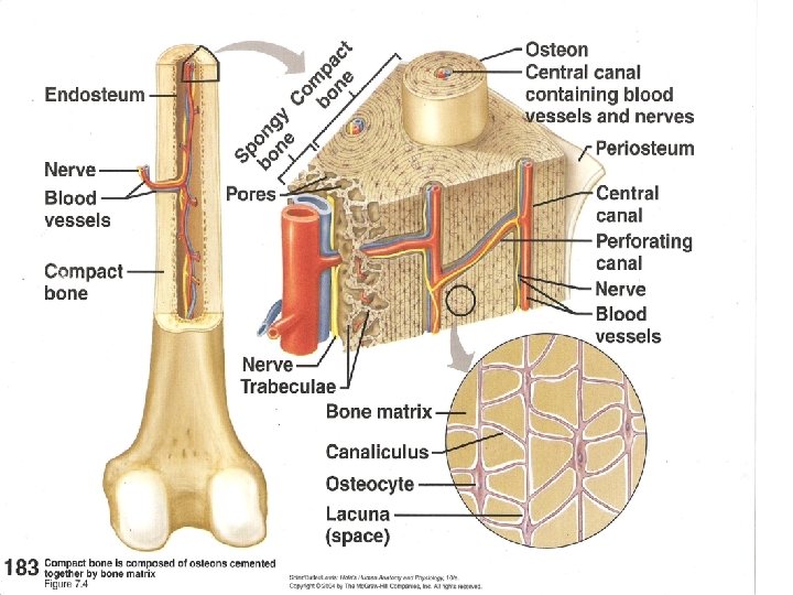
OsteonHaversion Systemfunctional unit of bone Central osteonic canal
 canal - for B. V. , and")
Osteon/Haversion Systemfunctional unit of bone Central (osteonic) canal - for B. V. , and nerves Lamellae – sheets/layers of matrix Lacunae – space for a bone cell

Canaliculi – tiny interconnecting channels that allow the osteocytes to communicate w/ each other (pass waste and nutrients) Perforating canal – allows the vessel in the osteonic canal to connect with the source blood vessel in the medullary cavity [Volkman’s] Circumferential lamellae – layers of bone tissue just beneath the periosteum

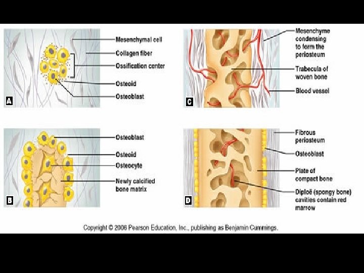
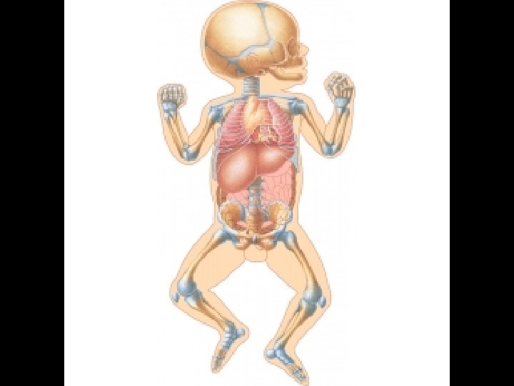
Bone Growth & Development OSSIFICATION=Formation of bone During first eight weeks of embryonic dev’t
 A. osteoblasts cluster B.")
1. Intramembranous bones – (flat bones of skull, mandible, clavicle) A. osteoblasts cluster B. secrete matrix which forms thin plates and becomes spongy bone C. o‘blasts become trapped in lacunae (become o‘cytes); periosteum forms D. o‘blasts in the periosteum form a layer of compact bone over the spongy bone
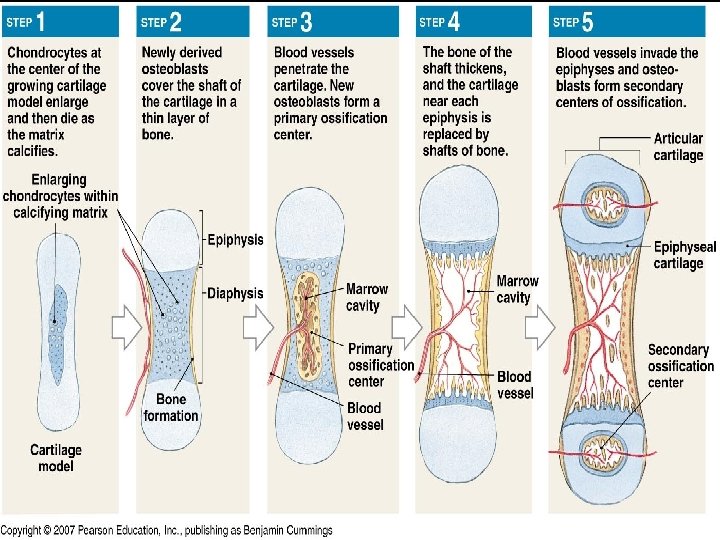
 CHONDROCYTES-cartilage")
2. Endochondral Bones (All but the clavicle, flat bones of skull and mandible) CHONDROCYTES-cartilage cells A. chondroblasts produce hyaline cartilage forms the shape of the bone it will become (template) B. chondroblasts become surrounded by cartilage – now known as chondrocytes C. B. V. penetrate – bring inc. # of o’blasts, some chondros die, others turn into o’blasts.

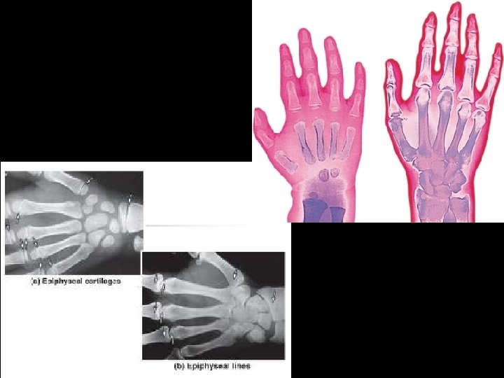
• D. Cartilage is destroyed, beginning in the center of diaphysis – forms primary (10) ossification center - cartilage is replaced w/ bone • E. Secondary (20) ossif. centers form in the epiphyses. Almost all cartilage is replaced with bone – some remains … • F. Artic. cart. remains on the outside of epiphyses at joint surfaces • G. Thin line of cartilage remains (epiphyseal/ growth plate) at border of epiph. and diaph.
- Slides: 12