Osteology2 Department of Anatomy Luzhou medical college Edited
 Department of Anatomy Luzhou medical college Edited by professor Xiao")


- Slides: 67
Osteology(2) Department of Anatomy Luzhou medical college Edited by professor Xiao
Review the contents of the last lesson 1. The suppose axes of the human body are? 2. The sections or planes of the human body on the basis of the axis? 3. Which types of the development of the bone? (membranous and cartilaginous ossification) 4. The characteristics of the long bone? ¡ (Epiphyses surface) , Epiphysial cartilage, Epiphysial lines, Articular 5. Which bone exists the red marrow in life? 6. The periosteum (Endosteum) are two layers (outer is the fibrous and inner the membrenous )(Osteoblast, Osteoclast)
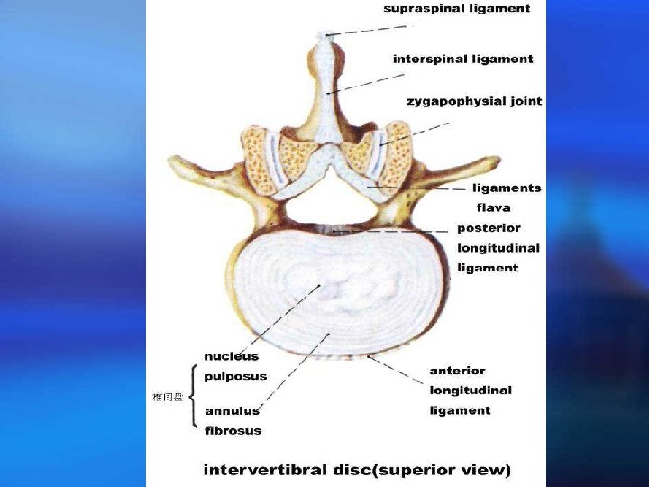
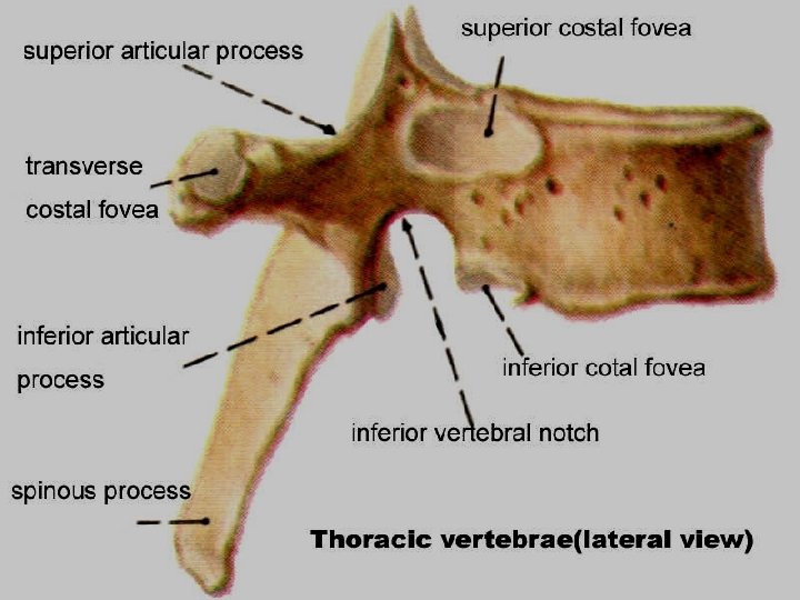
The Bones of Trunk ¡ The vertebrae ¡ In the child, 33 ¡ The general features ¡ Vertebral body ¡ Vertebral arch ¡ Vertebral foramen ¡ Vertebral canal ¡ Pedicles and laminae of vertebral arch ¡ Intervertebral foramen ¡ Processes : ¡ Spinous ¡ Transverse ¡ articular Vertebral body Superior costal fossa Spinous process The thoracic vertebra
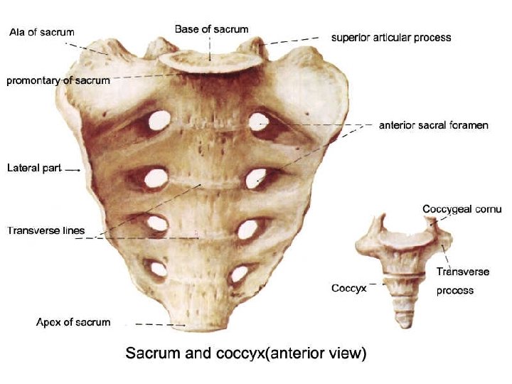
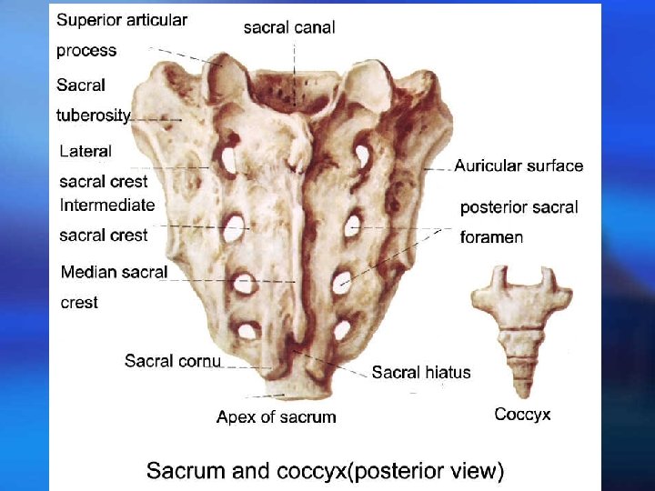
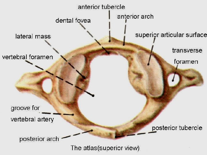
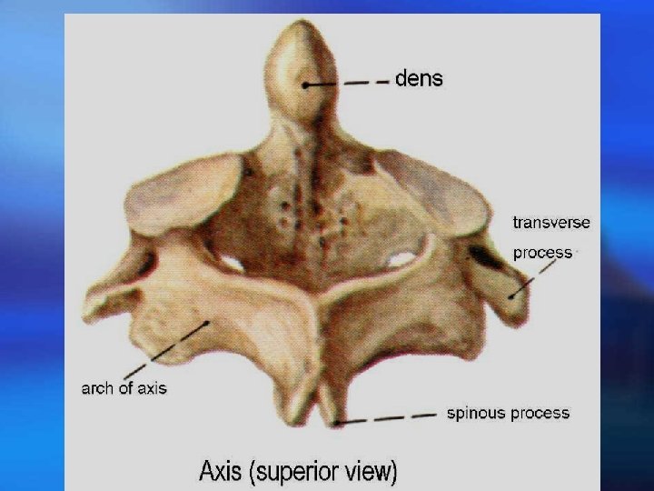
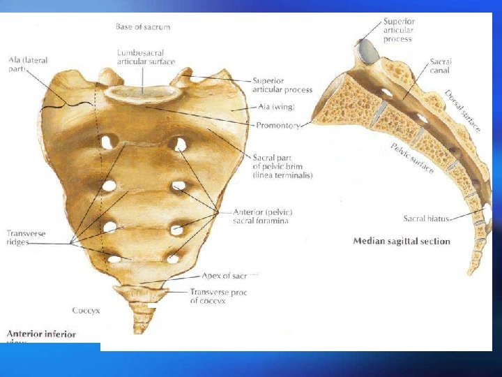
The main characteristics of vertebrae in each region 1. Thoracic vertebrae: costal facets on each side of their body 2. The cervical vertebrae: transverse foramen. Atlas: Anterior arch and posterior arch and a lateral mass. Fovea dentis, groove for vertebral artery. Axis: dens or odontoid process Vertebra prominens: nonbifid spine is long and easily felt. 3. Lumbar vertebrae: The spines are strong, square and horizontal 4. sacrum: It is made up of five fused vertebrae and roughly triangular. There are three surfaces Promontory of sacrum, anterior and posterior foramina, lateral masses, ala, median sacral crest, intermediate and lateral crests
Dorsal sacral foramina, sacral canal, sacral hiatus, sacral cornu or horns, auricular surface, sacral tuberosity. 5. coccyx: It is made up of four, more or less, coccygeal vertebrae
T h e The sternum Flat bone Internal plate External plate diploe
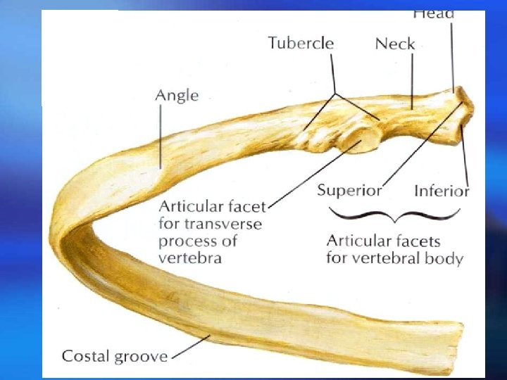
RIBS ¡ Two parts: costal bone costal cartilage ¡ General features of the costal bone ¡ Costal bone: one body, two extremities, two surfaces and two borders ¡ Head(articular facet of head, articular facet of costal tubercle, neck, shaft and tubercle. ¡ Costal groove ¡ Angle of rib ¡
First and second ribs
Thoracic cage The composition True ribs False ribs Floating ribs Intercostal space
The bones of the limbs ¡ The appendicular skeleton includes the bones of upper limbs and those of the lower limbs. The bones of the upper and lower limbs are constructed after a common type, but the different functions for which they have become adapted in man, because of the erect standing in human being, have led to structural differences of a very definite kind.
¡ The upper limbs are released from weight bearing and become the organs of labour with greater and delicated mobility, so, the bones of upper limbs are lighter and smaller in shape and size. The bones of lower limbs are very heavy and strong so as bear the weight of the body and to provide movement of the whole body. Each limb has a girdle, which connects it to the trunk, and three segments. In the upper limb, these are the shoulder girdle, the upper arm, the forearm and the hand. In the lower limb, those are the pelvic girdle, the thigh, the leg and the foot.
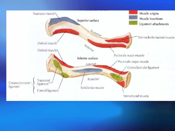
The bones of upper limb ¡ The shoulder ¡ Girdle ¡ The clavicle ¡ Lies almost horizontally on each side of the neck, extending from the sternum to the acromion of the scapula
Scapula : anterior aspect view
Scapula : posterior aspect view
The bones of the free upper limb Those include the bone of the arm, the bones of the forearm and the bones of hand (the carpal bones, metacarpal bones and phalanges) The bone of arm
The humerus is the longest and largest bone of the upper limb. It extends from the scapula to the elbow joint and has a body and two ends
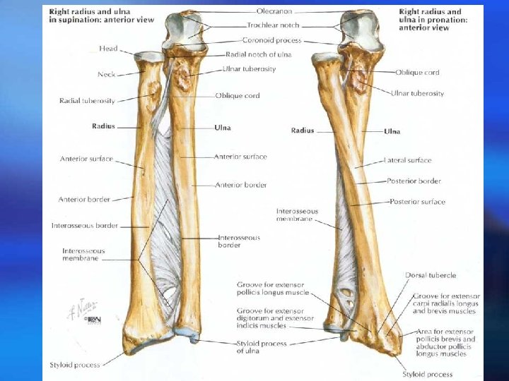
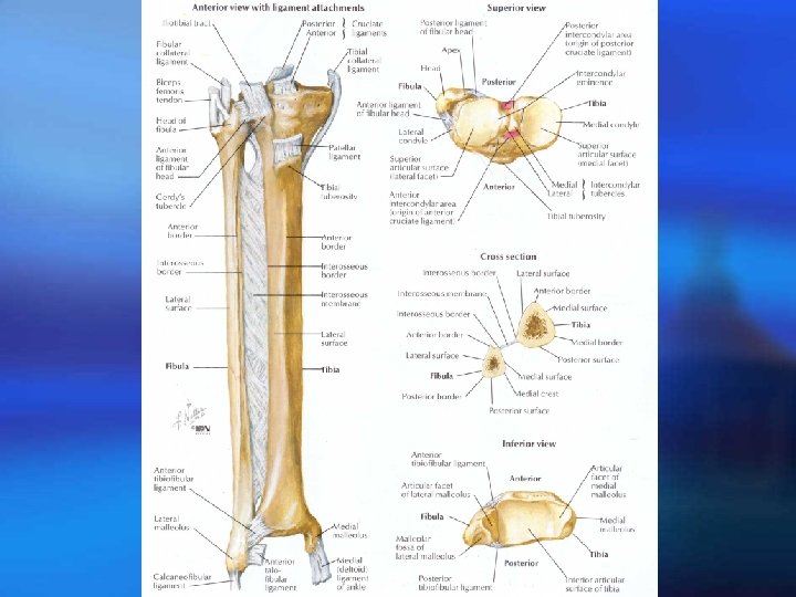
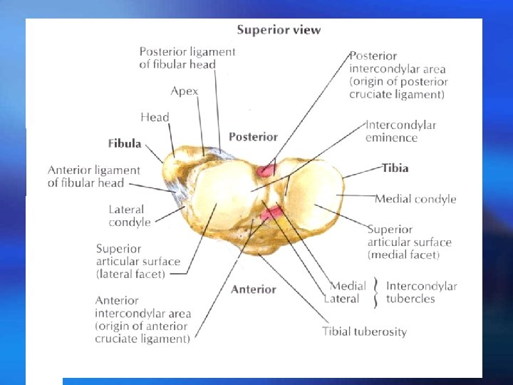
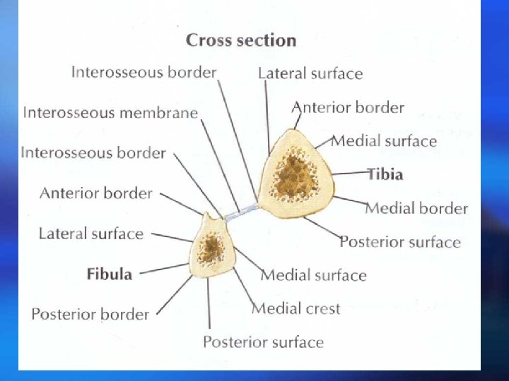
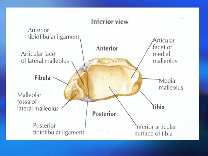
The bones of forearm The radius is the lateral bone of the two in the forearm. It has a shaft and two ends. The proximal end A head, neck and tuberosity; head is disc-shaped and its upper surface is a shallow cup—articular fovea for articulation with the capitulum of the humerus. Its articular circumference articulates with radial notch of the ulna. Shaft of radius: interosseous border Distal end Ulnar notch, styloid process, carpal articular surface
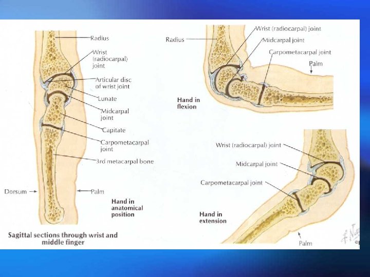
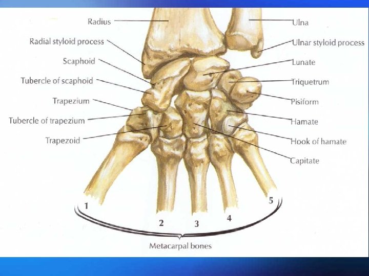
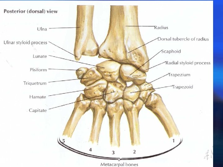
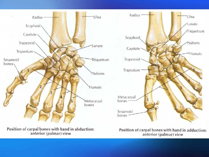
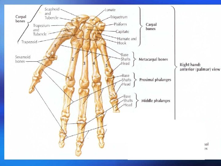
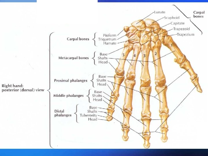
The bones of hand ¡ Three parts: the carpal bones, the metacarpal bones and phalanges ¡ The carpal bones ¡ There are eight short bones, arranged in two rows of four ¡ Proximal row from lateral to medial include : ¡ Distal row from lateral to medial include:
The metacarpal bones ¡ Which are five in number, connect with the carpal bones above and the phalanges below. ¡ Base ¡ Shaft ¡ Distal head ¡ The first metacarpal bones is short and stout; its base is saddle shaped for articularion with the trapezium
The phalanges ¡ They are long bones ¡ They are known as the proximal, middle and the distal phlanx ¡ Base ¡ Head ¡ Distal end
The bones of lower limbs ¡ The lower limb which is similar to the upper, is connected to the trunk by a girdle, the pelvix girdle, the free lower limb is divided into three segements, the thigh, the leg and the foot.
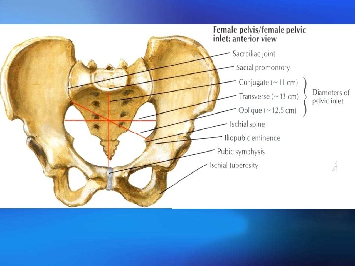
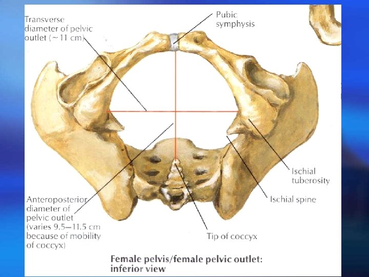
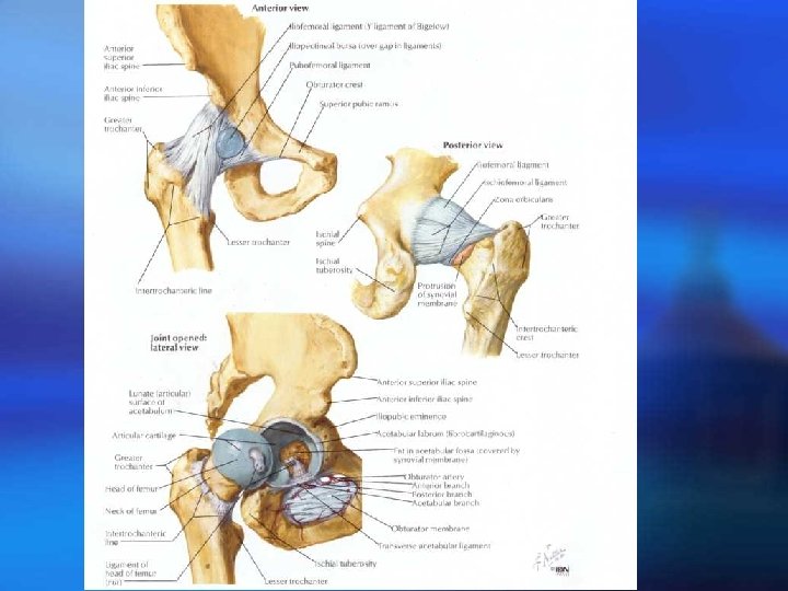
The pelvic girdle is formed by a hip bone on each side. They articulate posteriorly with the sacrum and meet below and in front at the pubic symphysis Bony pelvis =the pelvis girdle Sacrum coccyx
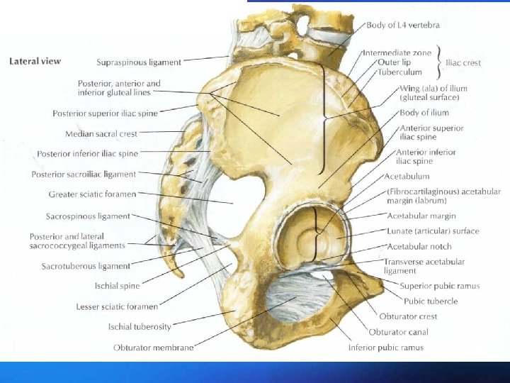
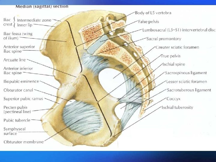
Hip bone
Hip bone
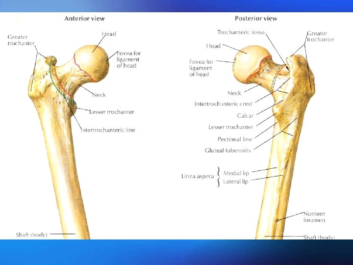
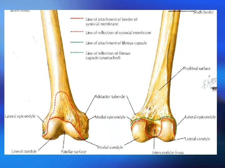
Femur
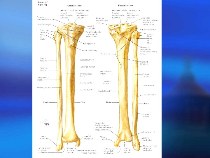
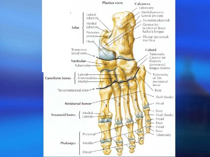
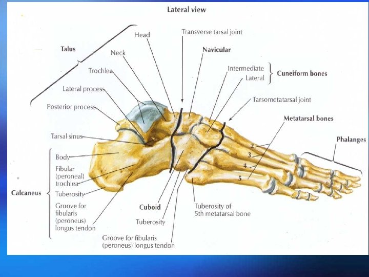
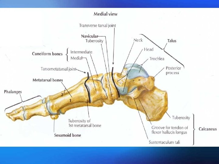
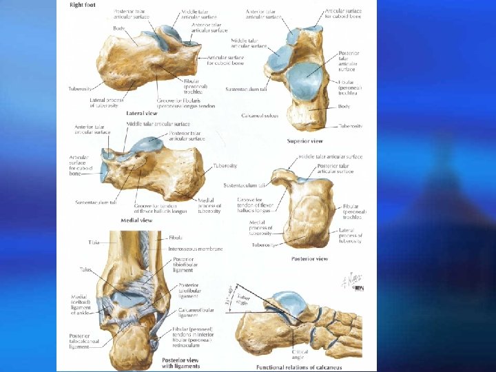
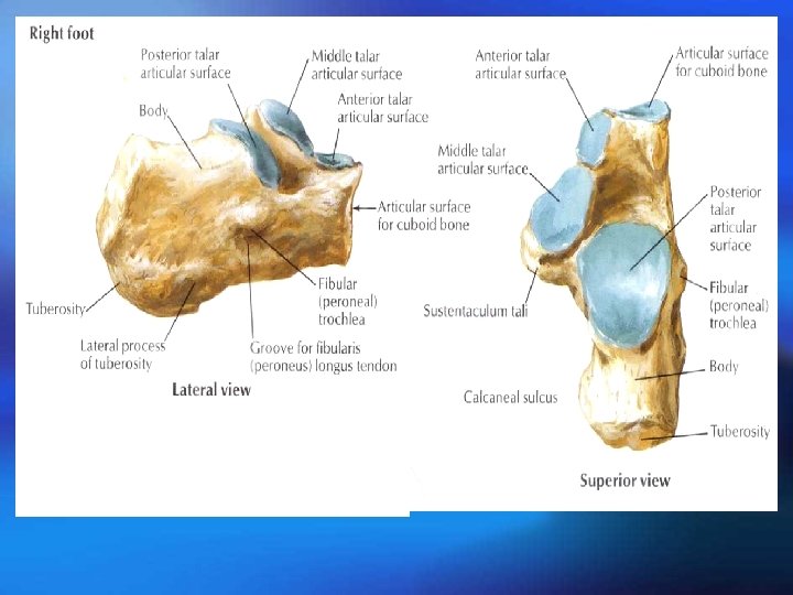
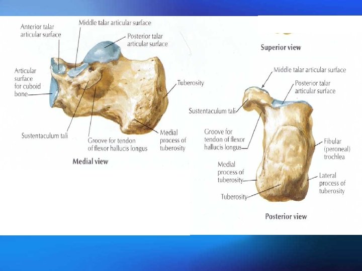
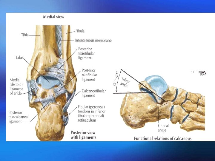
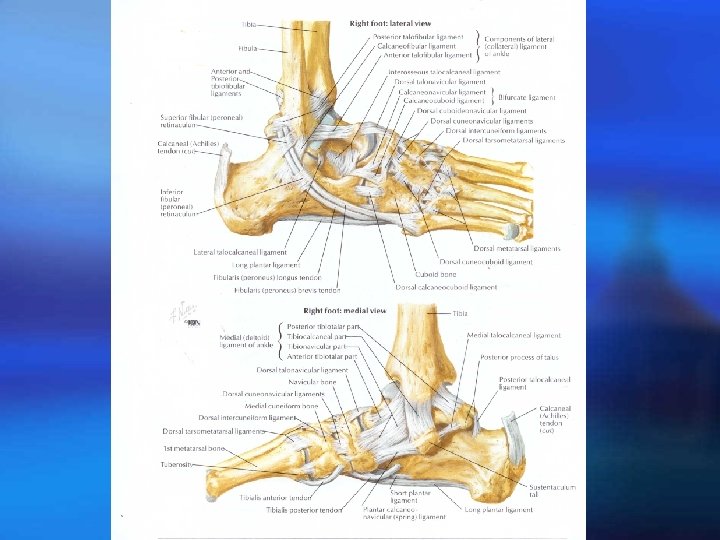
Bones of foot