Osteochondromagenesis Loss of heterozygosity modeled via Cremediated inversion


 for reverse orientation of")


- Slides: 34
Osteochondromagenesis: Loss of heterozygosity modeled via Cre-mediated inversion of the second exon of Ext 1 in chondrocytes Department of Orthopaedics and Rehabilitation University of Iowa Hospitals & Clinics Kevin B. Jones, MD Charles Searby, BS Gail Kurriger, BS James Martin, Ph. D Peter J. Roughley, Ph. D Jose A. Morcuende, MD, Ph. D Joseph A. Buckwalter, MD, MS Val C. Sheffield, MD, Ph. D Connective Tissue Oncology Society London, UK Friday, 14 November 2008
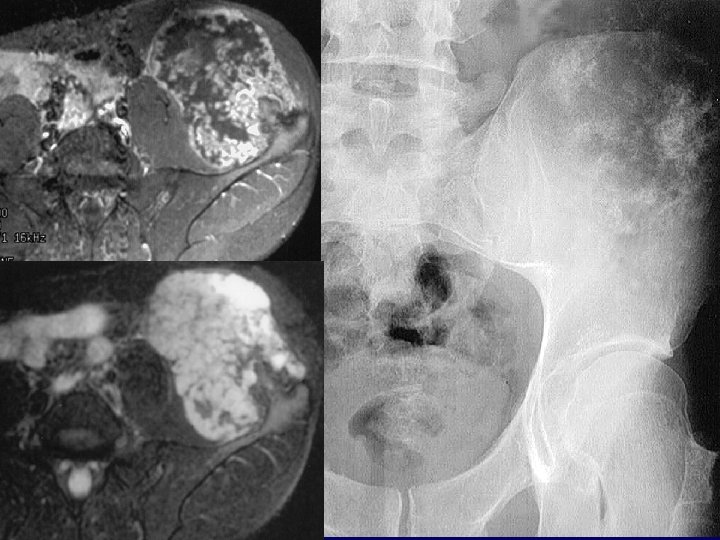
Human EXT 1: What is known clinically. . . Hereditary Multiple Exostosis • • Autosomal dominant inheritance Shortened long bones Multiple osteochondromas Homozygosity lethal
EXT 1 & EXT 2: Tumor Supressor Genes? • Loss of heterozygosity in chondrocytes of cartilage cap of HME osteochondromas • Both alleles somatically mutated in some solitary osteochondromas Raskind et al. 1995; Bovee et al. 1999; Hecht et al. 1995 and 1997.
Ext 1 Knock-Out Mice by Lin et al. 2000 Mouse Ext 1 has 99% Homology to Human EXT 1 PROBLEMS: • Homozygous Ext 1 -knock-out mice do not survive to birth • Heterozygous Ext 1 -knock-out mice very rarely form osteochondromas Simlar with Ext 2 knock-out mice, Sitckens et al. 2005.
Of Mice and Men
Of Mice and Men
~60 kg Human flesh ~30 g Mouse flesh ~2000 X the number of cells
Haploinsufficiency or Loss of Heterozygosity?
Methods
lox. P Lox. P CRE Cre Routine or cis orientation of lox. P sites results in fragment excision
trans orientation of lox. P sites results in reversible fragment inversion Lox. P Pxol CODING SEQUENCE CRE Cre ECNEUQES GNIDOC Pxo. L lox. P
You do the math. . . • Cre-mediated inversion yields 40% reverse orientation per trans-floxed allele • Homozygosity for trans-floxed alleles should yield 40% loss of total copies of functional gene across tissue – – 20% of cells will remain unaffected and fwd/fwd 20% of cells will end up fwd/fwd after inversion 40% of cells will end up fwd/rev after inversion 20% of cells will end up rev/rev after inversion
Targeting Construct Intron 1 lox. P exon 2 lox. P neo lox. P intron 2 • Exon 2 contains two of the very few diseasecausing missense mutations at highly conserved amino acid residues 339 and 340. • Exon 2 inversion not only disrupts the sequence, but introduces a stop codon in the reading frame exon 3
Cre-Recombinase Driver • Doxycycline-inducible Collagen type II promoter-Cre • Doxycycline adminstered via maternal drinking water during second week of life Gover and Roughley et al. , 2006
Results
PCR Testing with Multiple Primers Intron 1 lox. P exon 2 lox. P neo lox. P intron 2 exon 3 • All permutations of flipping were present in every cartilage containing tissue.
excised neo exon 2 excised neo reversed neo 2 exon excised neo reversed exon 2 neo excised neo reversed exon 2
Effects of Cre-mediated inversion RT-PCR demonstrated the expected 40% reduction in Ext 1 transcripts across homozygous tissue exposed to Cre
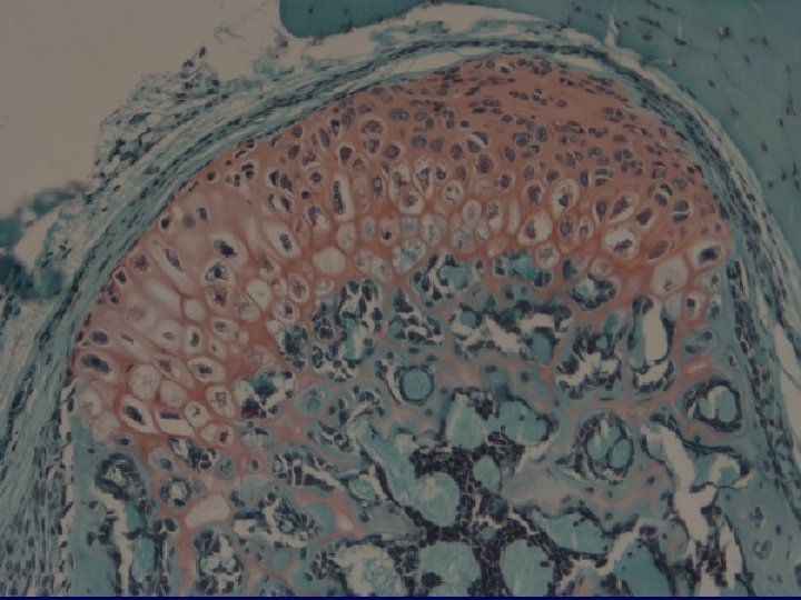
Phenotype: Osteochondromas?
Distal femur physis 6 week old Ext 1 fl/fl with doxy-col 2 -Cre
Proximal tibia 6 week old Ext 1 fl/fl with doxycol 2 -Cre
Genotype # of mice with osteochondromas/ # of mice over 6 weeks old _______________________ fl/fl+doxy-col 2 a 1 -Cre 12/12 fl/fl without Cre 0/5 wt/fl+doxy-col 2 a 1 -Cre 0/7 wt/fl without Cre 0/1 wt/wt without Cre 0/8 ________________
Comparison Traditional Knockout Trans-floxed Conditional Knockout • 50% tissue reduction in Ext 1 alleles • Very rare biallelic loss • 40% tissue reduction in Ext 1 alleles • 20% biallelic loss • Osteochondromas very rare • Osteochondromas rampant
Discussion Loss of heterozygosity is the mechanism of osteochondromagenesis in the setting of hereditary multiple exostoses
Hypothesis Disruption of both Ext 1 alleles in a physeal chondrocyte is sufficient to form an osteochondroma
Alternate Hypothesis Disruption of both Ext 1 alleles in some physeal chondrocytes is sufficient to derange signals and form osteochondromas
Hypothesis vs. Alternate Hypothesis Are osteochondromas clonal (at least consistent) for reverse orientation of exon 2? On-going work with laser capture micro-dissection and in situ hybridization
Conclusion Osteochondromageneis resulted from lowprevalence biallelic disruption of Ext 1 in chondrocytes, modeling loss of heterozygosity with a unique twist on the standard conditional knock-out
Project Funding Support • OREF Resident Research Award 2003 • Department of Orthopaedics and Rehabilitation, University of Iowa • Val C. Sheffield Genetics Laboratory Department of Orthopaedics and Rehabilitation University of Iowa Hospitals & Clinics
Osteochondromagenesis: Loss of heterozygosity modeled via Cre-mediated inversion of the second exon of Ext 1 in chondrocytes Department of Orthopaedics and Rehabilitation University of Iowa Hospitals & Clinics Kevin B. Jones, MD Charles Searby, BS Gail Kurriger, BS James Martin, Ph. D Peter J. Roughley, Ph. D Jose A. Morcuende, MD, Ph. D Joseph A. Buckwalter, MD, MS Val C. Sheffield, MD, Ph. D Connective Tissue Oncology Society London, UK Friday, 14 November 2008