Ossifying fibroma a benign neoplasm Presenter Dr sabreena

Ossifying fibroma : a benign neoplasm Presenter : Dr sabreena mukhtar

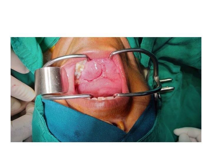
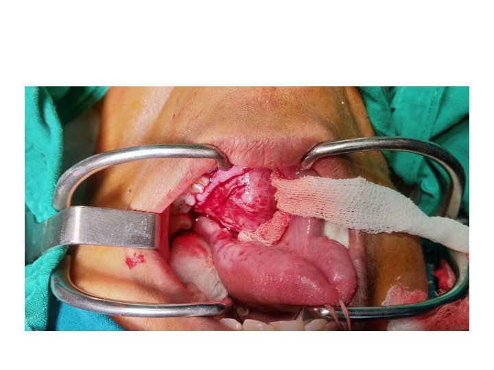
Case History • A 46 yr old female • Chief complaints – Swelling over floor of mouth x 1 yr • Gradual onset , progressive, not associated with pain – Difficulty in swallowing – Difficulty in articulation – No h/o trauma, weight loss, previous surgery, tooth fall

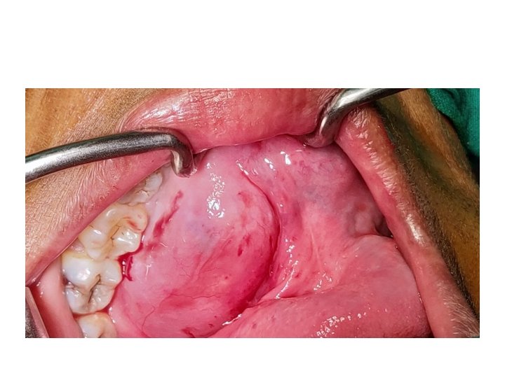
Examination • Inspection – 5 x 4 cm smooth, round swelling arising from floor of mouth, pushing tongue upwards, more towards left, pinkish in color.
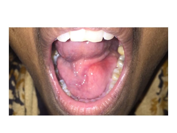

• Palpation – Consistency bony hard, smooth surface, non compressible, non tender, non mobile, – No glow on trans illumination – No palpable neck lymph nodes.

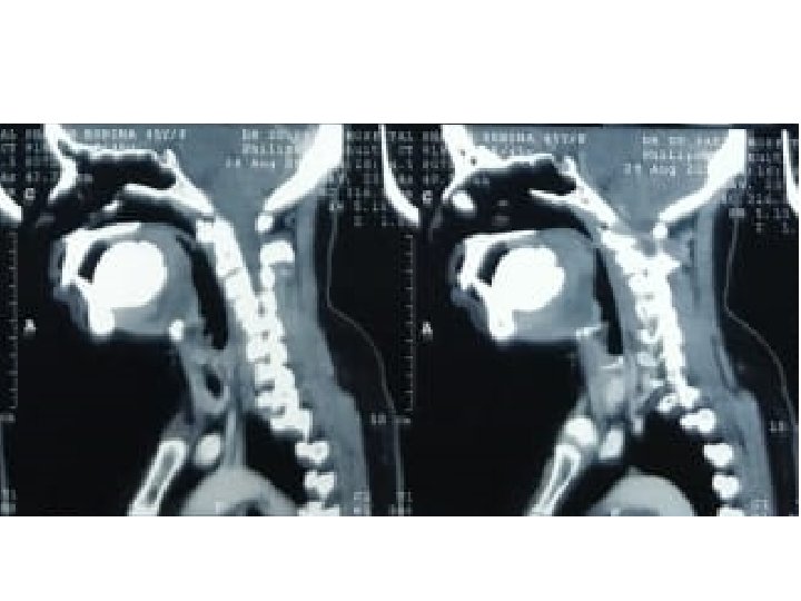
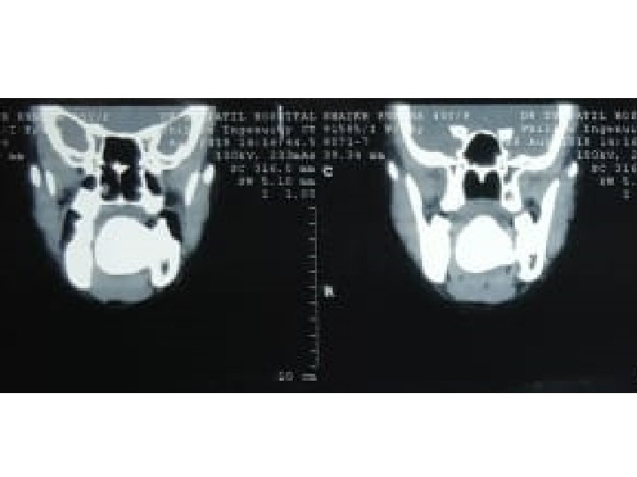
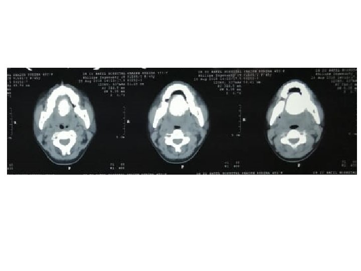
Imaging • USG neck – Normal no significant findings • Orthopantogram Bony mass arising from left mandible • CT Head & Neck – A lobulated well define bond density lesion noted in the oral cavity at the floor of mouth in left sublingual space, measuring 3. 6 x 4 x 3. 3 cm. – Lesion seen attached to left medial aspect of mandible at the mylohyoid line – No extension noted – No soft tissue component

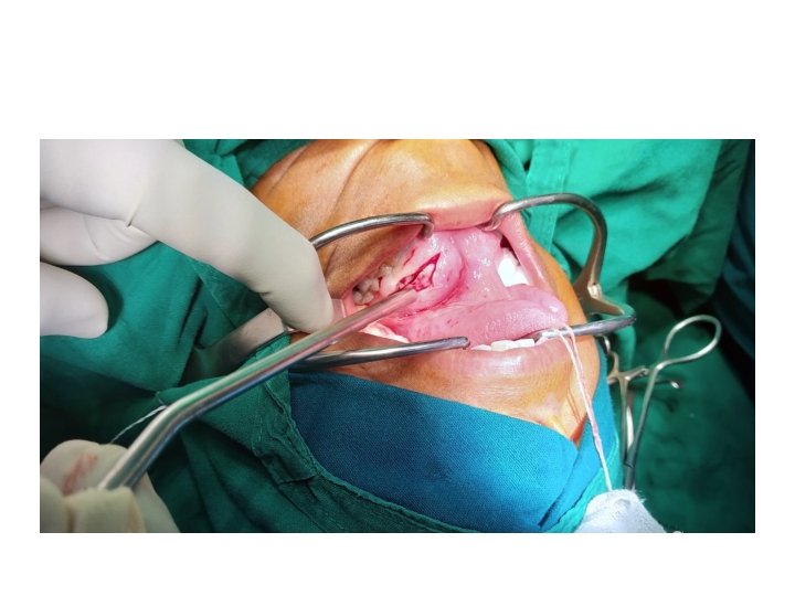
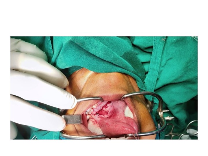
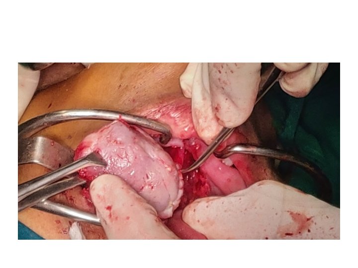
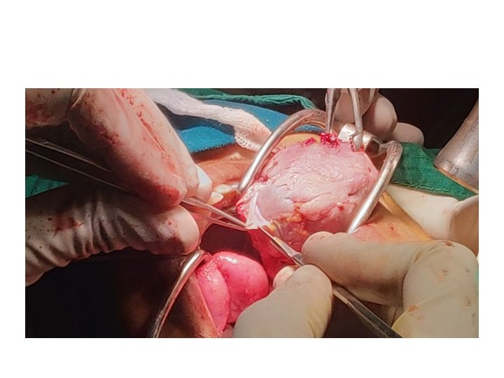
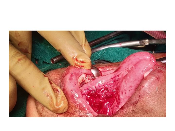
Procedure • Treatment Plan: • Total surgical excision Under General anaesthesia.
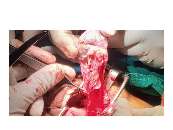

Histopathology • Fibrous tissue with bony bits • Foci of oval to spindle cells with intervening fibrous tissue and calcification • Areas of ossification noted Impression – Fibroma with calcification • Follow up of patient was done after 6 months where patient appeared symptom free without any clinical evidence of recurrence


Discussion • Ossifying Fibroma • It is a odontogenic benign tumor of the jaw a type of fibro osseous lesion may have odentogenic, traumatic developmental origon. • A mutation in a tumor supressor gene HRPT 2 a protein product known as para fibronin leads to tumor formation. • Clinically of two types: • ossifying fibroma • Cementifying fibroma Central ossifying fibroma is more common in females than males and involves mandible more than maxilla.

• Tumor shows a female preponderance with a ratio of 5: 1 • Mostly seen in mandible 70 -90%(pre molar and molar region) followed by maxilla , ethmoid and orbital regions • It presents as a painless swelling. • Conventional radiographs specialized imaging techniques like CT and CBCT help to delineate from other similar lesions of jaw.

• Differential diagnosis include other radio opaque lesions such as fibrous dysplasia, calcifying epithelial odontogenic tumor, calcifying epithelial odontogenic cyst, osteogenic sarcoma. • They have locally aggressive behavior with high recurrence rate particularly in partial and incomplete excisions, with complete removal being the gold standard treatment. • Prognosis is good without any metastasis.

• Ossifying fibromas are entities with different morphological features that can be mistaken for other benign osseofibrous lesions. This similarity and over lapping micro characteristics with other similar lesions make pre operative diagnosis a challenge. Multidisciplinary approach, comprehending clinical, radiological and pathological aspects and an accurate histopathological report post operatively are mandatory for the correct diagnosis and appropriate treatment.
- Slides: 25