Osmotic Fragility of Red Blood Cells DEFINITION it

Osmotic Fragility of Red Blood Cells

DEFINITION - it is a test that measures the resistance to hemolysis of red blood cells (RBC) exposed to hypotonic solutions RBC are exposed to a series of saline (Na. Cl) solutions with increasing dilution The sooner hemolysis occurs, the greater is osmotic fragility of RBC

The osmotic fragility : is the resistance to hemolysis of red blood cells when exposed to hypotonic saline. It is employed as a screening test for hemolytic anemia.
 solution – 0. 9 % RBC burst in hypotonic")
Isotonic Na. Cl (physiological) solution – 0. 9 % RBC burst in hypotonic (< 0. 9 % Na. Cl), and shrink (crenate) in hypertonic solutions (> 0. 9 % Na. Cl) In hypotonic medium a membrane rupture occurs, allowing hemoglobin (Hb) to exit from the cells By measuring Hb concentration, the % of hemolysis at different Na. Cl concentrations can be calculated

INCREASED OSMOTIC FRAGILITY - Hereditary spherocytosis - Acquired spherocytosis Hereditary spherocytosis is a disorder characterized by a defective RBC membrane and decreased surface-to-volume ratio Characteristic round cells (spherocytes) are seen in blood smear and they are more fragile and break open in less hypotonic solutions than normal red blood cells
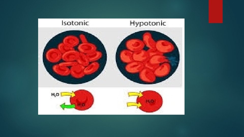

In hypotonic solutions water enters red blood cells Therefore, normal RBC with a biconcave shape swell and expand their volume On the other hand, spherocytes cannot absorb much extracellular liquid and break very easily

DECREASED OSMOTIC FRAGILITY - Thalassemia - Sickle cell anemia - Iron deficiency anemia


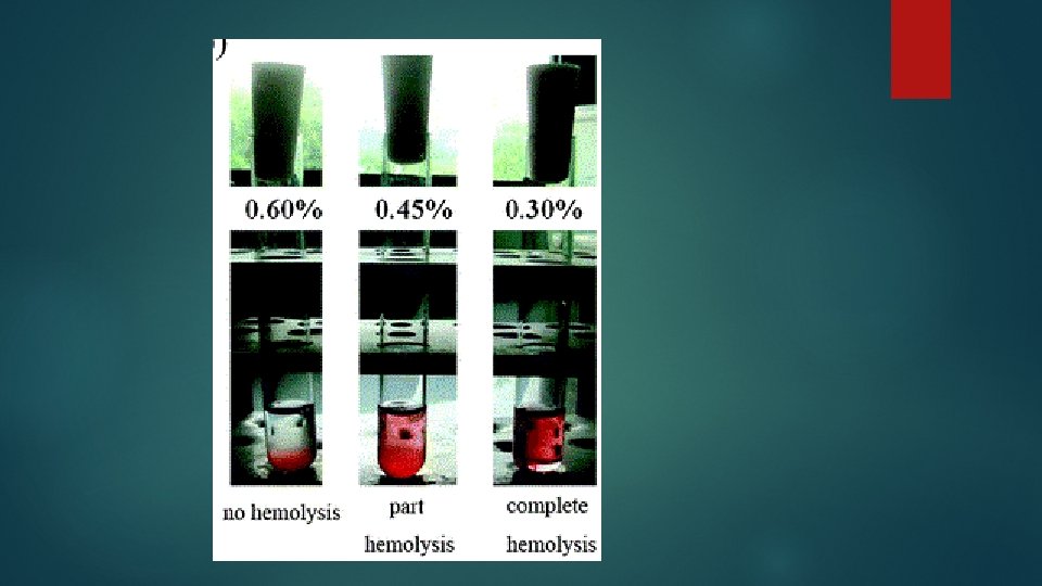
Normal range of fragility Normally, hemolysis begins in about 0. 48% saline. No cells hemolyze in solutions of 0. 5% saline and above. Hemolysis is complete at about 0. 36% saline Note When red cells become more fragile, hemolysis may begin at about 0. 64% saline and be complete at about 0. 44% saline

Apparatus and materials Ø Test tube rack with 7 clean dry glass test tubes, marking pencil and dropper Ø 3 ml syringe Ø 0. 9% sodium chloride solution and distilled water

Procedure 1. Number the test tubes from 1 to 7 with the marking pencil and put them in the rack. 2. Using the dropper, place the varying number of drops of normal saline and distilled water in each of the 7 test tubes as shown in the table below and mix

3. Draw 2 ml of blood from a suitable vein and gently eject one drop of blood into each of the 7 tubes and mix* 4. Leave the test tubes undisturbed for one hour then observe the extent of hemolysis in each tube

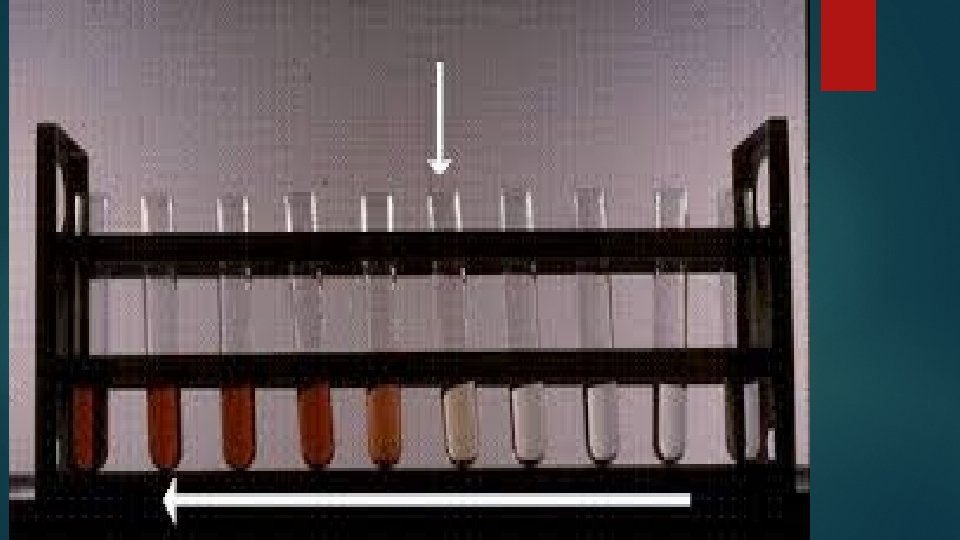
No. of test tube 1 2 3 4 5 6 7 No. of drops of Na. Cl 25 16 12 10 9 7 0 No. of drops of D. W 0 9 13 15 16 18 25 0. 64 0. 48 0. 4 0. 36 0. 28 0 Tonicity of Na. Cl 0. 9 (in%)
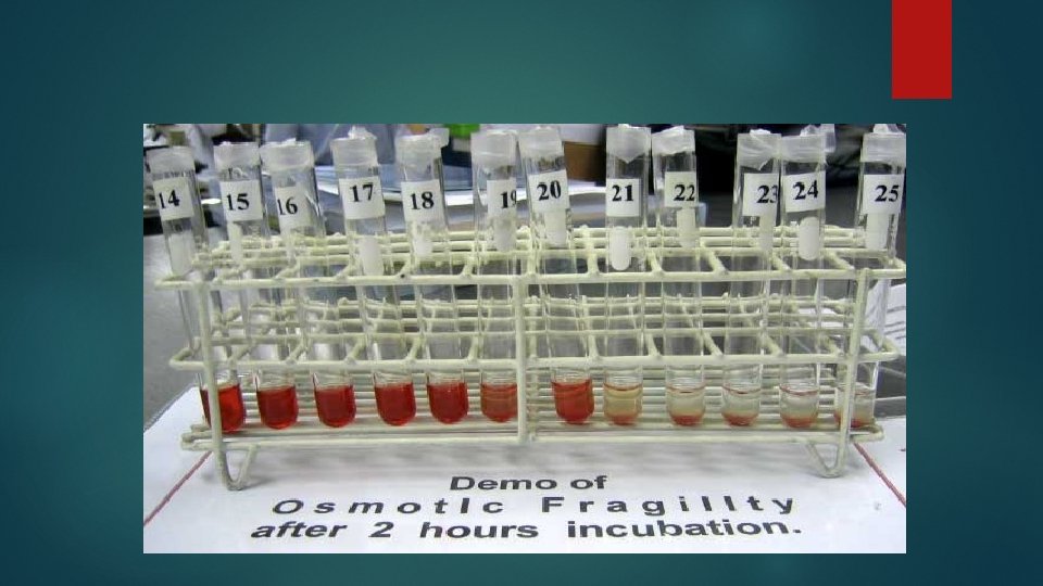

While judging the extent of hemolysis from the depth of red color of supernatant saline, tube#1 (normal saline) and tube#7 (distilled water) will act as controls, i. e. , no hemolysis in normal saline and complete hemolysis in distilled water

a. The test tubes in which no hemolysis occur, the RBCs will settle down and form a red dot at the bottom of the tube, leaving the saline above clear b. If there is some hemolyis, the saline will be tinged red with Hb with unruptured cells forming a red dot at the bottom. The color of the saline will be seen to be increasingly deeper with decreasing tonicity of saline c. The test tubes in which there is complete hemolysis, the saline will be equally deep red with no red cells at the bottom of these tubes.
 and record")
Results § Note the start of hemolysis (the onset of fragility) and record the test tube number. Express your results in % saline § Note the start of complete hemolysis

- Slides: 21