Osmoregulation By Angsuman Chanda Freshwater animals show adaptations

Osmoregulation By Angsuman Chanda • Freshwater animals show adaptations that reduce water uptake and conserve solutes • Desert and marine animals face desiccating environments that can quickly deplete body water • Osmoregulation regulates solute concentrations and balances the gain and loss of water • Excretion gets rid of metabolic wastes

Osmoregulation balances the uptake and loss of water and solutes • Osmoregulation is based largely on controlled movement of solutes between internal fluids and the external environment • Osmoregulators expend energy to control water uptake and loss in a hyperosmotic or hypoosmotic environment

Gain of water and salt ions from food and by drinking seawater Excretion of salt ions from gills Osmotic water loss through gills and other parts of body surface Excretion of salt ions and small amounts of water in scanty urine from kidneys Osmoregulation in a saltwater fish

Osmotic water gain through gills and other parts of body surface Uptake of water and some ions in food Uptake of salt ions by gills Osmoregulation in a freshwater fish Excretion of large amounts of water in dilute urine from kidneys

Classification of aquatic animals on basis of Osmotic Regulation Osmoconformer: ◦ internal osmolarity nears that of the external environment ◦ if external conditions change, internal osmolarity changes with it. Osmoregulators: ◦ Maintains internal osmolarity within an narrow range regardless of the external environment. ◦ Depending on conditions, the animal could have an osmolarity higher or lower than surrounding water.

Clasification on the basis of salt tolerance Stenohaline = animals that can only tolerate a narrow range of salt Concentrations Euryhaline = animals that can tolerate widely variant osmolarities No predetermined relationship between strategy and degree of tolerance.

Salt concentration in confirmers and regulators

Specialization in bird Nasal salt gland Nostril with salt secretions

Proteins Nucleic acids Amino acids Nitrogenous bases —NH 2 Amino groups Most aquatic animals, including most bony fishes Ammonia Mammals, most amphibians, sharks, some bony fishes Urea Many reptiles (including birds), insects, land snails Uric acid

Ammonia • Animals that excrete nitrogenous wastes as ammonia need lots of water • They release ammonia across the whole body surface or through gills

Urea • The liver of mammals and most adult amphibians converts ammonia to less toxic urea • The circulatory system carries urea to the kidneys, where it is excreted

Uric Acid • Insects, land snails, and many reptiles, including birds, mainly excrete uric acid • Uric acid is largely insoluble in water and can be secreted as a paste with little water loss

Excretory Processes • Most excretory systems produce urine by refining a filtrate derived from body fluids • Key functions of most excretory systems: – Filtration: pressure-filtering of body fluids – Reabsorption: reclaiming valuable solutes – Secretion: adding toxins and other solutes from the body fluids to the filtrate – Excretion: removing the filtrate from the system

Nephrons and associated blood vessels are the functional unit of the mammalian kidney • Kidneys, the excretory organs of vertebrates, function in both excretion and osmoregulation • The mammalian excretory system centers on paired kidneys, which are also the principal site of water balance and salt regulation • Each kidney is supplied with blood by a renal artery and drained by a renal vein • Urine exits each kidney through a duct called the ureter • Both ureters drain into a common urinary bladder

Posterior vena cava Renal artery and vein Kidney Renal medulla Renal cortex Renal pelvis Aorta Ureter Urinary bladder Urethra Ureter Excretory organs and major associated blood vessels Juxta. Cortical medullary nephron Afferent arteriole Glomerulus from renal Bowman’s capsule artery Proximal tubule Peritubular capillaries Renal cortex Collecting duct 20 µm Renal medulla To renal pelvis Nephron Section of kidney from a rat Kidney structure SEM Efferent arteriole from glomerulus Distal tubule Collecting duct Branch of renal vein Descending Loop limb of Henle Ascending limb Vasa recta Filtrate and blood flow

Structure and Function of the Nephron and Associated Structures • The mammalian kidney has two distinct regions: an outer renal cortex and an inner renal medulla • The nephron consists of a single long tubule and a ball of capillaries called the glomerulus

Filtration of the Blood • Filtration occurs as blood pressure forces fluid from the blood in the glomerulus into the lumen of Bowman’s capsule • The filtrate in Bowman’s capsule mirrors the concentration of solutes in blood plasma

Pathway of the Filtrate • From Bowman’s capsule, the filtrate passes through three regions of the nephron: the proximal tubule, the loop of Henle, and the distal tubule • Fluid from several nephrons flows into a collecting duct

From Blood Filtrate to Urine: A Closer Look • Filtrate becomes urine as it flows through the mammalian nephron and collecting duct • Secretion and reabsorption in the proximal tubule change the filtrate’s volume and composition • Reabsorption of water occurs as filtrate moves into the descending limb of the loop of Henle

• In the ascending limb of the loop of Henle, salt diffuses from the permeable tubule into the interstitial fluid. • The distal tubule regulates the K+ and Na. Cl concentrations of body fluids. • The collecting duct carries filtrate through the medulla to the renal pelvis and reabsorbs Na. Cl. • The mammalian kidney conserves water by producing urine that is much more concentrated than body fluids.

Proximal tubule Na. Cl Nutrients HCO 3– K+ H 2 O H+ NH 3 Distal tubule H 2 O Na. Cl K+ HCO 3– H+ CORTEX Descending limb of loop of Henle Filtrate H 2 O Salts (Na. Cl and others) HCO 3– H+ Urea Glucose; amino acids Some drugs Thick segment of ascending limb Na. Cl H 2 O OUTER MEDULLA Na. Cl Thin segment of ascending limb Key Active transport Passive transport Collecting duct Urea Na. Cl INNER MEDULLA H 2 O

Solute Gradients and Water Conservation • The action and precise arrangement of the loops of Henle and collecting ducts are largely responsible for the osmotic gradient that concentrates the urine. • Na. Cl and urea contribute to the osmolarity of the interstitial fluid, which causes reabsorption of water in the kidney and concentrates the urine.
 300 100 CORTEX Active transport Passive transport OUTER MEDULLA")
Osmolarity of interstitial fluid (mosm/L) 300 100 CORTEX Active transport Passive transport OUTER MEDULLA H 2 O Na. Cl 400 600 H 2 O INNER MEDULLA H 2 O Na. Cl 200 Na. Cl H 2 O 400 Na. Cl 900 300 H 2 O Na. Cl 300 H 2 O 600 H 2 O Urea 700 H 2 O Urea 900 H 2 O Urea 1200 600 1200

• The collecting duct conducts filtrate through the osmolarity gradient, and more water exits the filtrate by osmosis • Urea diffuses out of the collecting duct as it traverses the inner medulla • Urea and Na. Cl form the osmotic gradient that enables the kidney to produce urine that is hyperosmotic to the blood

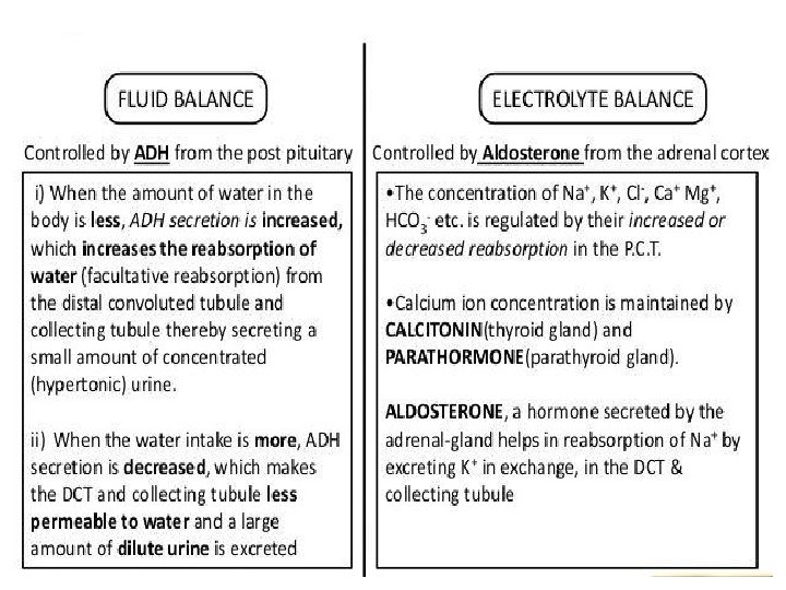
Regulation of Kidney Function • The osmolarity of the urine is regulated by nervous and hormonal control of water and salt reabsorption in the kidneys • Antidiuretic hormone (ADH) increases water reabsorption in the distal tubules and collecting ducts of the kidney. • Aldosterone is a hormone that regulates salt reabsorption in the kidney.

Elemination of wast product

Blood Osmolarity Feedback Endocrine System Control Osmoreceptors in hypothalamus pituitary ADH increased water reabsorption nephron high Blood Osmolarity Blood Pressure nephron adrenal gland increase thirst increased water & salt reabsorption Juxta. Glomerula Apparatus low nephron (JGA) renin aldosterone angiotensinogen
- Slides: 28