
Orthopaedic Aspects of Child Abuse NAT Abdulaziz Alomar
Orthopaedic Aspects of Child Abuse NAT Abdulaziz Alomar, MD, MSc FRCSC Assistant Professor and consultant Orthopaedic surgeon. KKUH, KSU
Child Abuse A major cause of disability and death among children. n Fractures are the second most common presentation of physical abuse after skin lesions. n One third of physically abused children will require orthopaedic treatment. n
Definition “at a minimum, any act or failure to act resulting in imminent risk of serious harm, death, serious physical or emotional harm, sexual abuse, or exploitation of a child by a parent or caretaker who is responsible for the child’s welfare. ”
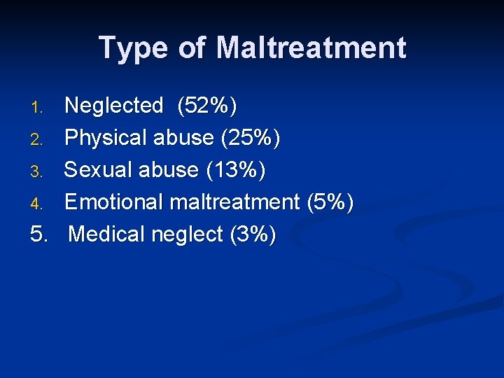
Type of Maltreatment Neglected (52%) 2. Physical abuse (25%) 3. Sexual abuse (13%) 4. Emotional maltreatment (5%) 5. Medical neglect (3%) 1.
Risk factors n n n n n First-born children. Unplanned children. Premature infants. Stepchildren. Handicapped children. Single-parent homes. Drug abusing parents. Parents who were themselves abused. Unemployed parents. Families of lower socioeconomic status.
The majority of maltreated children are abused by birth parents.
Who is at Risk? n Most children with NAT fractures age of < 3 years
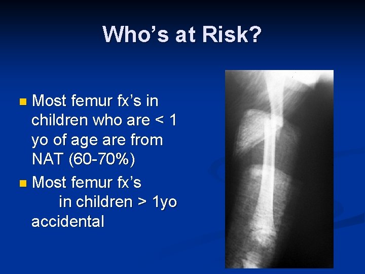
Who’s at Risk? n Most femur fx’s in children who are < 1 yo of age are from NAT (60 -70%) n Most femur fx’s in children > 1 yo accidental
Clinical Features n History: Delay in presenting. n History vague, lacking in detail, contradictory n Mechanism of injury insufficient to explain injuries n History of a fall n
Clinical Features n History : Less than 3 years old n Poor household environment, drug or physical abuse n Overly aggressive or passive n Behavioral problems n Handicapped child n Stepchild n Premature child n Subnormal growth n

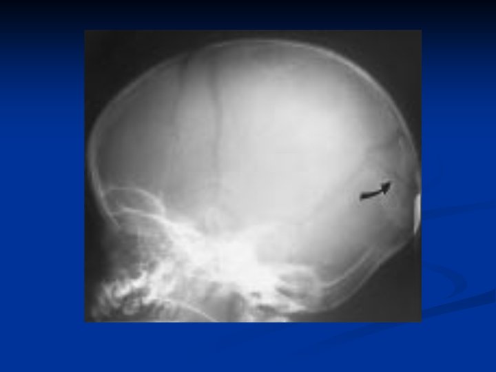
Non Orthopaedic Features n Skin: n Bruises (buttocks, perineum and genitalia, trunk, back of head and legs). n n n Multiple bruises in various stages of healing Burns (pattern may reflect mechanism of burn). Head and CNS: n n Skull fracture (multiple, bilateral, skull base, crossing nervous system suture lines, depressed fractures) Subdural hematoma, subarachnoid hemorrhage Retinal hemorrhage, retinal detachment Cognitive disabilities
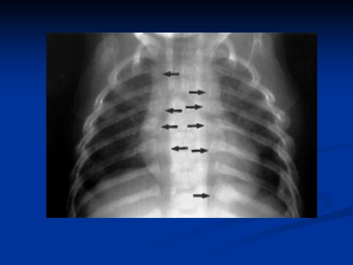
Non Orthopaedic Features n Chest, abdomen, and pelvis: Rib fractures (posterior, multiple), n Sternal fractures and pelvis. n Pneumothorax, hemothorax n Rupture of organ (liver, spleen, or pancreas laceration; bowel or bladder rupture) n Intramural bowel hematoma n Kidney contusion, retroperitoneal hemorrhage n Sexual abuse n
Orthopaedic Features Multiple fractures system n Fractures in various stages of healing n Metaphyseal corner fracture n Long-bone fracture in child <2 yr n Vertebral compression fractures, spinous process avulsion. n Scapular fracture n Epiphyseal separation n
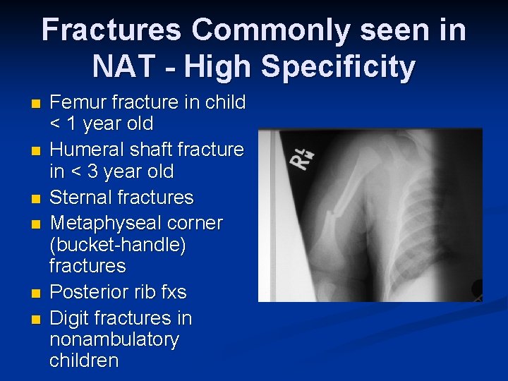
Fractures Commonly seen in NAT - High Specificity n n n Femur fracture in child < 1 year old Humeral shaft fracture in < 3 year old Sternal fractures Metaphyseal corner (bucket-handle) fractures Posterior rib fxs Digit fractures in nonambulatory children
50% to 69% of all fractures occurred in children less than 1 year of age. n 78% to 85% occurred in children less than 3 years of age. n
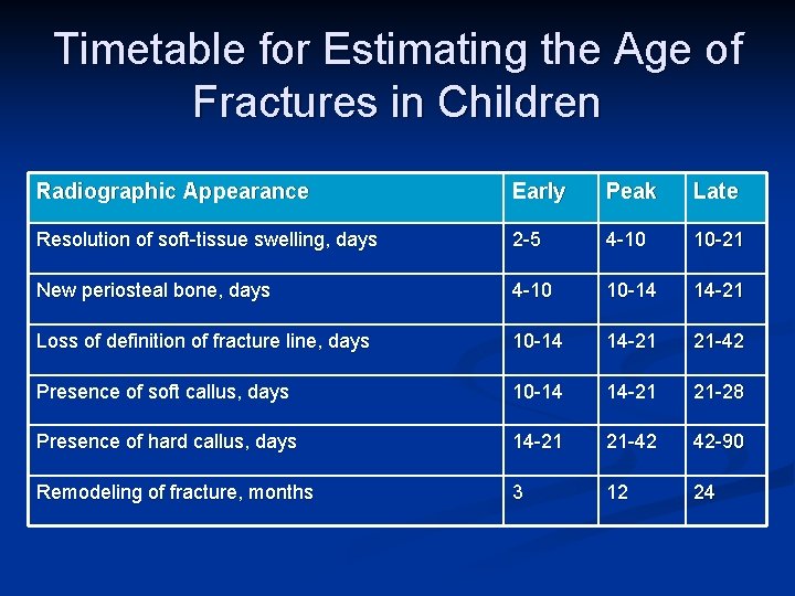
Timetable for Estimating the Age of Fractures in Children Radiographic Appearance Early Peak Late Resolution of soft-tissue swelling, days 2 -5 4 -10 10 -21 New periosteal bone, days 4 -10 10 -14 14 -21 Loss of definition of fracture line, days 10 -14 14 -21 21 -42 Presence of soft callus, days 10 -14 14 -21 21 -28 Presence of hard callus, days 14 -21 21 -42 42 -90 Remodeling of fracture, months 3 12 24
Radiographic W/U n Skeletal survey for children with suspicion of NAT n “Babygram” not sufficient as does not provide necessary detail to identify fractures
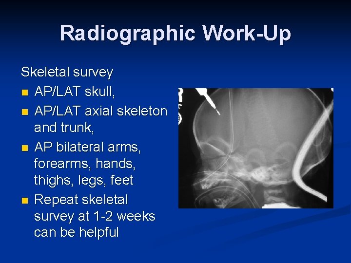
Radiographic Work-Up Skeletal survey n AP/LAT skull, n AP/LAT axial skeleton and trunk, n AP bilateral arms, forearms, hands, thighs, legs, feet n Repeat skeletal survey at 1 -2 weeks can be helpful
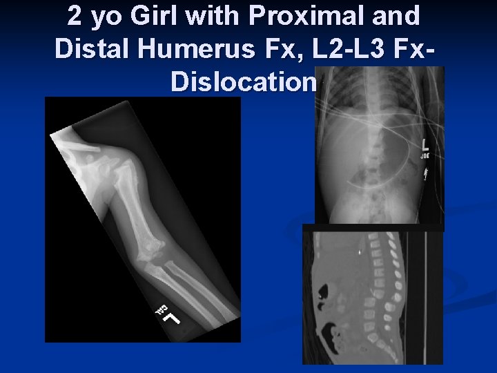
2 yo Girl with Proximal and Distal Humerus Fx, L 2 -L 3 Fx. Dislocation
Bone Scan n Usually reserved for highly suspicious cases with negative skeletal survey n Good at picking up rib fx’s and vertebral fx’s n Repeat bone scan at 2 weeks can identify occult injuries

Multiple Vs single # n Multiple fractures in various stages of healing are found in more than 70% of abused children less than 1 year of age and more than 50% of all abused children. Krishnan J, Aust N Z J Surg 1990 n n n 50% of the children had only a single fracture, 33% had two or three fractures, and 17% had more than three fractures. King et al, J Pediatr Orthop 1988 Most common orthopaedic presentation of children with NAT - 65% of children with fx’s Only 13% of children with fractures presented with multiple fractures in different stages of healing Loder, JPO 1991
Patterns of fractures n Spiral Vs transverse 48% to 71% of long-bone fractures in several large series are transverse #. n no difference in diaphyseal fracture pattern between fractures due to abuse and those resulting from accidental injury. n Beals , Pediatr Orthop 1983 n no difference in diaphyseal fracture pattern between fractures due to abuse and those resulting from accidental injury.
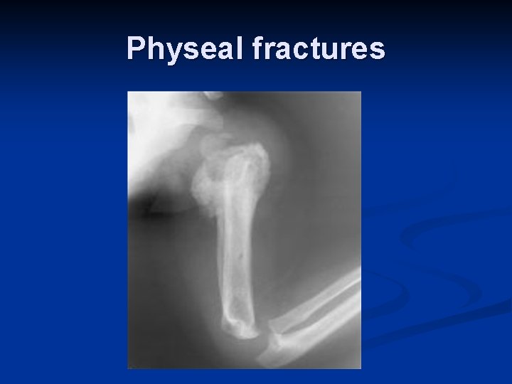
Physeal fractures
Specific bone n Clavicle: n n n physeal fractures: n n n Most commonly fractured bones in accidental childhood injury. Unusual in child abuse, detected in only 2% to 7% of abused children. Uncommon in the abused child. Transphyseal fractures of the distal humerus in children less than 1 year old. Spinal : (0% to 3%) n n n Asymptomatic compression fractures detected on skeletal survey. Fracture or avulsion of the spinous processes is fairly specific to abuse. Hyperflexion and hyperextension associated with violent shaking.
Specific bone n Femure Most femur fx’s in children who are < 1 yo of age are from NAT (60 -70%) n Most femur fx’s in children > 1 yo accidental n
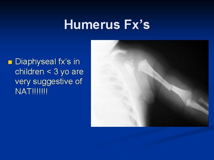
Humerus Fx’s n Diaphyseal fx’s in children < 3 yo are very suggestive of NAT!!!!!!!

Humerus Fx’s n Most common fx in some series n Supracondylar fx’s common in accidental trauma n Transphyseal fx’s - high association with NAT

Transphyseal Humerus n Common NAT in
Metaphyseal Vs diaphyseal Metaphyseal injuries are less common than diaphyseal fractures. n Metaphyseal lesions have high specificity and are considered to be a “classic” radiographic finding in physical abuse n
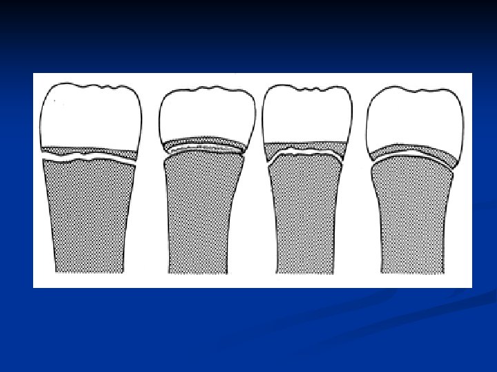
Metaphyseal or Bucket Handle Fx’s n Mechanism – traction and twisting n Planar injuries through the primary spongiosum n May be picked up at autopsy when not seen on x-ray

Corner Fractures n n Traction/rotation mechanism of injury Planar fracture through primary spongiosa, creates disklike fragment of bone/cartilage, thicker at periphery
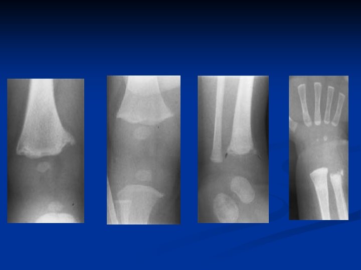
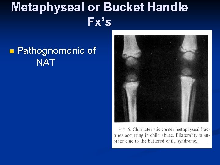
Metaphyseal or Bucket Handle Fx’s n Pathognomonic NAT of
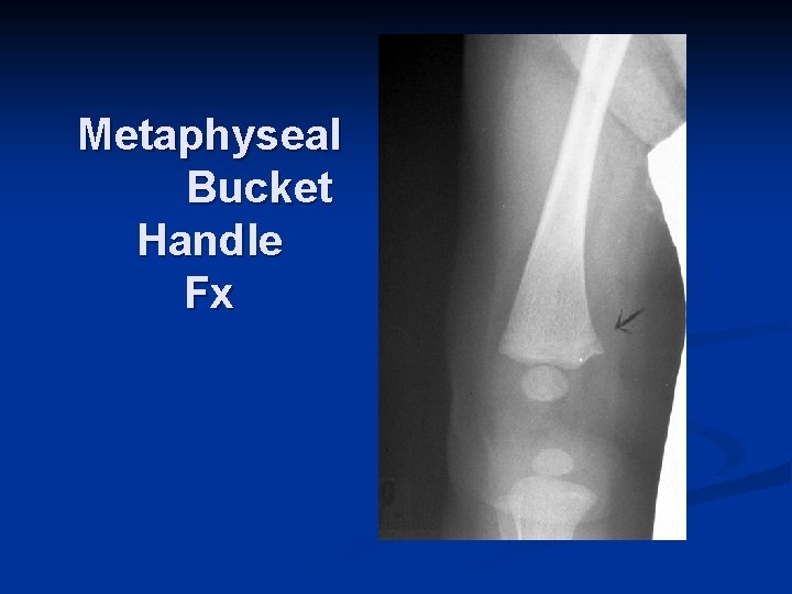
Metaphyseal Bucket Handle Fx
Differential Diagnosis Osteogenesis imperfecta n Accidental injury n Birth trauma n Rickets n Coagulation disorders n Leukemia n Congenital insensitivity to pain n
Management - NAT Suspected Professional, tactful, nonjudgmental approach in initial encounter and workup n Explain workup to parents as standard approach to specific ages/injury patterns n Early involvement of child protection team if available n Early contact/involvement of child’s primary care physician n
Management - Documentation Many cases result in medical records becoming part of legal record n Carefully document history, physical exam and radiographic findings n Document evidence supporting physical abuse n Document statement regarding level of certainty of abuse n
- Slides: 40