ORTHOMYXOVIRUSES ORTHOMYXOVIRUSES Affinity for mucins Myxamucus Adsorb to

ORTHOMYXOVIRUSES
 Adsorb to mucoprotein receptors on RBC’s")
ORTHOMYXOVIRUSES • • • Affinity for mucins (Myxa-mucus) Adsorb to mucoprotein receptors on RBC’s Myxovirus – Orthomyxo & Paramyxoviruses Orthomyxo: Genus Influenza – Type A, B &C Species of Type A– H 1 N 1(A 1 human, Hsw N 1 - swine flu) – H 2 N 2 (A 2) – H 3 N 2 (A 3, A Hong Kong) – H 5 N 1 ( bird flu/avian influenza)

Differences between Ortho & Paramyxoviruses Property Orthomyxo Paramyxo Size 80 -120 nm 100 -300 nm Shape Spherical Pleomorphic Genome ss. RNA; 8 segments ss. RNA; 1 segment Ribonucleo Nucleus protein synthesis Cytoplasm

Property Orthomyxo Paramyxo Genetic Recombination Common Absent DNA dependent Required for RNA synthesis multiplication not required Rate of antigenic High Change Low Hemolysin Present Absent

INFLUENZA

History: Known Flu Pandemics Name of pandemic Date Deaths Asiatic Flu 1889 -1890 1 million Spanish Flu 1918 -1920 40 -100 million Asian Flu 1957 -1958 1 - 1. 5 million Hong Kong Flu 1968 -1969 0. 75 - 1 million

1918 Flu Pandemic American Red Cross nurses tend to flu patients in temporary wards set up inside the Oakland municipal Auditorium.

Method of Infection and Replication: • The flu virus binds onto sugars on the surfaces of epithelial cells such as nose, throat, and lungs of mammals and intestines of birds.

‘FLU’ • True influenza – influenza virus A or influenza virus B (or influenza virus C infections - much milder) • Febrile respiratory disease with systemic symptoms caused by a variety of other organisms often inaccurately called ‘flu’
 with abrupt disease onset •")
Influenza - the Disease • Acute respiratory infection (ARI) with abrupt disease onset • Clinically it cannot be distinguished from other ARI • Complications - primary influenza pneumonia or secondary bacterial pneumonia, and myocarditis, particularly in children, elderly people and people with underlying chronic diseases

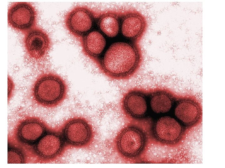
Influenza Virus - Structure • • 80 – 120 nm, Spherical Single-stranded RNA Virus, 8 segments RNA polymerase Nucleocapsid is enclosed by an inner protein layer called matrix M protein • Outer lipid layer – derived from host cell membrane

M protein
 lipid")
ORTHOMYXOVIRUSES HA - hemagglutinin NA - neuraminidase helical nucleocapsid (RNA plus NP protein) lipid bilayer membrane polymerase complex M 1 protein type A, B, C : NP, M 1 protein sub-types: HA or NA protein

Antigenic classification • 3 influenza types: A, B, C ( Based on RNP & M protein) – type A: moderate/severe epidemics, affects humans and animals – type B: milder epidemics, affects only humans – type C: no epidemics; sporadic cases in humans • Influenza subtypes of type A determined by hemagglutinin (H) and neuraminidase (N)

ANTIGENIC VARIATION Antigenic drift - minor changes in either • • haemagglutinin or neuraminidase or both Gradual change; regular; frequent intervals Mutation in HA & NA genes Periodic epidemics Cross reacts with antisera to predecessor virus

• Antigenic shift – Major changes in HA & NA or both • New subtypes • Abrupt & Drastic change • Results from gene re-assortment between human and animal strains • Major epidemics & Pandemics • Antibodies to predecessor virus cannot neutralize new variants
 and/or")
Antigenic Changes of Influenza Virus • Type A: major change of hemagglutinin (H) and/or neuraminidase (N) resulting in new subtypes and pandemics – – – – 4 pandemics in 19 th century 1918: subtype H 1 N 1: severe pandemic, 20 million deaths 1957: subtype H 2 N 2: severe pandemic 1968: subtype H 3 N 2: moderate pandemic 1977: subtype H 1 N 1: mild pandemic 1989; subtype H 1 N 1; swine flu 2009; subtype H 1 N 1; – Minor antigenic change within the same subtype, associated with epidemics • Type B: no subtypes, only minor antigenic changes, associated with epidemics
; • all human influenza A")
Reservoir and Transmission • Reservoir: humans, animals (type A); • all human influenza A viruses infect avian species, • few antigenic subtypes of influenza A infect man and other animals (pigs and horses); • evidence that the viruses which caused pandemics originated from animals: swine strain (1918), avian strains (1957, 1968) • Transmission: airborne, through respiratory droplets • Seasonality: temperate regions: winter, tropics: often in the rainy season • Incubation period: 1 -3 days • Communicability: 1 -2 days before to 4 -5 days after onset

where do “new” HA and NA come from? • 13 types HA • 9 types NA – all circulate in birds • pigs – avian and human

Where do “new” HA and NA come from?

Where do “new” HA and NA come from - can ‘new’ bird flu directly infect humans? Bird flu H 5 N 1?

Pathogenesis • Infection spreads from person to person via respiratory secretions, by airborne droplets or by contact with contaminated hands or surfaces. • Virus attaches to and infects the respiratory mucosal epithelial cells. • Progeny virions are soon produced and spread to adjacent cells, where the replicative cycle is repeated.

• Influenza viruses cause cellular destruction and desquamation of superficial mucosa of the respiratory tract. • Viral damage to the respiratory tract epithelium lowers its resistance to secondary bacterial invaders. • Prominent systemic symptoms associated with influenza probably reflect the production of cytokines.

NORMAL TRACHEAL MUCOSA 3 DAYS POST-INFECTION 7 DAYS POST-INFECTION

• DECREASED CLEARANCE • RISK BACTERIAL INFECTION • VIREMIA RARE
 • PRIMARY INFLUENZA VIRUS PNEUMONIA • SECONDARY")
PULMONARY COMPLICATIONS • CROUP (YOUNG CHILDREN-barking cough) • PRIMARY INFLUENZA VIRUS PNEUMONIA • SECONDARY BACTERIAL INFECTION – Streptococcus pneumoniae – Staphlyococcus aureus – Hemophilus influenzae
 • cardiac")
NON-PULMONARY COMPLICATIONS • myositis (rare, > in children, > with type B) • cardiac complications • recent studies report encephalopathy • liver and CNS – Reye’s syndrome • peripheral nervous system – Guillian-Barré syndrome

LAB DIAGNOSIS Microscopy – Smears from nasopharyngeal secretions, nasal swab, centrifuged deposit of throat garglings • Direct immunofluorscence – Demonstration of viral antigens Culture - throat garglings • Amniotic cavity of chick embryo - amniotic fluid and allantoic fluid is tested for HA using Guinea pig & fowl RBC’s at RT & 4 • C • Monkey kidney cell cultures

Haemagglutination Guinea pig RBC’s fowl RBC’s Type A + _ Type B + + Type C - +

Serology • CFT • HAI test – Serum diluted serially – Influenza virus suspension with 4 HAunits added – Fowl RBC’s added – Highest dilution inhibiting HA – HAI titre

Treatment • Neuraminidase inhibitors are a new class of drugs used for the treatment of influenza. • They act by inhibiting the release of the virus from infected cells. This limits the infection by reducing the cell to cell spread of the virus. • They are about 70 – 90 % effective against influenza
 – is administered by inhalation. • Oseltamivir (Tami flu) –")
• Zanamivir (Relenza) – is administered by inhalation. • Oseltamivir (Tami flu) – is given orally. • Amantadine & Rimantadine inhibit viral uncoating of Influenza A virus. • They are effective only against influenza A virus and were widely used in the prevention and treatment of influenza A. But now, due to the development of resistance, their use is not recommended.
 – It is an inactivated")
Prevention using Influenza Vaccines • Trivalent Inactivated Vaccine (TIV) – It is an inactivated vaccine prepared from the influenza viruses grown in embryonated eggs. • It is given by intramuscular injection in 2 doses separated by a month.
 – It is a cold adapted vaccine")
• Live Attenuated Influenza Vaccine: (LAIV) – It is a cold adapted vaccine containing temperature sensitive (ts) mutants of influenza A and B viruses. • These mutant strains can replicate in the cooler temperature (33 o. C) of the nasal mucosa where it induces the production of secretory Ig. A antibodies, but not in the warmer (37 o. C) of the lower respiratory tract. • It is administered by nasal spray.

Current Research: • The Influenza Genome Sequencing Project creating a library of influenza sequences to study why one strain is more lethal than another. • Research into new vaccines. • Study the infection in other animals, especially birds. Viral strains between species can occur.

THANK YOU
- Slides: 37