Organs and Organ Systems Respiratory System Respiratory System


- Slides: 57
Organs and Organ Systems Respiratory System
Respiratory System
Points to ponder l l l What are the parts and functions of the upper and lower respiratory system? What is the mechanism for expiration and inspiration? How is breathing controlled by the nervous system and through chemicals? Where and how is exchange of gases accomplished? What are some common respiratory infections and disorders? What do you know about tobacco and health?
Overview of the respiratory system Copyright © The Mc. Graw-Hill Companies, Inc. Permission required for reproduction or display. Nasal cavity filters, warms, and moistens air Pharynx passage way where pathway for air and food cross Upper Respiratory Tract Glottis space between the vocal chords; opening to larynx Larynx (voice box) ; produces sound Trachea (wind pipe) ; passage of air to bronchi Bronchus passage of air to lungs Lower Respiratory Tract Bronchioles passage of air to alveoli Lung contains alveoli (air sacs); carries out gas exchange Diaphragm skeletal muscle; functions in ventilation Figure 9. 1 The human respiratory tract.
What is the pathway that air follows? nose pharynx larynx trachea bronchus bronchioles alveoli
What constitutes the upper respiratory tract? Copyright © The Mc. Graw-Hill Companies, Inc. Permission required for reproduction or display. l Nose sinus nasal cavity l Pharynx hard palate nares sinus tonsil Pharynx nasopharynx uvula mouth l Larynx tongue tonsils epiglottis larynx trachea Figure 9. 2 The upper respiratory tract. oropharynx laryngopharynx esophagus
The nose l The nose opens at the nostrils/nares and leads into the _____. l Hairs and mucus in the nose _____ the air. l The nasal cavity has a lot of capillaries that warm and moisten the air. l Specialized cells act as odor receptors. l _____ drain into the nasal cavities that can lead to a runny nose.
The pharynx l The _____ is a funnel-shaped cavity commonly called the throat. l It has 3 portions based on location: nasopharynx, oropharynx, and laryngopharynx. l _____ provide a lymphatic defense during breathing at the junction of the oral cavity and pharynx.
9. 2 The Upper Respiratory Tract The larynx l The larynx is a triangular, cartilaginous structure that passes air between the ______ and _______. l It is called the ______ and houses vocal cords. l There are 2 mucosal folds that make up the vocal cords with an opening in the middle called the ______.
The larynx Copyright © The Mc. Graw-Hill Companies, Inc. Permission required for reproduction or display. base of tongue epiglottis vocal cords glottis (left): © CNRI/ Phototake Figure 9. 4 The vocal cords.
What constitutes the lower respiratory tract? Copyright © The Mc. Graw-Hill Companies, Inc. Permission required for reproduction or display. l Trachea Nasal cavity filters, warms, and moistens air Pharynx passage way where pathway for air and food cross Upper Respiratory Tract l Bronchial tree Glottis space between the vocal chords; opening to larynx Larynx (voice box) ; produces sound Trachea (wind pipe) ; passage of air to bronchi Bronchus passage of air to lungs l Lungs Lower Respiratory Tract Bronchioles passage of air to alveoli Lung contains alveoli (air sacs); carries out gas exchange Diaphragm skeletal muscle; functions in ventilation Figure 9. 1 The human respiratory tract.
The trachea l The trachea is a tube, often called the windpipe, that connects the larynx with the _______. l It is made of connective tissue, smooth muscle, and cartilaginous rings. l The trachea is lined with ______ and ______ that help to keep the lungs clean.
The trachea Copyright © The Mc. Graw-Hill Companies, Inc. Permission required for reproduction or display. cilia goblet cell epithelial cell particle movement mucus air tracheal lumen (bottom half): © ED Reschke Figure 9. 5 The cells lining the trachea.
18. 1 Effects of Smoke on the Respiratory System l On average, a resting human: l l l Breathes once every 12 seconds Takes a breath with a volume of about 500 milliliters Sends about 1 liter of air per minute into the lungs
Diaphragm l The respiratory system is separated from the digestive organs by the diaphragm. l l When the diaphragm contracts, it flattens. Simultaneously, the rib cage lifts up and out. Volume of chest cavity increases, pressure lowers and air rushes in. When diaphragm relaxes, chest cavity loses volume, pressure increases, and air leaves.
Diaphragm § Inhalation brings air into the lungs and exhalation brings air out of the lungs.
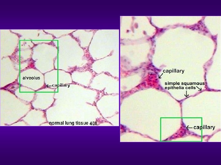
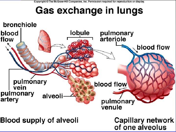
Lungs l Healthy lungs are spongy, pink sacs that fill the chest cavity. l Lungs are attached to the chest wall by a doublelayered membrane the pleura. l Air enters the lungs through bronchi. l Bronchi branch into bronchioles. l Bronchioles finally end at alveoli—small, vascularized sacs. l Alveoli are the functional unit of the lung and where gas exchange occurs.
Lungs l On average, lungs contain about 300 million alveoli, and these contain the respiratory surface through which the body acquires oxygen and eliminates CO 2 waste. l The total area of the respiratory surface in a pair of lungs is about the same area as a tennis court.
Lungs l Each alveolus is surrounded by a net of capillaries – tiny, thin-walled blood vessels that connect the gases exchanged with the body.
Blood Transports Gases Between Lungs and Tissues
Gas Exchange l Gas exchange is the primary function of the lungs: O 2 from the environment is exchanged for CO 2 from the body. l Gas exchange occurs by simple diffusion between the alveoli and the capillaries.
The Role of Hemoglobin in Gas Exchange l CO 2 easily diffuses from blood to air; O 2 requires help to enter the blood. l l Hemoglobin – respiratory pigment Hemoglobin produces color when it binds with oxygen. A single hemoglobin is made up of 4 different protein chains, each with an iron atom. l Iron binds to the oxygen and carries it in the blood. A red blood cell contains about 250 million hemoglobin molecules; it can carry 1 billion oxygen molecules.
The Role of Hemoglobin in Gas Exchange l Hemoglobin is efficient at binding O 2, but even more effective at binding carbon monoxide. l l Even small amounts of carbon monoxide can tie up a lot of hemoglobin. Carbon monoxide causes oxygen shortages in tissues. Carbon monoxide is especially damaging to fetuses and embryos. Lower than average birth weights associated with smoking mothers are due to oxygen deprivation.
10. 4 Mechanism of Breathing Two phases of breathing/ventilation 1. Inspiration – an active process of inhalation that brings air into the lungs 2. Expiration – a typically passive process of exhalation that expels air from the lungs 28
10. 4 Mechanism of Breathing Boyle’s Law • Ventilation is governed by Boyle’s Law. • At a constant temperature the pressure of a given quantity of gas is inversely proportional to its volume. Figure 10. 7 The relationship between air pressure and volume. 29
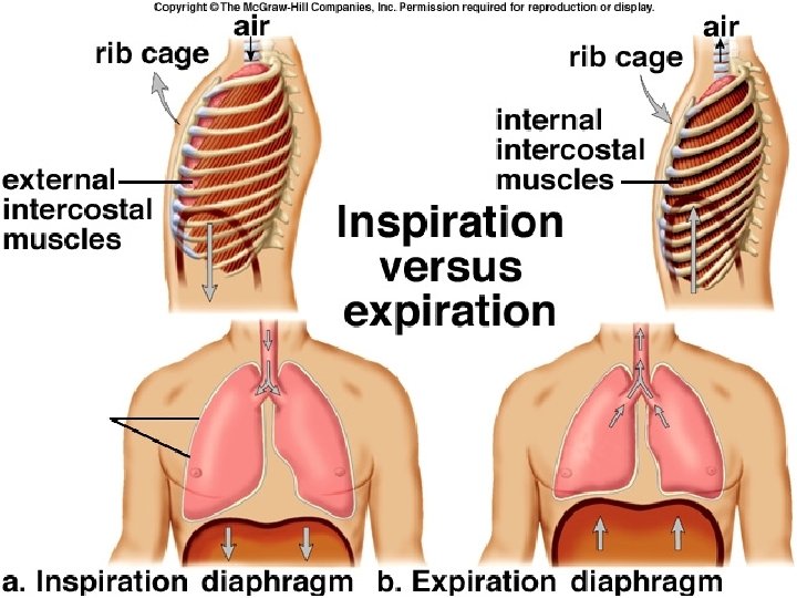
10. 4 Mechanism of Breathing Inspiration • The diaphragm and intercostal muscles contract. • The diaphragm flattens and the rib cage moves upward and outward. • Volume of the thoracic cavity and lungs increase. • The air pressure within the lungs decreases. • Air flows into the lungs. 30
10. 4 Mechanism of Breathing Inspiration trachea Rib cage moves up and out. External intercostal muscles pull the ribs outward. lungs Diaphragm contracts and moves down. air in lung rib cage When Pressure in lungs decreases, air comes rushing in. a. Inspiration Figure 10. 8 a The thoracic cavity during inspiration. 31
10. 4 Mechanism of Breathing Expiration • The diaphragm and intercostal muscles relax. • The diaphragm moves upward and becomes domeshaped. • The rib cage moves downward and inward. • Volume of the thoracic cavity and lungs decreases. • The air pressure within the lungs increases. • Air flows out of the lungs. 32
10. 4 Mechanism of Breathing Expiration Rib cage moves down and in. Internal intercostal muscles pull the ribs inward during forced expiration. Diaphragm relaxes and moves up. air out When pressure in lungs increases, air is pushed out. b. Expiration Figure 10. 8 b The thoracic cavity during expiration. 33
10. 4 Mechanism of Breathing Different volumes of air during breathing • Tidal volume – the small amount of air that usually moves in and out with each breath • Vital capacity – the maximum volume of air that can be moved in plus the maximum amount that can be moved out during one breath • Inspiratory and expiratory reserve volume – the increased volume of air moving in or out of the body • Residual volume – the air remaining in the lungs after exhalation 34
10. 4 Mechanism of Breathing Visualizing the vital capacity 5, 800 maximum expiration Average Lung Volume (ml) 4, 800 maximum inspiration inspiratory reserve volume vital capacity 3, 600 2, 900 2, 400 tidal volume total Lung capacity expiratory reserve volume 1, 200 residual volume 0 Figure 10. 9 Measuring the vital capacity of the lungs. residual volume © Veronique Burger/Science Source 35
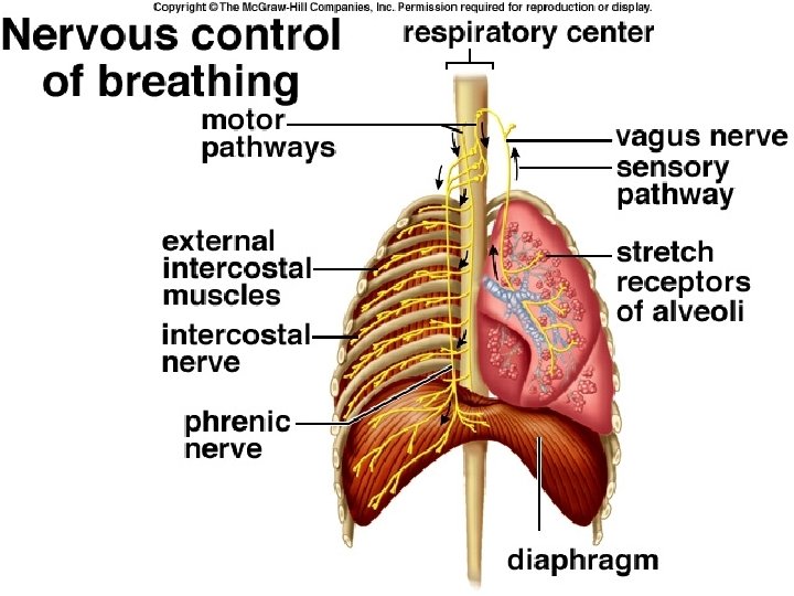
10. 5 Control of Ventilation How is breathing controlled by the nervous system? • Nervous control – Respiratory control center in the brain (medulla oblongata) sends out nerve impulses to contract muscle for inspiration. – Sudden infant death syndrome (SIDS) is thought to occur when this center stops sending out nerve signals. 36
10. 5 Control of Ventilation How is breathing controlled by the nervous system? brain Respiratory center: region of the brain that automatically regulates breathing Intercostal nerves stimulate the intercostal muscles to contract. External intercostal muscles help expand the thoracic cavity by contracting. Phrenic nerve stimulates the diaphragm to contract. Diaphragm helps expand the thoracic cavity by flattening when it contracts. Figure 10. 10 The control of breathing by the respiratory center. 37
10. 5 Control of Ventilation How is breathing chemically controlled? • Chemical control – Two sets of chemoreceptors sense the drop in p. H: one set is in the brain and the other in the circulatory system. – Both are sensitive to carbon dioxide levels that change blood p. H due to metabolism. 38
10. 6 Gas Exchanges in the Body Exchange of gases in the body • Oxygen and carbon dioxide are exchanged. • The exchange of gases is dependent on diffusion. • Partial pressure is the amount of pressure each gas exerts (PCO 2 or PO 2). • Oxygen and carbon dioxide will diffuse from the area of higher to the area of lower partial pressure. 39
10. 6 Gas Exchanges in the Body External respiration • Exchange of gases between the lung alveoli and the blood capillaries. • PCO 2 is higher in the lung capillaries than the air, thus CO 2 diffuses out of the plasma into the lungs. • The partial pressure pattern for O 2 is just the opposite, so O 2 diffuses into the red blood cells in the lungs. 40
10. 6 Gas Exchanges in the Body External respiration Carbon dioxide transport: H+ + HCO 3 H 2 CO 3 Oxygen transport: Hb + O 2 carbonic anhydrase H 2 O + CO 2 Hb. O 2 41
10. 6 Gas Exchanges in the Body The movement of oxygen and carbon dioxide in the body alveolus plasma H+ + HCO– 3 External respiration Hb H+ CO 2 pulmonary capillary HCO– 3 RBC H 2 CO 3 CO 2 Hb O 2 H 2 O RBC O 2 Hb CO 2 pulmonary capillary O 2 alveolus CO 2 exits blood CO 2 a. plasma O 2 enters blood O 2 lung pulmonary artery pulmonary vein heart tissue cells systemic vein systemic artery HCO– 3 H+ + HCO– 3 plasma systemic capillary RBC Figure 10. 11 Movement of gases during external and internal respiration. CO 2 Hb H+ H 2 CO 3 CO 2 H 2 O Internal respiration RBC systemic capillary O 2 O 2 Hb Hb CO 2 tissue fluid CO 2 enters blood b. tissue cell tissue fluid O 2 exits blood 42
10. 6 Gas Exchanges in the Body Internal respiration • The exchange of gases between the blood in the capillaries outside of the lungs and the tissue fluid. • PO 2 is higher in the capillaries than the tissue fluid, thus O 2 diffuses out of the blood into the tissues. 43
10. 6 Gas Exchanges in the Body Internal respiration Oxyhemoglobin gives up oxygen: Hb. O 2 Hb + O 2 Most CO 2 is carried as a bicarbonate ion: CO 2 + H 2 O carbonic anhydrase H 2 CO 3 H+ + HCO 3 - 44
Smoke Particles and Lung Function l Normal function of the lungs: l l l Cough is first response to lung irritants Small particles don’t trigger cough; they become trapped in mucus lining the respiratory tract Cilia move trapped particles to nose and mouth Mucus is coughed out or swallowed The majority of the damage to lungs is caused by particulates in smoke, which damage the surfaces of the lungs. l Children and infants are particularly vulnerable
Smoke Particles and Lung Function l l l Particles can interfere with the lung’s defense systems Particulates increase mucus production, but damage cilia leading to bronchitis Asthma is an allergic reaction where bronchioles constrict and mucus production increases. l l Particulates are known to exacerbate asthma. The EPA estimates that environmental tobacco smoke, or ETS, will cause 26, 000 additional cases of asthma.
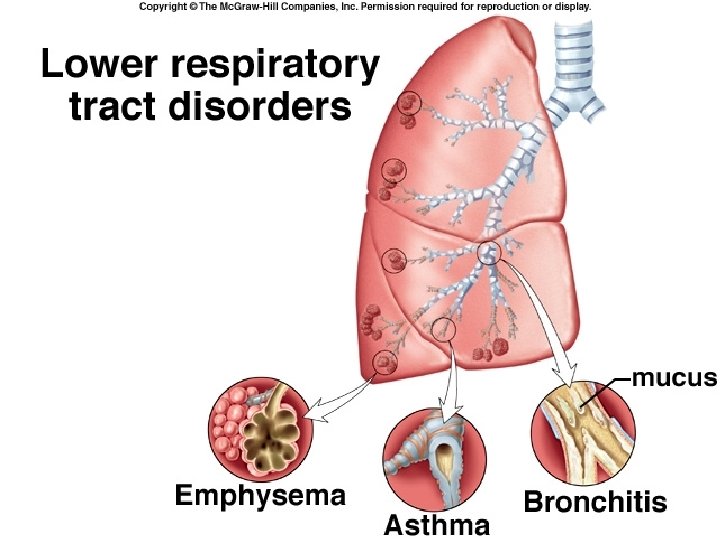
Emphysema l Emphysema is caused by scar tissue formation due to chronic bronchitis and asthma. l Alveoli can become damaged and merge into fewer and larger sacs. This drastically reduces surface area for gas exchange. l The damage is permanent; lung tissue is not regenerated. l
Lung Cancer l Many of the particulates in cigarette smoke contain chemicals known to be carcinogens l l Particulates can stay on lung surfaces for long periods of time Risk of mutation remains long after cigarette has been smoked leading to cancer
Diseases of the Respiratory System l Respiratory rate: 10 to 14 inhalations/minute. l In one day, an average human: l Breathes 20, 000 times l Inhales 35 pounds of air l Most of us breathe in air that is heavily contaminated with solid particles, ozone, sulfur oxide, carbon monoxide, nitrogen oxides, and many other damaging chemicals. l Breathing contaminated air can cause a number of diseases including asthma, bronchitis, emphysema, and lung cancer.
Diseases of the Respiratory System l Cigarette smoke is one of the worse air pollutants. l Over 1 million people start smoking every year. l Kills about 350, 000 people every year in U. S. l Contains 4000 different chemicals. l Each cigarette smoked subtracts about 5 minutes from life expectancy.
Diseases of the Respiratory System l Cigarette smoke paralyzes cilia in airways, preventing them from removing debris and from protecting delicate alveoli. l Frequent coughing is the only way airways can clean themselves. l Cigarette smoke also causes fetal damage, which can result in miscarriage, premature birth, low birth weight, and poor development.
Diseases of the Respiratory System l l Asthma: Condition in which breathing is impaired by constriction of bronchi and bronchioles, cough, and thick mucus secretions. The severity and incidence of asthma has risen dramatically in recent years, especially in children. May be fatal if not treated. Causes: Attacks may be precipitated by inhalation of allergens (e. g. : pollen, cats, and cockroach proteins), pollutants, infection, or emotional stress. Treatment: Alleviates symptoms (e. g. : immunosuppressors, bronchodilators), but is not a cure.
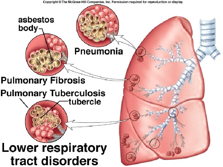
l l Diseases of the Respiratory System Bronchitis: Inflammation of the mucous membranes of the bronchi. May present with cough, fever, chest or back pain, and fatigue. Causes: Associated with smoking, pollution, and bacterial or viral infections. Pneumonia: Acute inflammation of the lungs. Symptoms include high fever, chills, headache, cough, and chest pain. Causes: Bacterial, fungal, or viral infections. Treatment: Antibiotics or other antimicrobials.
l Diseases of the Respiratory System Emphysema: Permanent and irreversible destruction of alveolar walls, resulting in loss of lung elasticity and gas exchange surface. Symptoms include shortness of breath, difficulty exhaling, cough, weakness, anxiety, confusion, heart failure, lung edema (swelling), and respiratory failure. Causes: Smoking, pollution, old age, and infections. Treatment: Oxygen to help breathing. No cure.
l Diseases of the Respiratory System Lung Cancer: Cancerous growth that invades and destroys lung tissue. Very high fatality rate. Symptoms include bloody sputum, persistent cough, difficulty breathing, chest pain, and repeated attacks of bronchitis or pneumonia. Causes: Smoking (50% of all cases) and pollution (radon, asbestos). Smokers are 10 times more likely to develop lung cancer than nonsmokers. Treatment: Surgery is most effective, but only 50% of all lung cancers are operable by time of detection. Other treatments include radiation and chemotherapy.