ORGANOGENESIS OF THE UROGENITAL SYSTEMS Indication the formation


 4 Metanephros develops at Mesonephric duct Urachus somites")
 4 The ureteric bud forms the duct system 4")


.")

 n Primary sex determination at fertilisation genetic sex: XY, XX (ii). Y")


- Slides: 14
ORGANOGENESIS OF THE UROGENITAL SYSTEMS.
Indication the formation of the nephric systems the formation of the genital ducts the formation of the gonads
Overview of organogenesis of the urogenital organs Cr. Urinary and reproductive systems are closely associated in topography, D V function and development. Ca. Two systems have common origin from the urogenital ridge(UGR) and have homologous structures. Internal genital duct system is derived from the foetal urinary system Malformation of one system affects the other. Mesonephros Gonad (nephrogenic The UGR is longitudinal swelling in (genital ridge) Plate) dorsolateral side of the abdomen UGR--> UGR formed mostly from mesoderm Lateral UGR(nephrogenic plate) forms Mesonephric urinary organs and internal genital ducts. Duct Ventromedial UGR is genital ridge, ridge Paramesonephric forms gonads duct The Urogenital ridge
Sequence of development of the metanephros(1) 4 Metanephros develops at Mesonephric duct Urachus somites from 2 precursors --> ureteric bud(UB) and metanephrogenic mass (MM) Bladder 4 Formation UB and MM is by interactions between the tissues. 4 UB forms the duct system 4 MM forms nephrons 4 The ureteric bud branches as it grows towards the metanephrogenic mass(B) Metanephrogenic mass A Int. mesoderm Ureteric bud Collecting ducts Cr D Ca. Metanephros Ureter B
Morphogenesis of the ureteric bud) 4 The ureteric bud forms the duct system 4 The metanephrogenic mass forms the Kidney of dog cortex medulla renal pelvis nephrons by nephrogenesis. 4 branching pattern of the ureteric bud is species specific. collecting 4 Branching of simple unipyramidal ureter duct kidneys(dog, horse) the proximal end kidneys collecting duct dilates into a renal pelvis with collecting ducts at the tip 4 In multipyramidal kidneys(ox) ureter bifurcates into 2 major calyces and several cortex minor calyces then collecting ducts minor calyx ureter major calyx medulla Kidney of ox
growth of internal and external genital organs Before the sixth week of pregnancy, there is no functional difference between the male and female genitals. After that time, primitive cells from the yolk sac to the intermediate mesoderm migrate to genital region and primitive genitalia. Following the gender differentiation of the fetus after the sixth week of pregnancy,
the primary and non-specific genitals follow different paths according to the effect of the SRY gene on Y chromosome, which plays a key role in determining the sex of the fetus by activating several genes including SF 1, SOXG and DAX 1, which helped to production of SERTOLI CELLS, and the substance that inhibits the appearance of female genital organs, called MIS or Mullerian Inhibiting Substance.
Under the influence of this inhibitory substance, the female ducts are inhabit but the male ducts will growth which include vase deference, epididymis and seminal vesicles by stimulant the male hormone (testosterone) which produced by LEYDIG CELLS in the testis under the influence of HCG from the placenta. n n
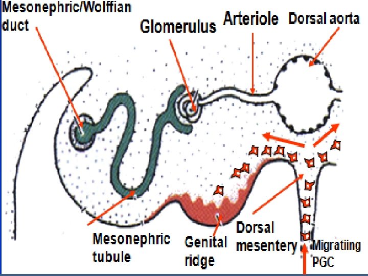
Gonadogenesis 4 Gonadogenesis occurs at the genital ridge initiated by 2 simultaneous events: (i). Formation of gonadal cords 4 Epithelium from degenerate mesonephric nephrons invade genital ridge, and form epithelial cords (ii). Migration of primordial germ cells (PGC) are endodermal cells, migrate from the yolk sac into the gonad. The gonad has a central medulla and a peripheral cortex, surrounded by epithelium. Epithelial PGC, forming gonadal cords. Gonadal differentiation begins.
SEX DETERMINATION Mullerian/ Paramesonephric duct Gonadal ridge DAX Wnt 4 Ovary SRY Testis gonad Oestrogen Female genital ducts Anti-Mullerian hormone (Sertoli cells) Regression of Mullerian duct Testosterone (Leydig cells) • Differentiation of Wolffian duct into Male genital ducts • Descent of testis
n (i) n Primary sex determination at fertilisation genetic sex: XY, XX (ii). Y chromosomes encodes testisdetermining factor SRY gonadogenesis secretion of foetal hormones by interstitial cells[(Sertoli and Leydig, theca cells] secondary sex(phenotypic sex)
Wolffian duct Definitive gonads: Testis Rete testis A Dorsal mesentery 4 Germ cells and Sertoli cells Testicular concentrate in the testicular cords Mullerian duct Fibrous capsule Interstitium 4 The cortex develops a thick Efferent ducts fibrous capsule 4 Testicular cords form loops (seminiferous tubules) and Wolffian duct interconnect with mesonephric tubules to form efferent ducts B Mullerian duct Seminiferous tubules
Ovary formation. Wolffian duct In the absence of the Y chromosome in primordial germ cells. Mullerian duct 4 Ovarian cords concentrate in the cortex 4 The medulla degenerates. The remnants form the vascular, lymphatic and nervous tissues. 4 Meiosis begins and the epithelia surround germ cells forming primordial B follicles. Wolffian duct A Dorsal mesentery Interstitium Ovarian cords Germinal epithelium Oocyte Primordial follicle Gilbert 2006)