Organization of the Cell Cell Theory Cells are


 Theodor Schwann- German zoologist (1839)")


 German scientists Max")


")


- Slides: 160
Organization of the Cell
Cell Theory Cells are the basic living units of organization and function in all organisms and all cells come from other cells
Cell Theory The players: Matthias Schleiden- German botanist (1838) Theodor Schwann- German zoologist (1839) Rudolph Virchow- German professor of pathology (1855)
Schleiden and Schwann The first to point out that all plants and animals are composed of cells. 1838
Rudolph Virchow The first to observe cells dividing 1855
History of the Microscope • Robert Hooke examined a thin piece of cork using a compound microscope- noticed the boxes in the thin slice and called them “cells” 1665 ?
History of the Microscope Anton van Leeuwenhoek viewed living cells with 200 magnification single lenses of his own construction. His important discoveries include bacteria, protists, blood cells, and sperm cells. 1670 s Dutch Scientist
Van Leeuwenhoek’s Microscope
1800
1860
1880
1890
1899
1908
1930
1951
1970
2004 Nikon ‘confocal’ microscope and, “No, I don’t know how much it costs. ”
Electron Microscope Invented in 1930 s by (believe it or not) German scientists Max Knott and Ernst Ruska
Transmission Electron Microscope • 2 -D Image • Image not living • 10, 000 X to 100, 000 X • Electron beam passes through the specimen • Specimen is thinly sliced
Scanning Electron Microscope • 3 -D imaging • Image not living • 1, 000 X-10, 000 X magnification • Image is coated with a thin film of metal and the electron beams are collected as they bounce off of the specimen
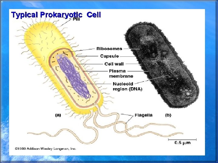
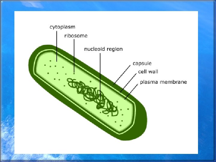
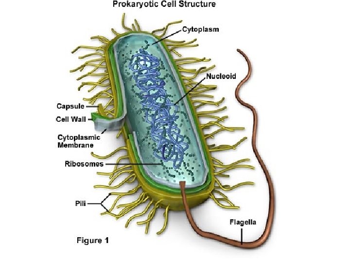
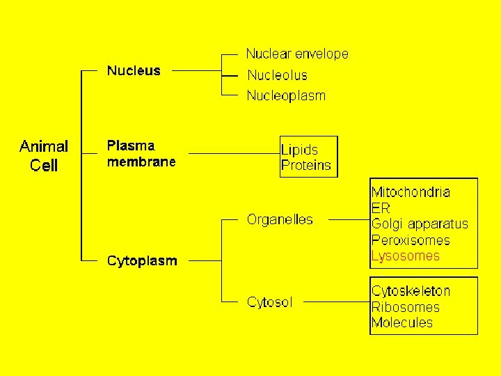
Prokaryotic Cells Bacteria are prokaryotic cells. All other known organisms consist of …. . Eukaryotic Cells
Prokaryotic Cells • Structurally simpler than eukaryotic cells • Nuclear material not enclosed in a membrane • Ribosomes smaller than Euk. • Lack of membrane bound organelles
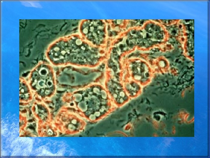
Cheek cells bacteria
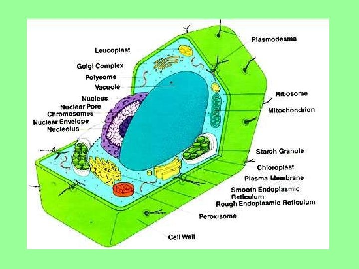
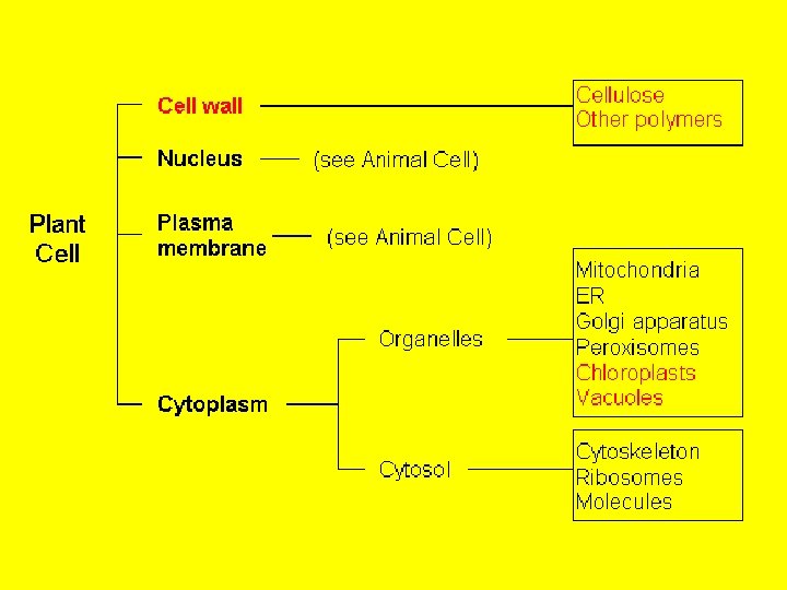
Eukaryotic Cells • • • Membrane bound organelles Cell Nucleus Ribosomes Endoplasmic reticulum Golgi complex Lysosomes Peroxisomes Vacuoles Mitochondria Chloroplasts
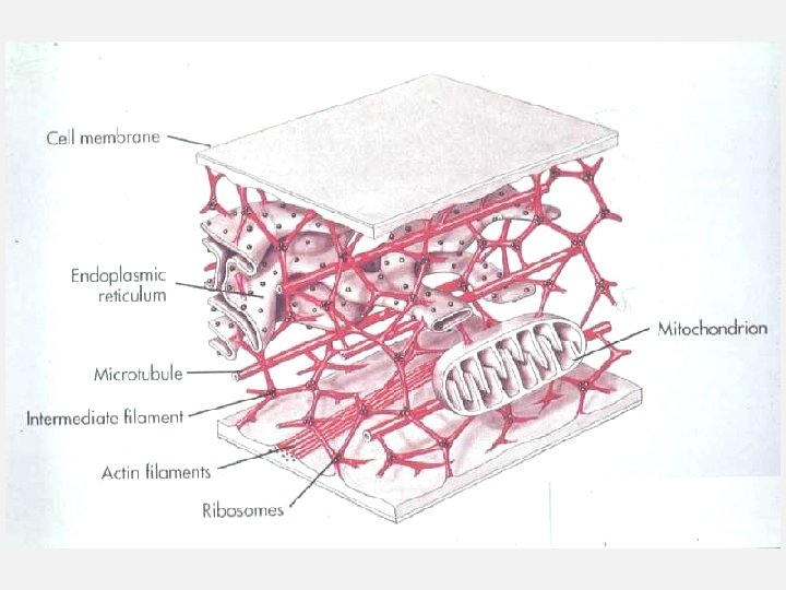
Membrane Bound Organelles The ‘stuff’ outside the nucleus and inside the cell membrane, suspended in cytoplasm
Membrane Bound Organelles Just to name a few ribosomes mitochondria Endoplasmic reticulum Vacuoles Peroxisomes Golgi complex Plastids Lysosomes
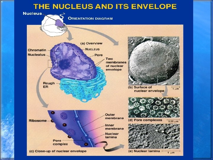
Cell Nucleus Contains nucleolus and chromosomes (DNA)
The Nucleus
Cell Nucleus • Typically in the center of the cell • Most cells have a single nucleus
Nuclear Envelope • Controls traffic between the nucleus and the cytoplasm • Pores in the nuclear membrane allow materials to pass in and out of the • nucleus
Nucleus – a closer look
Nuclear Envelope
EM View of Envelope
A closer look at the envelope
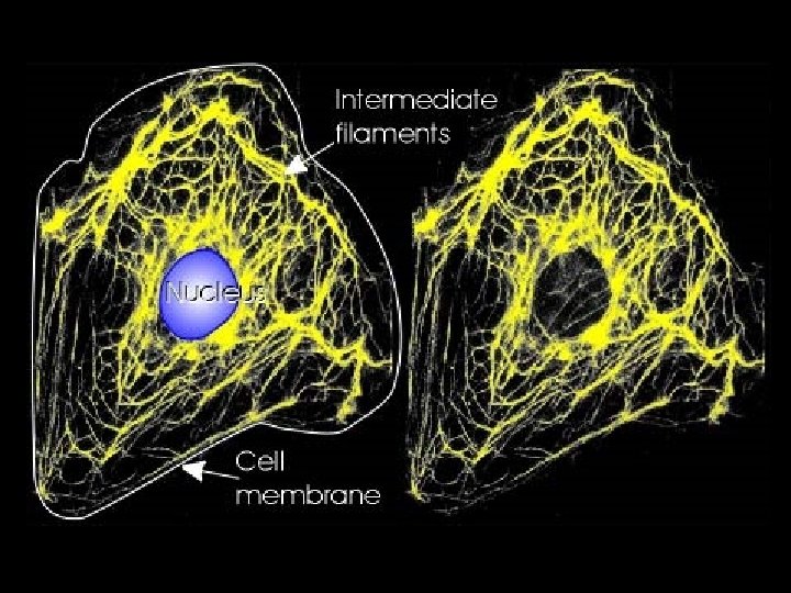
Nuclear Lamina • Inside the nucleus • Formed by intermediate filaments • Important in the timing of the disorganization of the membrane during cell division and the ensuing redevelopment
Lamina
Chromatin • When dividing, DNA takes the form of chromosomes • When not dividing, the DNA takes a looser form called chromatin
Loose Chromatin
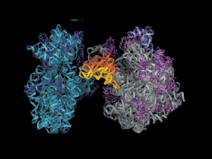
Ribosomal Subunits • Eukaryotic ribosomal subunits are assembled in the nucleolus • Ribosomes are composed of two subunits
Ribosomes • Ribosomes manufacture proteins • Ribosomes may be free or may be attached to the endoplasmic reticulum
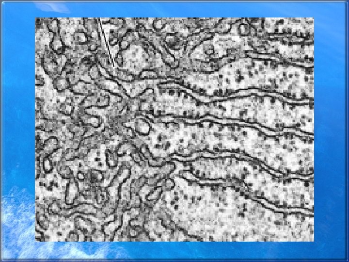
Endoplasmic Reticulum
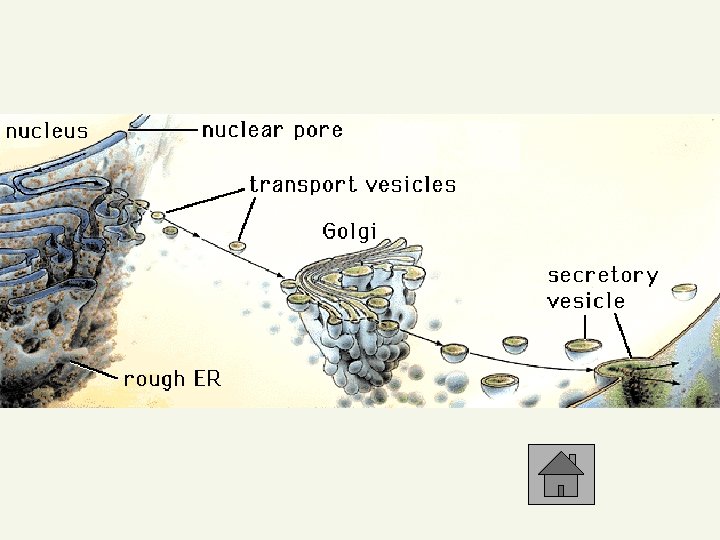
Endoplasmic Reticulum • Major manufacturing center- proteins • Extends from the nuclear membrane into the cytoplasm • Lumen- the space enclosed by the ER- typical intracellular membrane
The Cytosol side of the ER may be studded with ribosomes
Rough ER • Site of protein synthesis • Proteins formed may be transferred to other sites within the cell in transport vesicles
Transport vesicles
Smooth ER • Lacks ribosomes • Lipid production • Detoxifying chemical agent
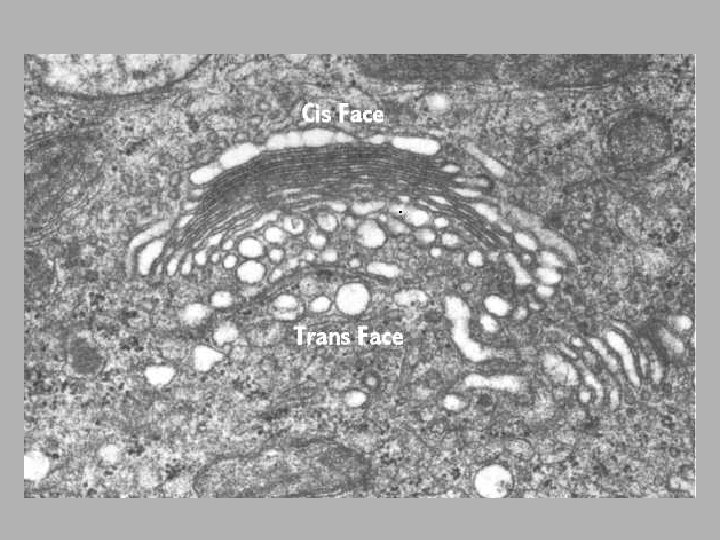
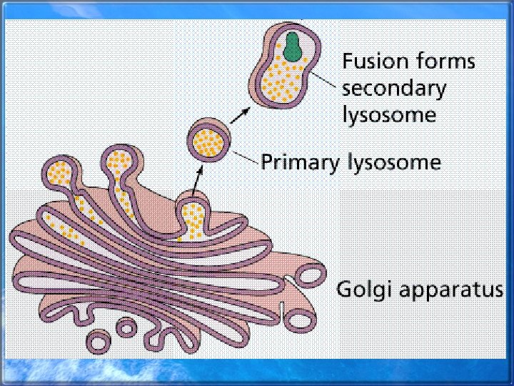
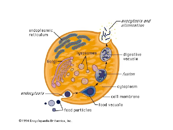
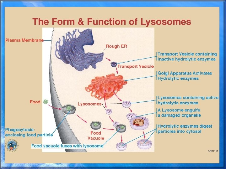
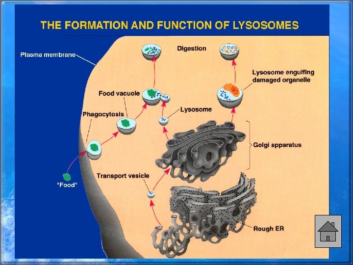
Golgi Complex • Cis face functions in receiving materials • The Trans face is directed toward the plasma membrane • Function: processing, sorting and modifying proteins • The process product is then passed to other organelles or to the plasma membrane • Manufactures lysosomes
Golgi Complex- Cis and Trans Face University of texas medical school
Convex shape
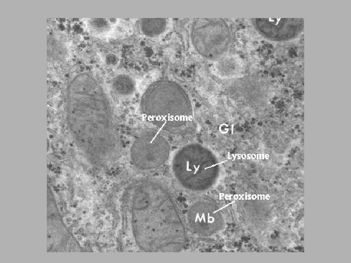
Lysosomes • Compartments for digestion • Small sacs filled hydrolytic enzymes • Primary lysosomes bud from the Golgi complex • Involved in apoptosis (programmed cell death) – Inappropriate apoptosis may be involved in many different catastrophic illnesses
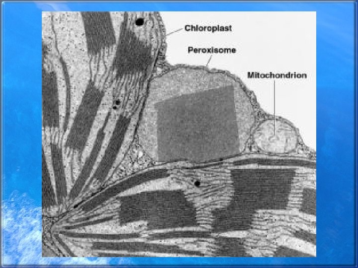
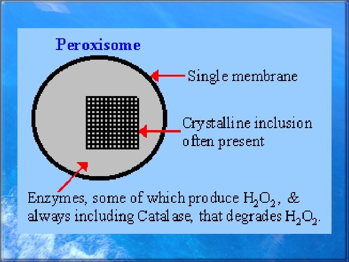
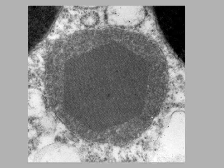
Peroxisomes • Metabolize small organic compounds • Transfer hydrogen from various compounds to oxygen, forming hydrogen peroxide • Catalase splits hydrogen peroxide rendering it harmless
Peroxisomes • Common in cells that synthesize, store, or degrade lipids • Plant cells have specialized peroxisomes called glyoxysomes
Peroxisome Functions: • metabolism of free oxygen radicals; • synthesis of cholesterol and ether lipids; • bile acid formation; • catabolism of long chain fatty acids; • catabolism of purines, prostaglandins, leucotriens; • alcohol detoxification in liver
Some interesting facts about peroxisomes are: • Human congenital diseases associated w/ absence of peroxisomes and/or dysfunction of their enzymes • many chemicals (drugs, industrial pollutants) induce a marked proliferation of peroxisomes; • prolonged Tx w/ most proliferators induce malignant hepatic tumors
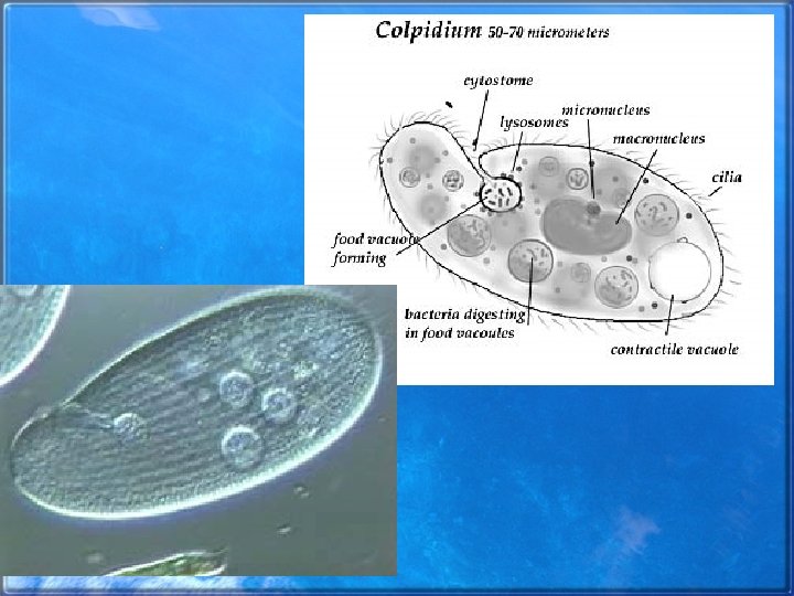
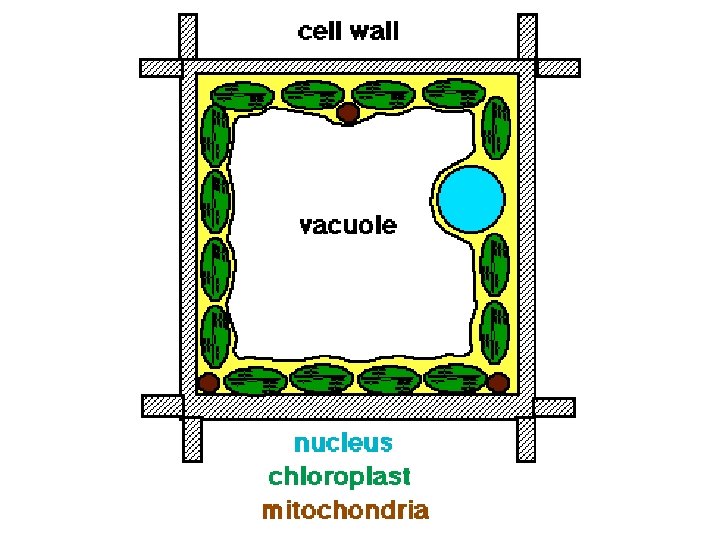
Vacuoles • Large, fluid filled sacs • Carry out variety of functions – In plants & fungi, vacuoles carry out many of the functions of the lysosome – Allow plants to increase in size • Bound by a membranous tonoplast • May store toxins or pigments • Protists have vacuoles that are involved in digestions and secretion
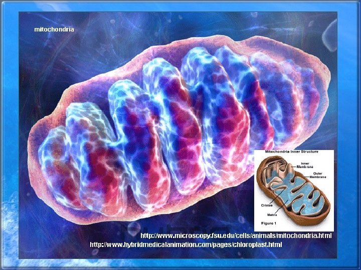
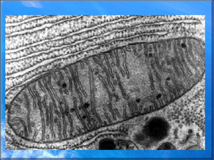
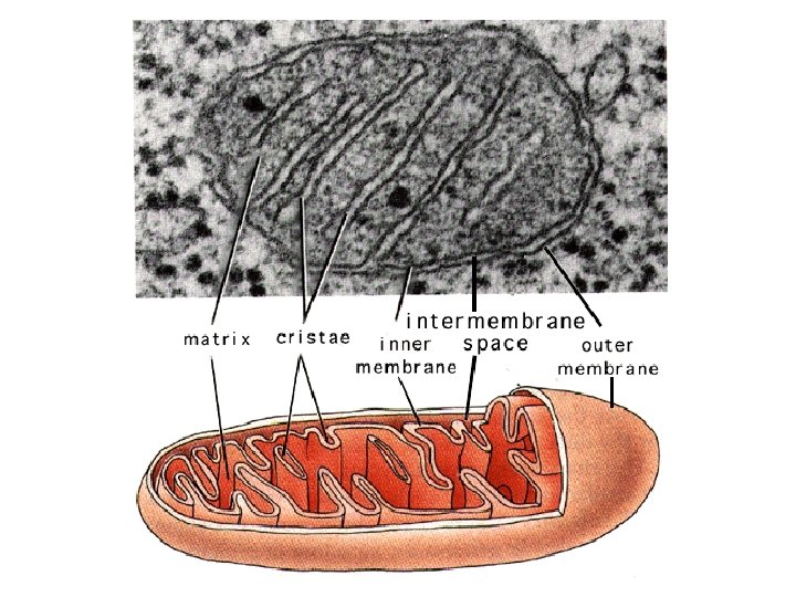
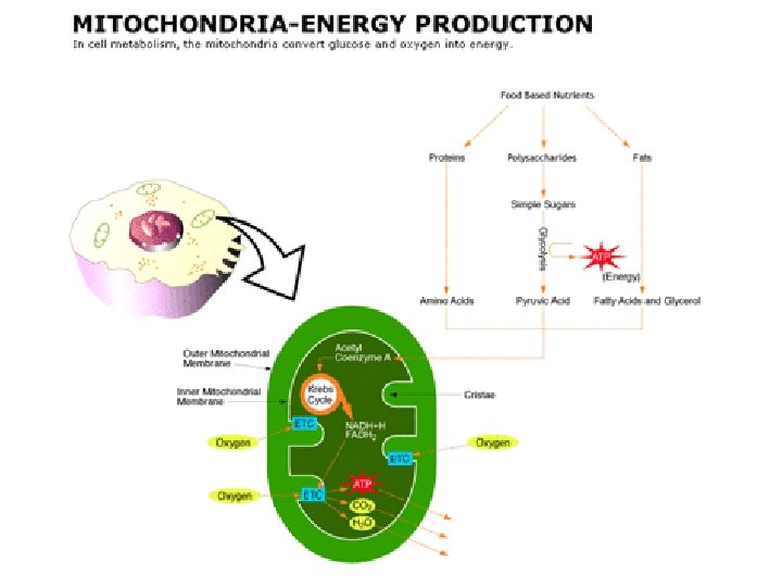
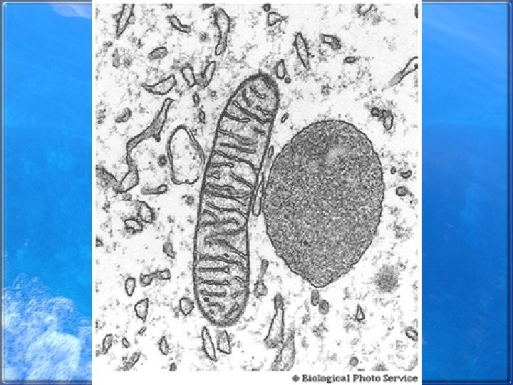
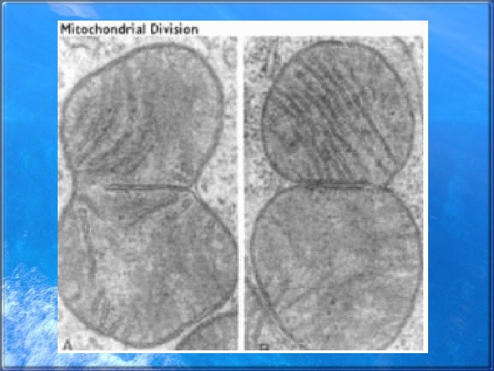
Mitochondria • Energy converting organelle- site of aerobic respiration • Double membrane bound • Matrix- inside of the inner membrane • Cristae- the foldings of the inner membrane, providing a large surface area • Mutations in mitochondrial DNA have been linked to several genetic diseases • Mitochondria also affect health by leaking electrons, which form free radicals, into the cell
Outer membrane lets many Molecules through, but inner membrane is very selective
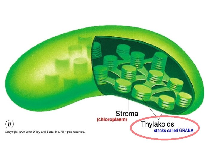
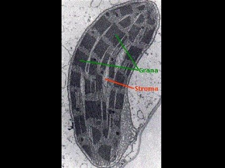
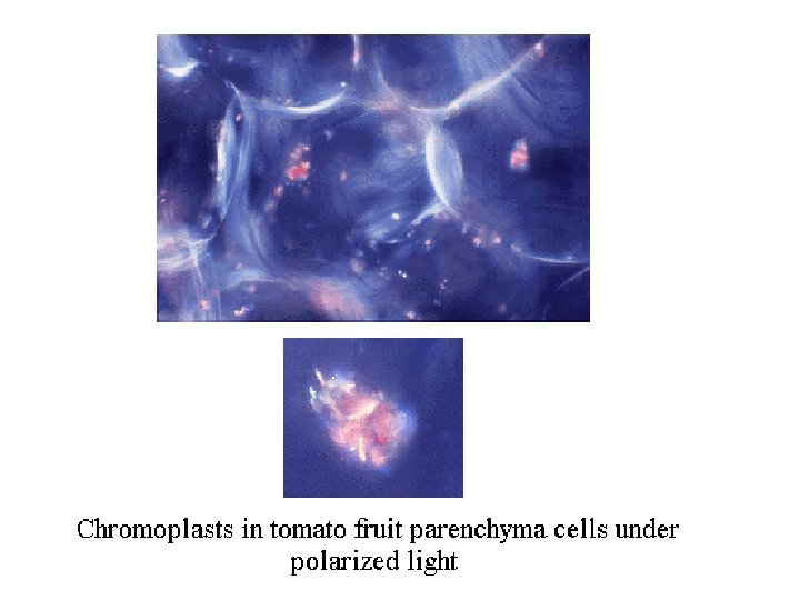
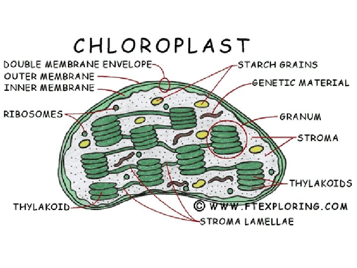
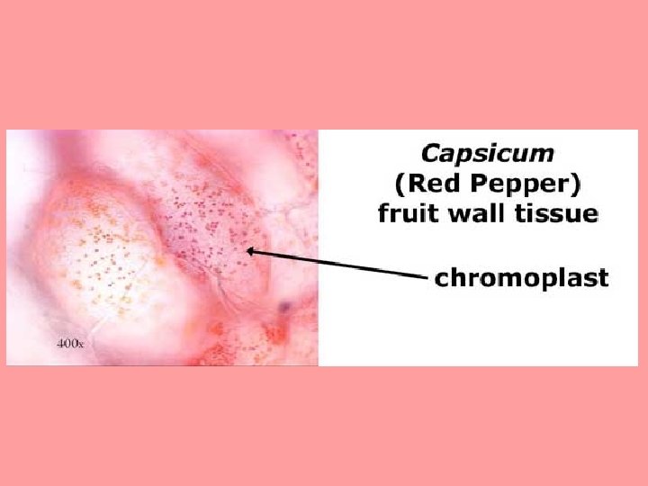
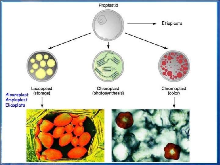
Chloroplasts • Convert light energy into chemical energy through photosynthesis • Pigments like chlorophylls are specialized for photosynthesis • Double membrane bound • Develop from proplastids • Chromoplasts contain pigments and are common in petals and ripe fruit • Leukoplasts lack pigments and may store starch
Fluid filled area Contains enzymes responsible for producing carbohydrates from carbon dioxide and water
Thylakoids are involved with producing ATP. This is where the chlorophyll is.
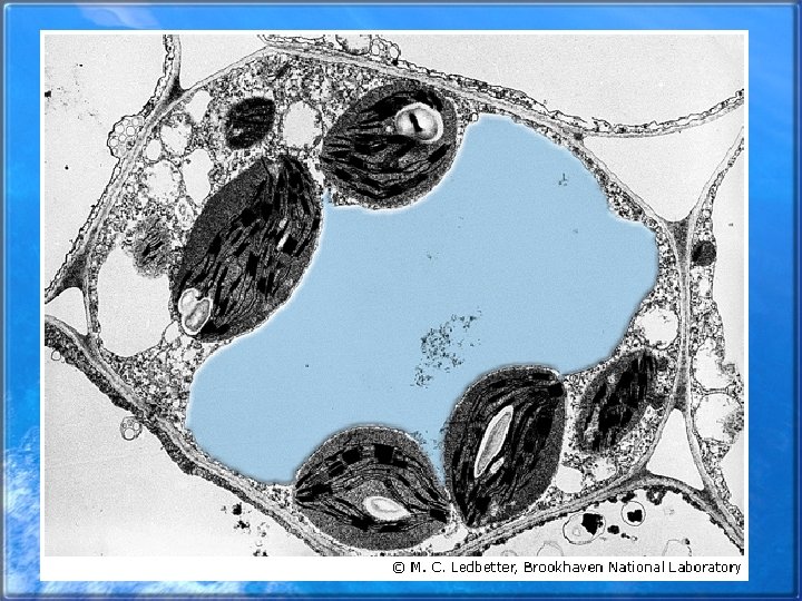
Would you recognize these structures as chloroplasts? How?
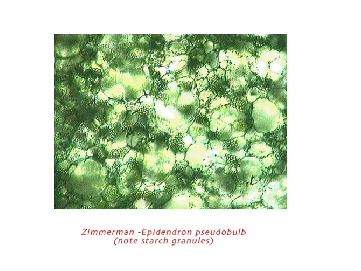
Proplastids that will turn into amyloplasts
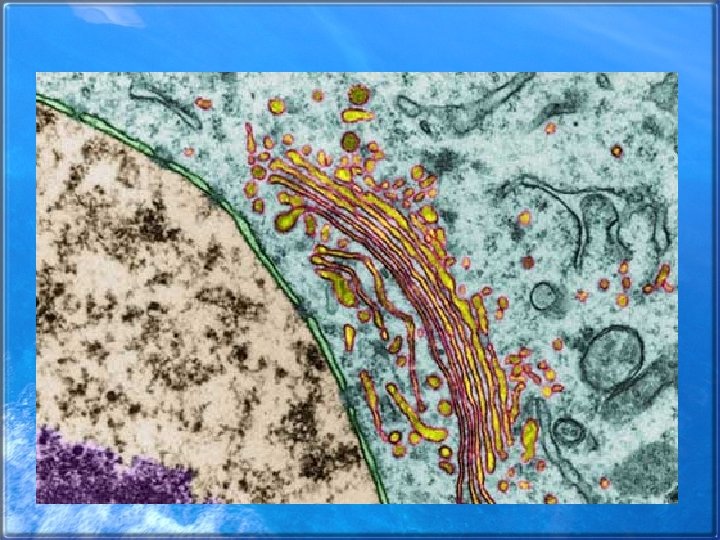
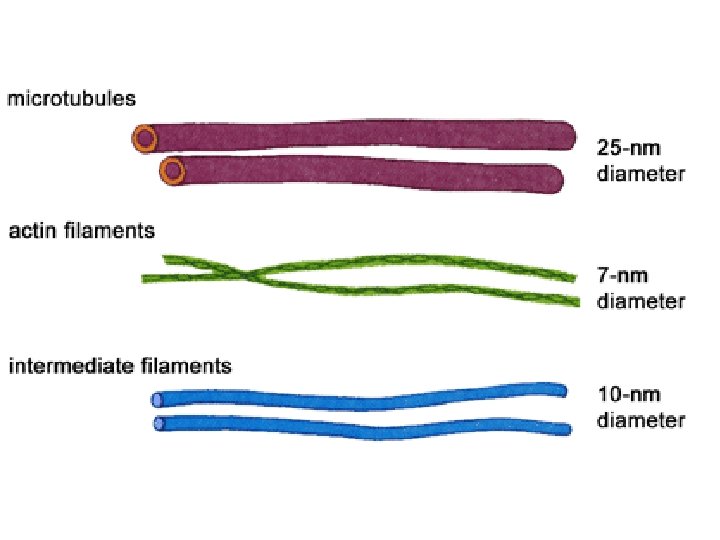
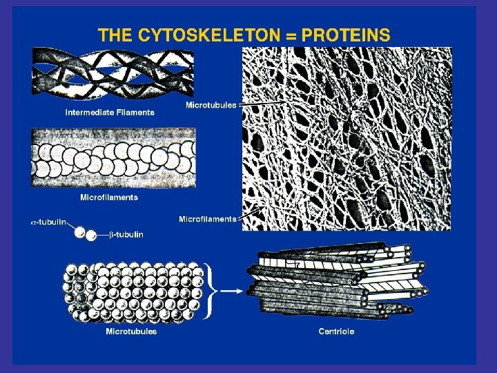
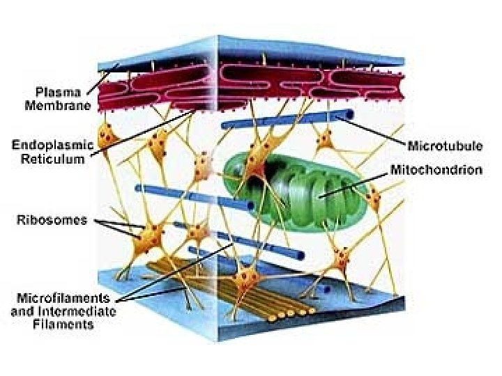
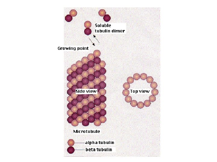
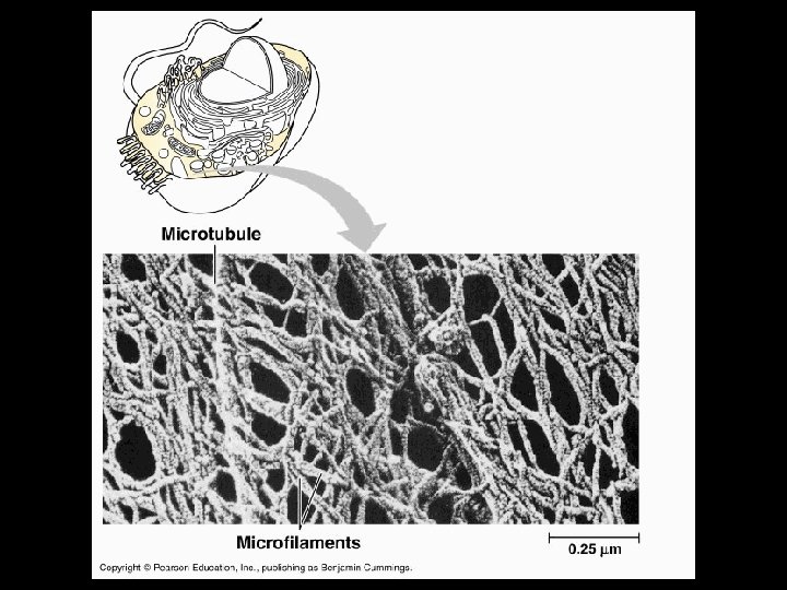
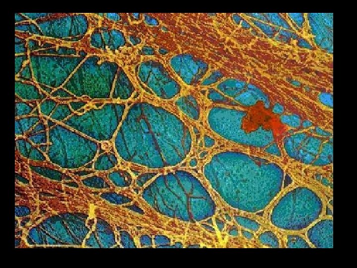
Cytoskeleton • Provides for cell shape and allows movement • Classifying elements of cytoskeleton by size – Microfilaments- smallest – Intermediate filaments – Microtubules- largest
Hollow cylinders
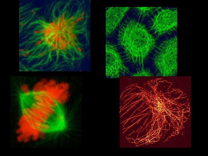
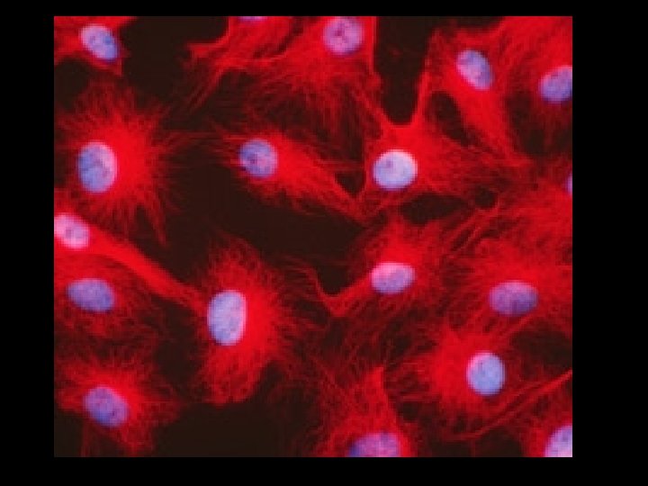
Microtubules are in green. Actin is in red. DNA is blue.
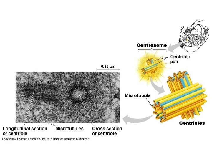
MTOC Microtubule Organizing Center- Centrosome
http: //www. cellsalive. com/mitosis. htm
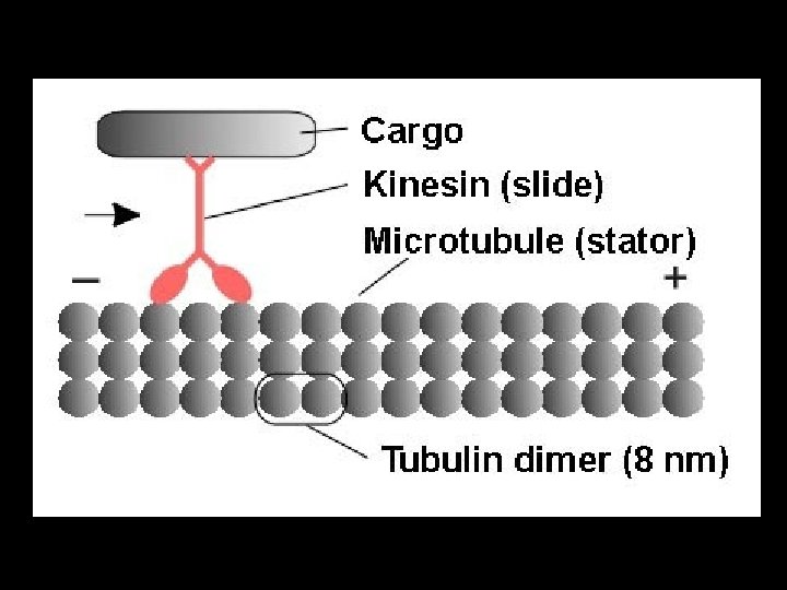
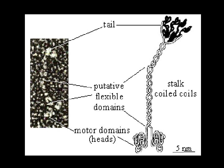
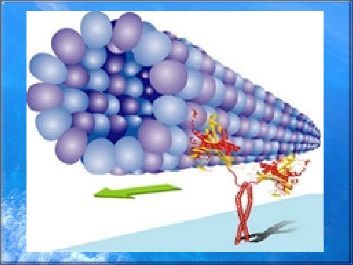
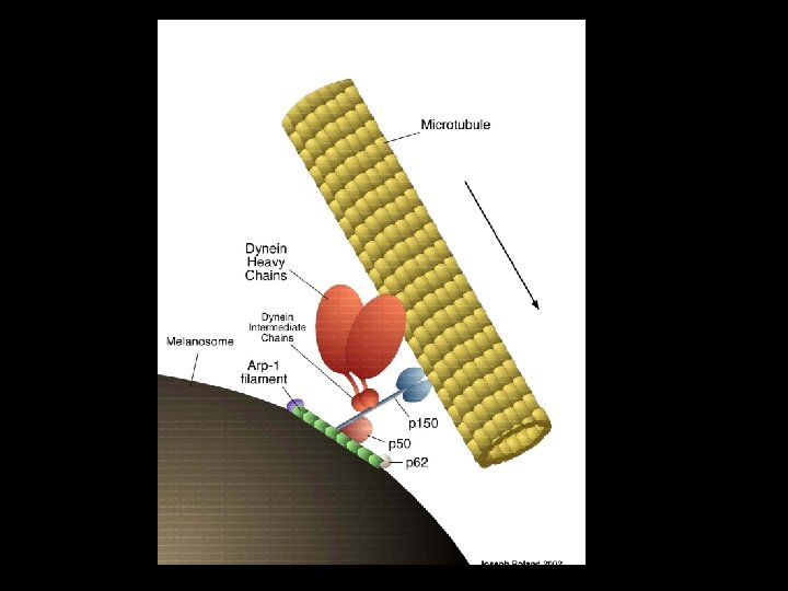
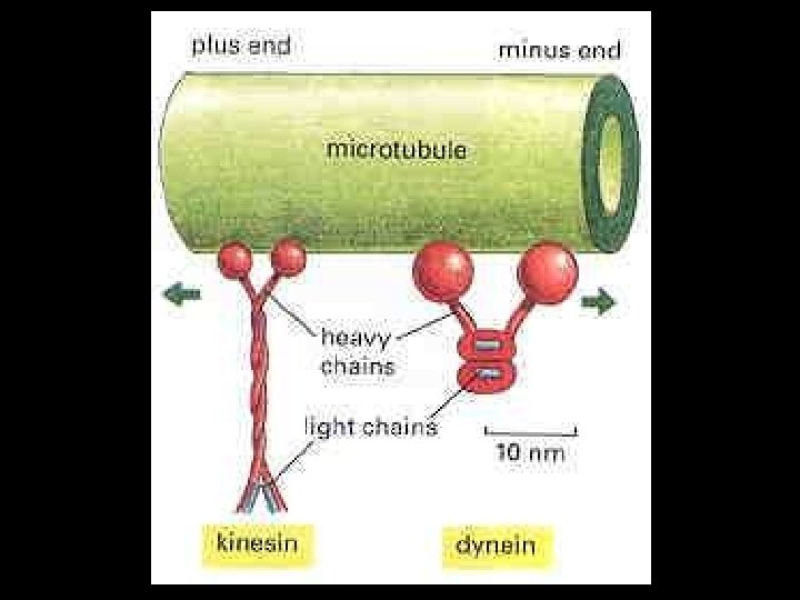
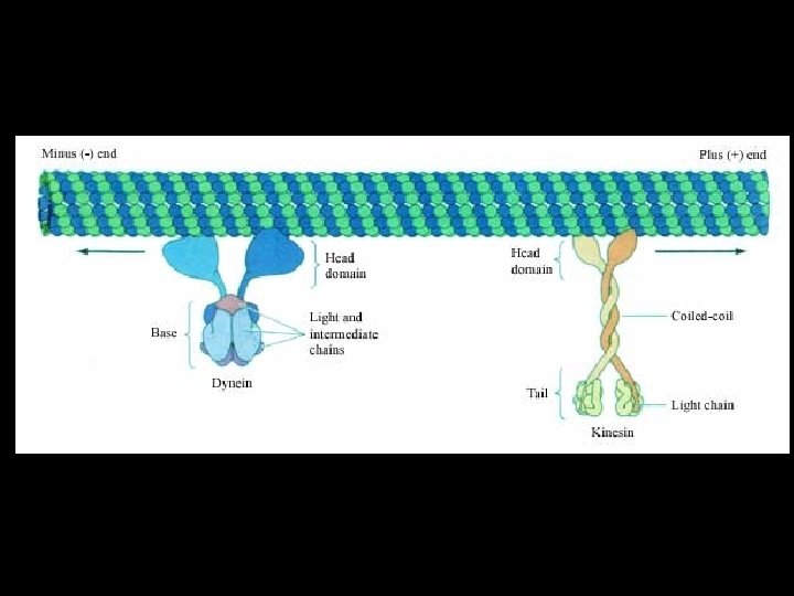
Dynein
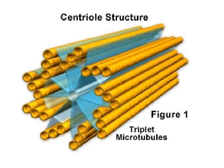
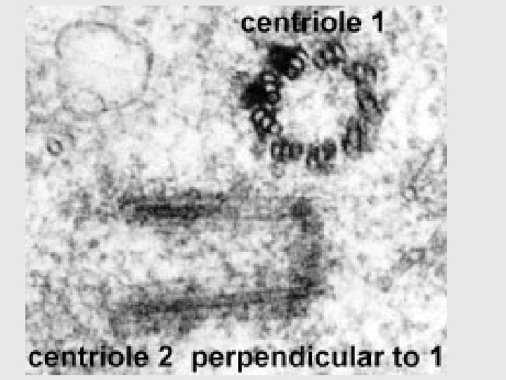
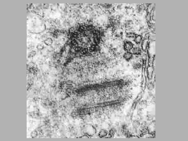
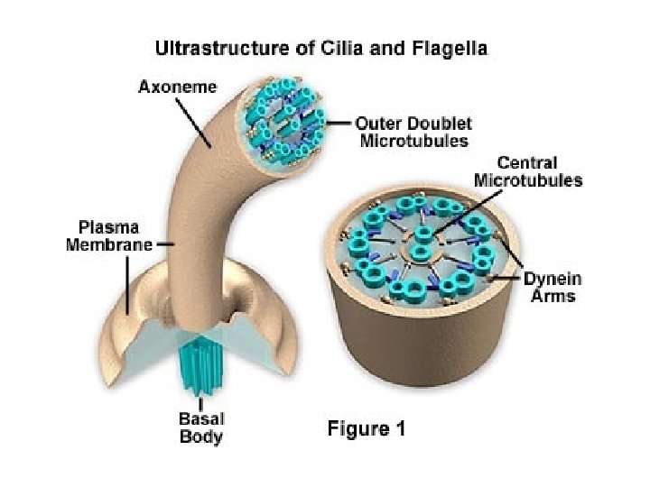
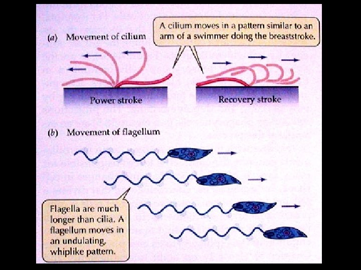
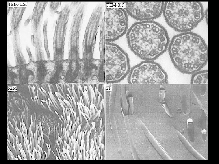
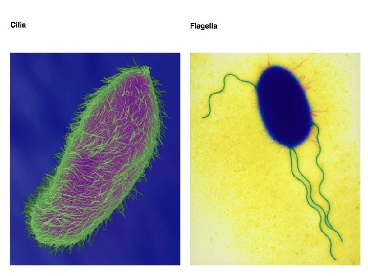
Cilia and Flagella • • Composed of microtubules Cilia- numerous and short Flagella- longer and fewer in number Move the cell or move substances over the surface of the cell • Both have 9+2 arrangement of microtubules • Basal body and centrioles have 9 X 3 arrangement
9 + 2 arrangement of cilia
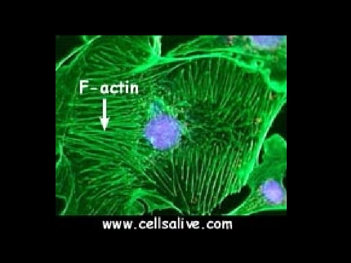
Microfilaments
microtubules
actin
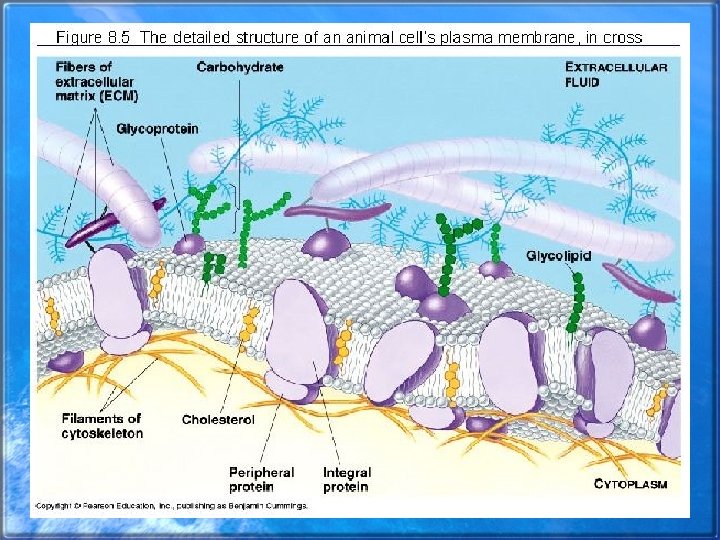
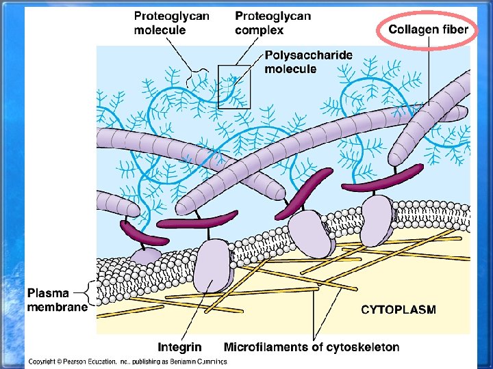
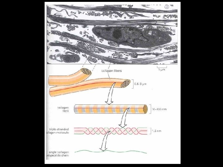
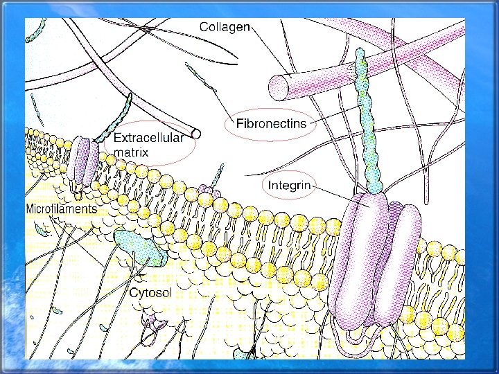
Extracellular Matrix • ECM • Secreted gel surrounding cell • Composed of collagen which forms very tough fibers • Integrins- main membrane receptors for the ECM • Help the cell signaling pathways and help regulate various functions of the cell
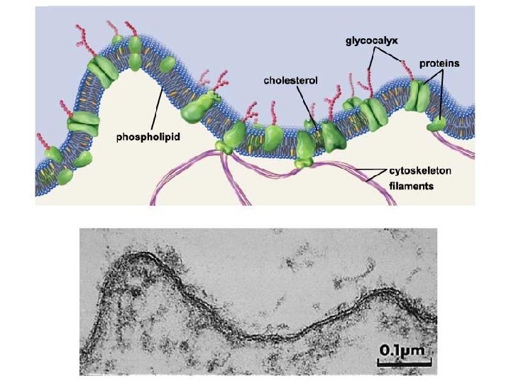
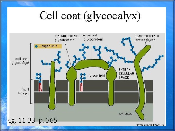
Glycocalyx- Cell Coat • Surround most eukaryotic cells • Formed by polysaccharide side chains • May act as recognition sites
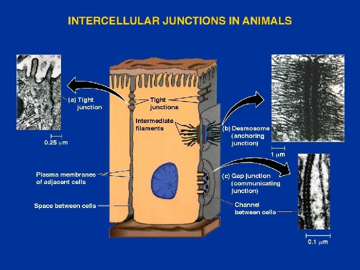
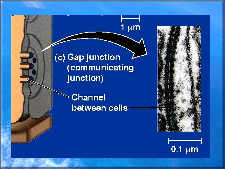
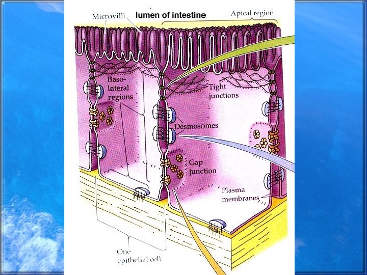
Junctions between Cells • • Form strong connections Prevent passage of materials Establish communication Animal cell junctions – Anchoring (desmosomes and adhering junctions – Tight junctions – Gap junctions • Plant Cell junction – plasmodesmata
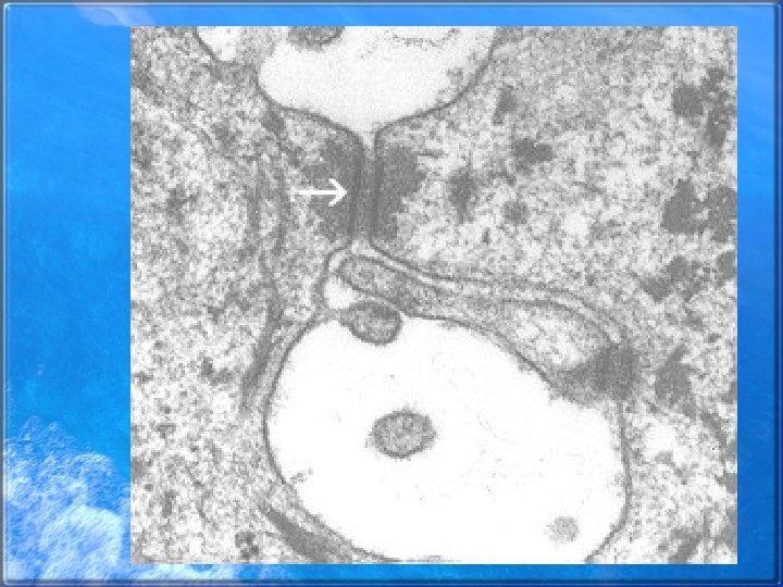
Desmosomes • Points of attachment for some animal cells • Hold cells subject to mechanical stress together • Composed of intermediate filaments, which span the gap between two cells
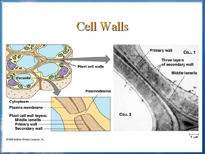
Cell Walls • Surround plant, fungal, and bacterial cells • Primary cell wall can expand as the cell grows • Secondary cell wall forms between the primary cell wall and the cell membrane • Middle lamella glues adjacent plant cells together
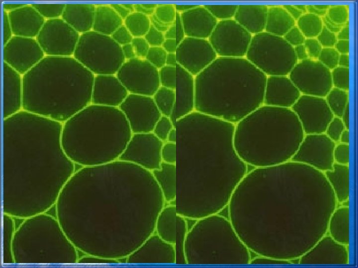
Middle Lamella