Organic Molecules Proteins Proteins Most structurally functionally diverse


")


 of one")


 structure • Order of amino acids in chain – amino acid sequence")


 structure • “Local folding” – folding along short sections of polypeptide –")
 structure • “Whole molecule folding” – interactions between distant amino acids •")
 structure • More than one polypeptide chain bonded together – only then")
 R groups hydrophobic interactions disulfide bridges (H & ionic bonds) 3°")


- Slides: 26
Organic Molecules: Proteins
Proteins • Most structurally & functionally diverse group • Function: involved in almost everything – enzymes (pepsin, DNA polymerase) – structure (keratin, collagen) – carriers & transport (hemoglobin, aquaporin) – cell communication • signals (insulin & other hormones) • receptors – defense (antibodies) – movement (actin & myosin) – storage (bean seed proteins)
Proteins • Structure H 2 O – monomer = amino acids • 20 different amino acids – polymer = polypeptide • protein can be one or more polypeptide chains folded & bonded together • large & complex molecules • complex 3 -D shape hemoglobin Rubisco growth hormones
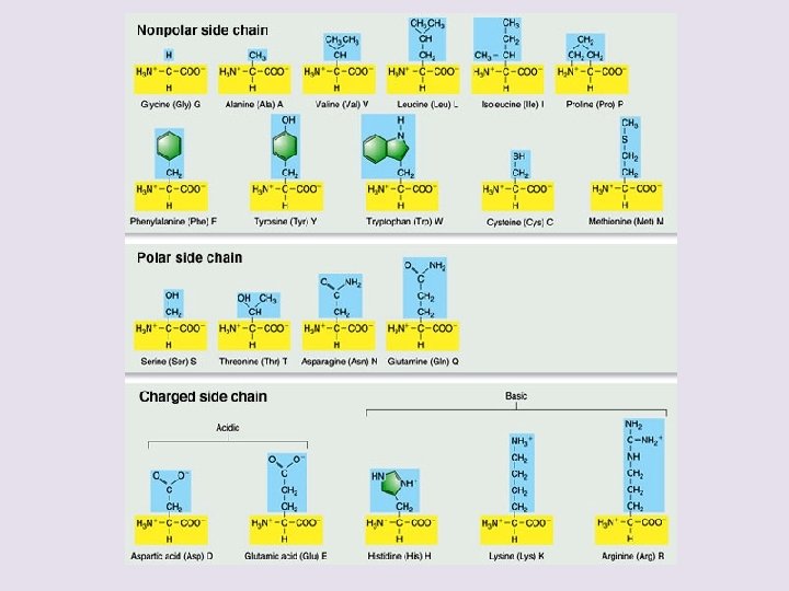
Amino acids • Structure – central carbon – amino group – carboxyl group (acid) – R group (side chain) • variable group • different for each amino acid • confers unique chemical properties to each amino acid - like 20 different letters of an alphabet - can make many words (proteins) Oh, I get it! amino = NH 2 acid = COOH
Effect of different R groups: Polar amino acids polar or charged & hydrophilic H+ donors H+ acceptors Why are these polar & hydrophillic?
Effect of different R groups: Nonpolar amino acids nonpolar & hydrophobic Why are these nonpolar & hydrophobic?
Sulfur containing amino acids • Form disulfide bridges S-S – covalent cross links betweens sulfhydryls – stabilizes 3 -D structure H-S – S-H
Building proteins • Peptide bonds – covalent bond between NH 2 (amine) of one amino acid & COOH (carboxyl) of another – C–N bond dehydration synthesis AP Biology H 2 O peptide bond
Building proteins • Polypeptide chains have direction – N-terminus = NH 2 end – C-terminus = COOH end – repeated sequence (N-C-C) is the polypeptide backbone • can only grow in one direction http: //www 2. nl. edu/jste/proteins. htm
Protein structure & function • Function depends on structure – 3 -D structure • twisted, folded, coiled into unique shape pepsin hemoglobin collagen
Protein Structure • Protein types include globular proteins which are usually enzymes and Fibrous proteins which usually serve for structure (eg. Hair) • Proteins Exhibit 4 levels of structure.
Primary (1°) structure • Order of amino acids in chain – amino acid sequence determined by gene (DNA)l; dictates all further levels of protein structure – slight change in amino acid sequence can affect protein’s structure & its function • even just one amino acid change can make all the difference!
Just 1 out of 146 amino acids! Sickle cell anemia I’m hydrophilic! But I’m hydrophobic!
Primary structure Normal hemoglobin Primary Val His Leu Thr Pro Glul Glu. . . structure 1 2 3 4 5 6 7 Secondary and tertiary structures Quaternary structure Function Red blood cell shape Figure 5. 21 Sickle-cell hemoglobin Val His Leu Molecules do not associate with one another, each carries oxygen. Normal cells are full of individual hemoglobin molecules, each carrying oxygen Quaternary structure Pro Val Glu 1 2 3 4 5 6 7 Secondary subunit and tertiary structures Hemoglobin A Thr 10 m Red blood cell shape Exposed hydrophobic region subunit Function 10 m . . . Hemoglobin S Molecules interact with one another to crystallize into a fiber, capacity to carry oxygen is greatly reduced. Fibers of abnormal hemoglobin deform cell into sickle shape. 15
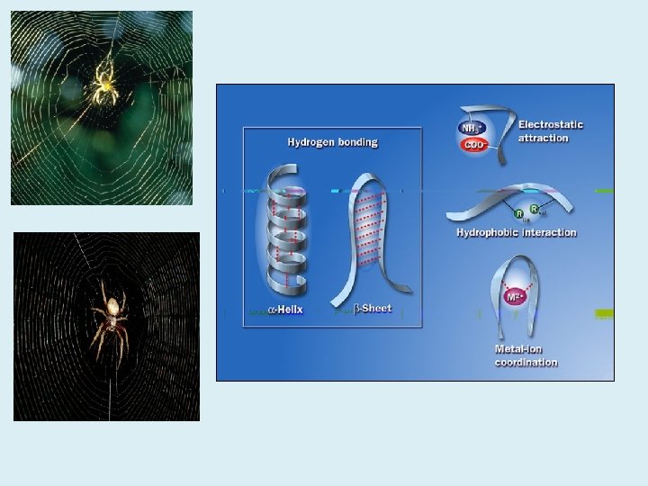
Secondary (2°) structure • “Local folding” – folding along short sections of polypeptide – interactions between adjacent amino acids – H bond: weak bonds between R groups – forms sections of 3 -D structure • -helix • -pleated sheet
Tertiary (3°) structure • “Whole molecule folding” – interactions between distant amino acids • hydrophobic interactions – cytoplasm is water-based – nonpolar amino acids cluster away from water • Covalent, H bonds & ionic bonds • disulfide bridges – covalent bonds between sulfurs in sulfhydryls (S–H) – anchors 3 -D shape
Quaternary (4°) structure • More than one polypeptide chain bonded together – only then does polypeptide become functional protein • p. H, changes or heat can disrupth bonds perm. denaturing the protein collagen = skin & tendons hemoglobin
Protein structure (review) R groups hydrophobic interactions disulfide bridges (H & ionic bonds) 3° multiple polypeptides hydrophobic interactions 1° amino acid sequence peptide bonds determined by DNA 4° 2° R groups H bonds
Protein denaturation • Unfolding a protein – conditions that disrupt H bonds, ionic bonds, disulfide bridges • temperature • p. H • salinity – alter 2° & 3° structure • alter 3 -D shape – destroys functionality • some proteins can return to their functional shape after denaturation, many cannot http: //highered. mcgraw-hill. com/sites/0072943696/student_view 0/chapter 2/animation__protein_denaturation. html
• Chaperonins – Are protein molecules that assist in the proper folding of other proteins Polypeptide Cap Correctly folded protein Hollow cylinder Chaperonin (fully assembled) Figure 5. 23 Steps of Chaperonin Action: 1 An unfolded polypeptide enters the cylinder from one end. 2 The cap attaches, causing the cylinder to change shape in such a way that it creates a hydrophilic environment for the folding of the polypeptide. 3 The cap comes off, and the properly folded protein is released. 23
Review Questions
A. What happens when a protein denatures? * 1. 2. 3. 4. 5. It loses its primary structure. It loses its secondary and tertiary structure. It becomes irreversibly insoluble and precipitates. It hydrolyzes into component amino acids. Its hydrogen bonds, ionic bonds, and peptide bonds are disrupted.
B. The R group or side chain of the amino acid serine is – CH 2 –OH. The R group or side chain of the amino acid alanine is –CH 3. Where would you expect to find these amino acids in globular protein in aqueous solution? 1. Serine would be in the interior, and alanine would be on the exterior of the globular protein. 2. Alanine would be in the interior, and serine would be on the exterior of the globular protein. 3. Both serine and alanine would be in the interior of the globular protein. 4. Both serine and alanine would be on the exterior of the globular protein. 5. Both serine and alanine would be in the interior and on the exterior of the globular protein.